

Effect of surgical intraocular pressure lowering on retinal structures - nerve fibre layer, foveal avascular zone, peripapillary and macular vessel density: 1 year results
- PMID: 31409906
- PMCID: PMC7042361
- DOI: 10.1038/s41433-019-0560-6
Effect of surgical intraocular pressure lowering on retinal structures - nerve fibre layer, foveal avascular zone, peripapillary and macular vessel density: 1 year results
Abstract
Objectives: To determine the effect of surgical intraocular pressure (IOP) lowering on peripapillary retinal nerve fibre layer thickness (RNFL), fovea avascular zone (FAZ), peripapillary and macular vessel density (VD) in glaucoma using with optical coherence tomography angiography (OCT-A).
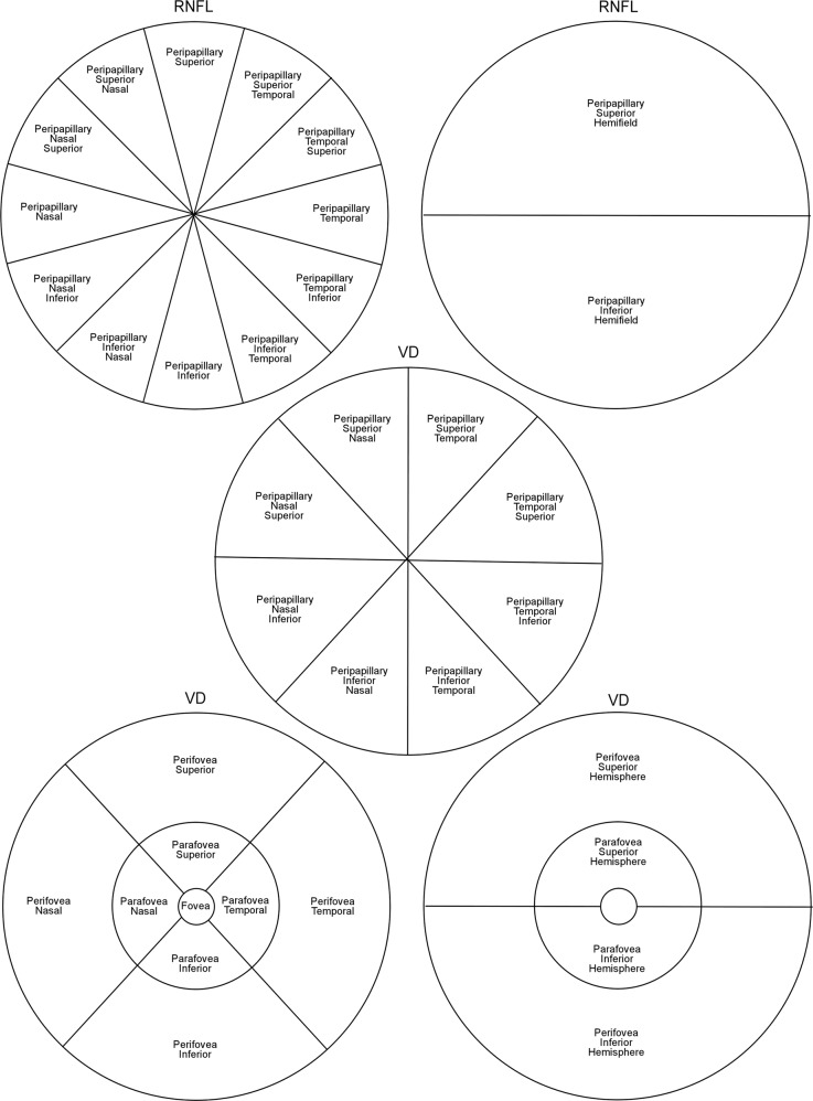
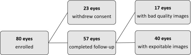
Methods: This was a prospective observational study performed at the Glaucoma Research Centre, Montchoisi Clinic, Lausanne. In total 40 eyes with open-angle glaucoma were included. OCT-A scans were performed before glaucoma surgery, and at 1-month, 3-month, 6-month, and 12-month post-operatively. AngioVue AngioAnalytic (Optovue Inc, Fremont, CA) software was used to analyse the RNFL, FAZ, peripapillary and macular VD. Changes were analysed using analysis of variance (ANOVA) models.
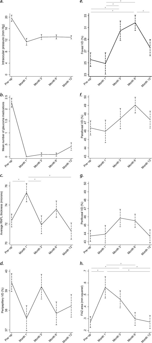
Results: Mean IOP dropped from 19.4 (±7.0) mmHg pre-surgery and stabilized at 13.0 (±3.1) mmHg at 12 months (p < 0.001). The number of topical medications reduced from 2.2 (±1.2) preoperatively to 0.4 (±0.8) at 1 year (p < 0.001). Peripapillary RNFL thickness was transiently increased at 1-month postoperatively (p = 0.03). Peripapillary VD fluctuated throughout the follow-up. Foveal VD showed increased perfusion at 3 and 6 months post-operatively with minimal changes at 1 month (p < 0.01). Glaucoma surgery had a significant effect initially on FAZ area (p = 0.03), FAZ perimeter (p = 0.02) and Acircularity Index (AI) (p = 0.04). By 12-months FAZ measurements had reversed to baseline values.
Conclusions: Peripapillary and macular microcirculations responded differently to surgically induced IOP reduction. Peripapillary microcirculation was IOP-independent within the studied range of surgically-controlled IOP. Macular microcirculation, however, exhibited a "delayed response" followed by near-normal reperfusion after glaucoma surgery. FAZ parameters could be potentially useful modalities to assess vascular reperfusion after glaucoma surgery as, amongst all studied parameters, the area was the most sensitive to surgically induced IOP changes.
Conflict of interest statement
The authors declare that they have no conflict of interest.
Figures
Comment in
-
The choice of analysis of variance models in repeated measurements analysis-on the effect of glaucoma surgery on retinal structures.Eye (Lond). 2020 Sep;34(9):1711. doi: 10.1038/s41433-019-0675-9. Epub 2019 Nov 13. Eye (Lond). 2020. PMID: 31723244 Free PMC article. No abstract available.
-
Effect of surgical intraocular pressure lowering on retinal structures-nerve fibre layer, foveal avascular zone, peripapillary and macular vessel density: 1 year results.Eye (Lond). 2020 Sep;34(9):1710. doi: 10.1038/s41433-019-0674-x. Epub 2019 Nov 13. Eye (Lond). 2020. PMID: 31723246 Free PMC article. No abstract available.
References
-
- Gordon MO, Beiser JA, Brandt JD, et al. The Ocular Hypertension Treatment Study: baseline factors that predict the onset of primary open-angle glaucoma. Arch Ophthalmol. 2002;120:714–20. - PubMed
-
- Kass MA, Heuer DK, Higginbotham EJ, et al. The Ocular Hypertension Treatment Study: a randomized trial determines that topical ocular hypotensive medication delays or prevents the onset of primary open-angle glaucoma. Arch Ophthalmol. 2002;120:701–13. - PubMed
-
- The AGIS Investigators. The Advanced Glaucoma Intervention Study (AGIS): 7. The relationship between control of intraocular pressure and visual field deterioration. Am J Ophthalmol. 2000;130:429–40. - PubMed
-
- Leske MC, Heijl A, Hussein M, et al. Factors for glaucoma progression and the effect of treatment: the early manifest glaucoma trial. Arch Ophthalmol. 2003;121:48–56. - PubMed
-
- Sommer A, Katz J, Quigley HA, et al. Clinically detectable nerve fiber atrophy precedes the onset of glaucomatous field loss. Arch Ophthalmol. 1991;109:77–83. - PubMed
Publication types
MeSH terms
LinkOut - more resources
Full Text Sources
