. 2019 Jul 15:13:1235-1246.
doi: 10.2147/OPTH.S203917. eCollection 2019.

360° ab-interno Schlemm's canal viscodilation in primary open-angle glaucoma

Affiliations

360° ab-interno Schlemm's canal viscodilation in primary open-angle glaucoma

Simon Ondrejka et al. Clin Ophthalmol. .

Abstract

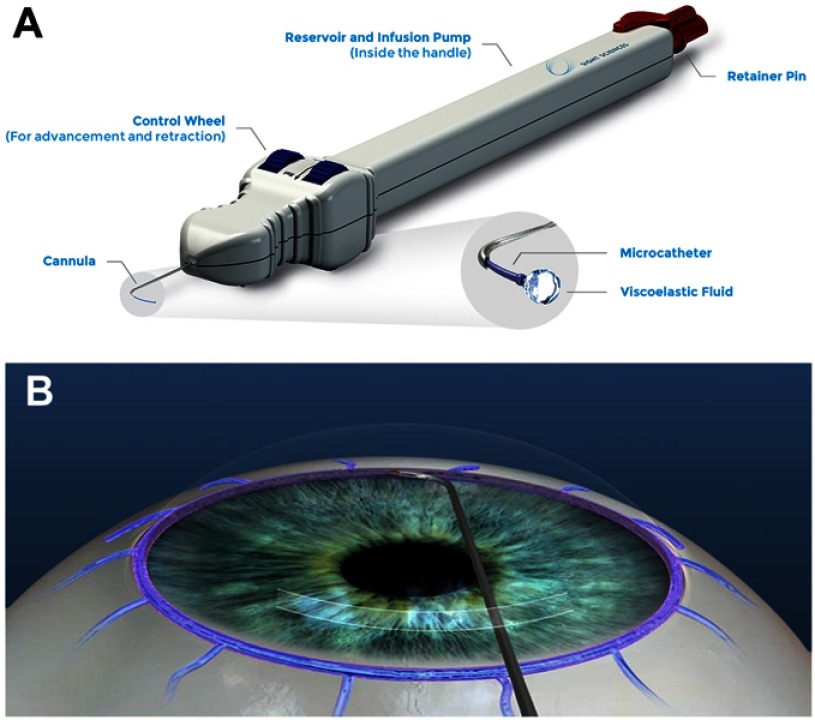
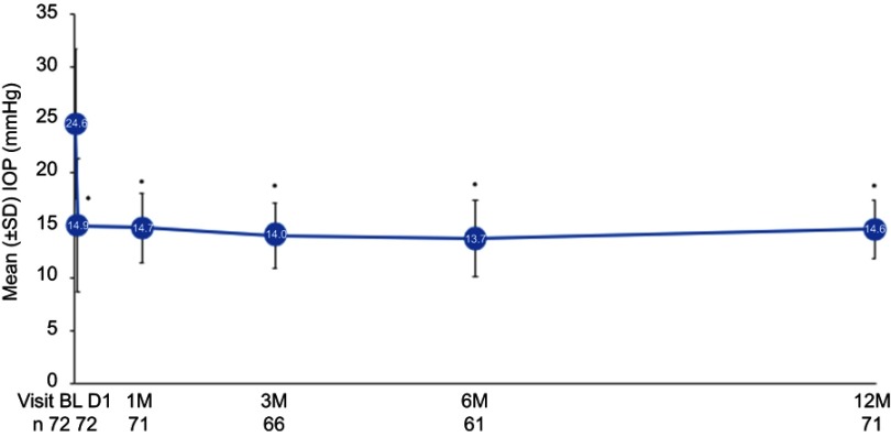
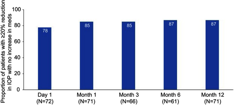
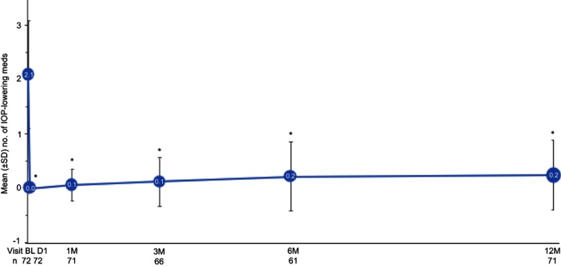
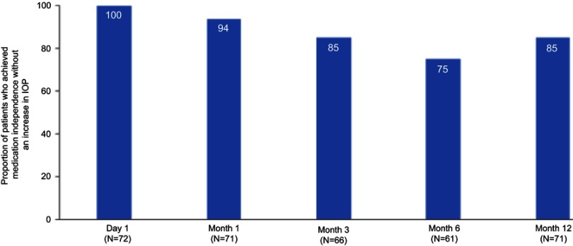
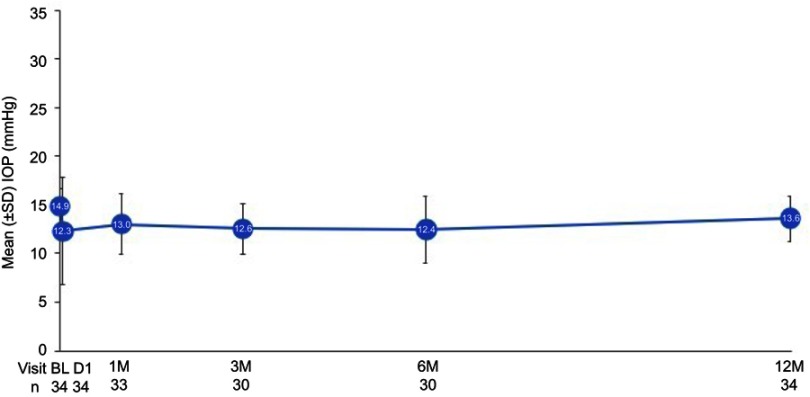
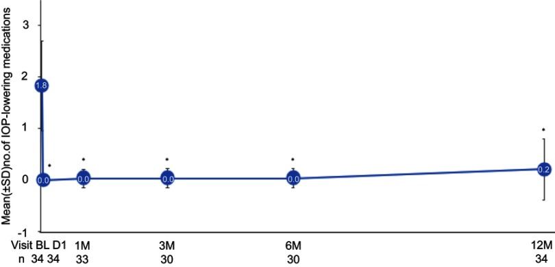
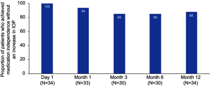
Purpose: To evaluate the safety and effectiveness of ab-interno microcatheterization and 360° viscodilation of Schlemm's canal (SC) using the VISCO360® Viscosurgical System in treatment of primary open angle glaucoma (POAG). Setting: Surgical center (Augencentrum Köln, Köln, Germany). Design: Retrospective analysis of 106 eyes from 71 consecutive patients. Methods: Ab-interno canal viscodilation (VISCO360®) with or without cataract extraction was performed in two groups of patients with mild-moderate POAG: Group 1 had a baseline intraocular pressure (IOP) ≥18 mmHg (n=72 eyes) and Group 2 had a baseline IOP <18 mmHg (n=34 eyes). IOP without washout was measured and number of IOP-lowering medications were documented at all visits. Effectiveness was determined by reduction in IOP and reduction in the number of IOP-lowering medications at 12±3 months from baseline. Safety was determined by the rate of adverse events (AEs) and secondary surgical interventions (SSI). Results: In Group 1, all eyes available at 12±3 months (n=72), had a 41.0% reduction in mean IOP (from 24.6±7.1 mmHg to 14.6±2.8 mmHg), 87% (n=62) of which showed an IOP reduction of ≥20% with no increase in IOP-lowering medications. In Group 2, all eyes (n=34) maintained their baseline IOP at all postoperative visits. In both groups, a significant decrease (>89%) in mean number of IOP-lowering medications was seen at 12 months with 86% of eyes completely off medication with no increase in IOP. The most common AE seen was hyphema (13%) and no eye required SSI during the study period. Conclusion: Ab-interno SC viscodilation (VISCO360) is safe and effective in lowering IOP and reducing hypotensive medications in patients with OAG.

Keywords: 360-degree trabeculotomy; POAG; VISCO360®; blebless, MIGS; canaloplasty.


Conflict of interest statement

The authors have no commercial or proprietary interest in any of the materials discussed in this article. The authors report no conflicts of interest in this work.

Figures

Figure 1
Figure 1
VISCO360® Viscosurgical System. (A) Illustration of the device showing a handpiece with a microcatheter, control wheel for advancing and retracting the microcatheter, viscoelastic reservoir/infusion pump, and a locking mechanism. (B) Schematic of circumferential viscodilation of Schlemm’s canal (SC) through clear corneal incision. VISCO360® cannula tip creates an opening into SC. A controlled volume of viscoelastic fluid is delivered as the microcatheter is retracted back. VISCO360 Viscosurgical System. Note: Images are sourced from Sight Sciences, http://sightsciences.com and used with permission.
Figure 2
Figure 2
Ab-interno 360° microcatheterization and viscodilation of Schlemm’s canal (SC) using the VISCO360® in a human eye. VISCO360® cannula (indicated by star) was positioned at the desired location in anterior chamber angle through clear corneal incision (A) and an opening into SC was created with the cannula tip (B), the finger wheel was used to gently advance the microcatheter (indicated by arrow) out of the cannula and into SC (C). As the microcatheter was retracted back out of SC and into the cannula, a controlled volume of viscoelastic fluid was delivered thereby dilating SC along its entire length (D).
Figure 3
Figure 3
Mean intraocular pressure (IOP) over time in Group 1. Effectiveness seen as a significant drop in mean IOP at postoperative Day 1 which remained similar across all postoperative visits until Month 12. *Statistically significant reduction (P<0.001). Error bars, SD. Abbreviations: BL, baseline; D, day; M, month; n, number of eyes available for analysis.
Figure 4
Figure 4
Reduction in intraocular pressure (IOP) by ≥20% with same or fewer medications in Group 1. The figure shows the proportion of eyes meeting the outcome measure with no increase in IOP-lowering medications over time. Abbreviation: N, number of eyes available for analysis.
Figure 5
Figure 5
Mean number of intraocular pressure (IOP)-lowering medications over time in Group 1. *Statistically significant reduction (P<0.001). Error bars, SD. Abbreviations: BL, baseline; D, day; M, month; n, number of eyes available for analysis.
Figure 6
Figure 6
Proportion of patients who achieved medication independence without an increase in intraocular pressure (IOP) as compared to baseline in Group 1. Abbreviation: N, number of eyes available for analysis.
Figure 7
Figure 7
Mean intraocular pressure (IOP) over time in Group 2. Effectiveness seen as no change in mean IOP across all postoperative visits up to Month 12 as compared to baseline (P>0.05). Error bars, SD. Abbreviations: BL, baseline; D, day; M, month; n, number of eyes available for analysis.
Figure 8
Figure 8
Mean number of intraocular pressure (IOP)-lowering medications over time in Group 2. *Statistically significant reduction compared to baseline (P<0.001). Error bars, SD. Abbreviations: BL, baseline; D, day; M, month; n, number of eyes available for analysis.
Figure 9
Figure 9
Proportion of eyes achieving medication independence without an increase in intraocular pressure (IOP) as compared to baseline in Group 2. Abbreviation: N, number of eyes available for analysis.

Similar articles

Cited by

References

    1. Quigley HA, Broman AT. The number of people with glaucoma worldwide in 2010 and 2020. Br J Ophthalmol. 2006;90(3):262–267. doi:10.1136/bjo.2005.081224 - DOI - PMC - PubMed
    1. Grieshaber MC, Pienaar A, Olivier J, Stegmann R. Canaloplasty for primary open-angle glaucoma: long-term outcome. Br J Ophthalmol. 2010;94(11):1478–1482. doi:10.1136/bjo.2009.163170 - DOI - PubMed
    1. Saheb H, Ahmed II. Micro-invasive glaucoma surgery: current perspectives and future directions. Curr Opin Ophthalmol. 2012;23(2):96–104. doi:10.1097/ICU.0b013e32834ff1e7 - DOI - PubMed
    1. Ahmed K II. MIGS and the FDA: what’s in a name? Ophthalmology. 2015;122(9):1737–1738. doi:10.1016/j.ophtha.2015.06.022 - DOI - PubMed
    1. Stegmann R. Visco-canalostomy: a new surgical technique for open angle glaucoma. Ann de Istituto Barraquer. 1995;25:229–232.

LinkOut - more resources