. 2019 Jul 15:13:1253-1265.
doi: 10.2147/OPTH.S176120. eCollection 2019.

Idiopathic epiretinal membrane surgery: safety, efficacy and patient related outcomes

Affiliations

Idiopathic epiretinal membrane surgery: safety, efficacy and patient related outcomes

Lorenzo Iuliano et al. Clin Ophthalmol. .

Abstract

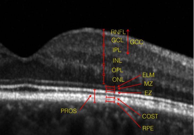
This review aims to give to the reader an overview selectively oriented on safety and efficacy of surgery, providing concise and direct answers about crucial questions of trainees and experts. Surgery for idiopathic epiretinal membrane (ERM) is a safe and effective procedure that can achieve long-term stable postoperative visual and anatomical improvement, with an overall low recurrence and complication rate. Young patients, with a short onset of symptoms and with better initial visual acuity achieve higher levels of visual outcome. The preoperative degree of metamorphopsia is the prognostic factor for their postoperative degree. Successful results may be obtained in eyes with specific optical coherence tomography criteria, such as thin ganglion cell layers, thin internal plexiform layer, longer photoreceptors outer segment, regular ellipsoid zone and cone outer segment tips line, and without ectopic inner foveal layer. Internal limiting membrane peeling demonstrates positive anatomical and functional outcomes, but final positions about its safety remain controversial.

Keywords: efficacy; idiopathic epiretinal membrane; outcome; prognostic factor; safety.


Conflict of interest statement

Professor Francesco Bandello reports personal fees from Allergan, Bayer, Boehringer-Ingelheim, Fidia Sooft, Hofmann La Roche, Novartis, NTC Pharma, Sifi, Thrombogenics, and Zeiss, outside the submitted work. The authors report no other conflicts of interest in this work.

Figures

Figure 1
Figure 1
Multicolor imaging of idiopathic epiretinal membrane. Multicolor imaging offers a detailed visualization of the macular area: multicolor montage (A), infrared reflectance (B), blue reflectance (C), green reflectance (D).
Figure 2
Figure 2
Two cases of swelling of the arcuate nerve fiber layer (SANFL). The two pictures (top and bottom) illustrate two cases of SANFL. The superimposed light-blue areas on the infrared reflectance imaging mark the region of hyperreflectant swollen arcuate nerve fiber layers. The blue arrows on the corresponding spectral-domain optical coherence tomography scans define the matching hyperreflecrive areas.
Figure 3
Figure 3
Dissociated optic nerve fiber layer (DONFL). Infrared reflectrance (A), blue light autofluorescence (B) and spectral-domain optical coherence tomography (C) of a post-surgical case of DONFL. The red arrow identifies a “dimple” in the inner retinal layer that corresponds to the hypofluorescent spot.
Figure 4
Figure 4
Spectral-domain optical coherence tomography layering of a normal eye. From inner to outer: retinal nerve fiber layer (RNFL), ganglion cell layer (GCL), inner plexiform layer (IPL), outer nuclear layer (INL), outer plexiform layer (OPL), outer nuclear layer (ONL), external limiting membrane (ELM, hyperreflective), myoid zone (MZ, hyporeflective), ellipsoid zone (EZ, hyperreflective), cone outer segment tips layer (COST, hyperreflective, otherwise known as “interdigitation zone”), retinal pigment epithelium (RPE, hyperreflective). The ganglion cell complex layer (GCC) encompasses together the RNFL, GCL and IPL. The photoreceptor outer segment (PROS) represents the distance form the ELM to the RPE.

Similar articles

Cited by

References

    1. Chang W-C, Lin C, Lee C-H, Sung T-L, Tung T-H, Liu J-H. Vitrectomy with or without internal limiting membrane peeling for idiopathic epiretinal membrane: a meta-analysis. PLoS One. 2017;12(6):e0179105. doi:10.1371/journal.pone.0179105 - DOI - PMC - PubMed
    1. Kim C-S, Kim K-N, Kim W-J, Kim J-Y. Intraoperative endolaser retinopexy around the sclerotomy site for prevention of retinal detachment after pars plana vitrectomy. Retina Phila Pa. 2011;31(9):1772–1776. doi:10.1097/IAE.0b013e31820b6129 - DOI - PubMed
    1. Melberg NS, Thomas MA. Nuclear sclerotic cataract after vitrectomy in patients younger than 50 years of age. Ophthalmology. 1995;102(10):1466–1471. - PubMed
    1. Pielen A, Guerra NIP, Böhringer D, et al. Intra- and postoperative risks and complications of small-gauge (23-G) versus conventional (20-G) vitrectomy for macular surgery. Eur J Ophthalmol. 2014;24(5):778–785. doi:10.5301/ejo.5000461 - DOI - PubMed
    1. Costarides AP, Alabata P, Bergstrom C. Elevated intraocular pressure following vitreoretinal surgery. Ophthalmol Clin N Am. 2004;17(4):507–512. doi:10.1016/j.ohc.2004.06.007 - DOI - PubMed

LinkOut - more resources