. 2019 Jul 17:13:2371-2379.
doi: 10.2147/DDDT.S203399. eCollection 2019.

MicroRNA-664 suppresses the growth of cervical cancer cells via targeting c-Kit

Affiliations

MicroRNA-664 suppresses the growth of cervical cancer cells via targeting c-Kit

Mingfen Lv et al. Drug Des Devel Ther. .

Abstract

Background: Cervical cancer is the second most common malignant cancer in women worldwide. Evidence indicated that miR-664 was significantly downregulated in cervical cancer. However, the mechanisms by which miR-664 regulates the tumorigenesis of cervical cancer remain unclear. Thus, this study aimed to investigate the role of miR-664 in cervical cancer.

Methods: Quantitative reverse transcription polymerase chain reaction was used to detect the level of miR-664 in tumor tissues and cell line. The dual luciferase reporter system assay and Western blotting were used to explore the interaction of miR-664 and c-Kit in cervical cancer.

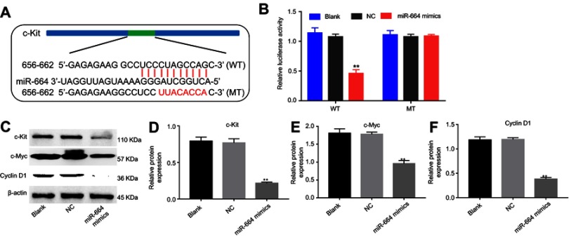
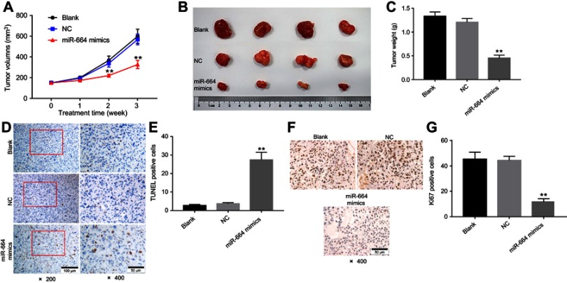
Results: The expression of miR-664 in patients with cervical cancer was dramatically decreased compared with that in adjacent tissues. MiR-664 mimics significantly inhibited proliferation in SiHa cells via inducing apoptosis. In addition, miR-664 mimics induced apoptosis in SiHa cells via increasing the expressions of Bax and active caspase 3 and decreasing the level of Bcl-2. Moreover, dual-luciferase assay showed that c-Kit was the directly binding target of miR-664 in SiHa cells; overexpression of miR-664 downregulated the expression of c-Kit. Meanwhile, upregulation of miR-664 significantly decreased the levels of c-Myc and Cyclin D in cells. Furthermore, miR-664 markedly inhibited tumor growth of cervical cancer in xenograft.

Conclusion: Our data indicated that miR-664 exerted antitumor effects on SiHa cells by directly targeting c-Kit in vitro and in vivo. Therefore, miR-664 might be a potential therapeutic target for the treatment of patients with cervical cancer.

Keywords: apoptosis; c-Kit; cervical cancer; microRNA-664.


Conflict of interest statement

The authors report no conflicts of interest in this work.

Figures

Figure 1
Figure 1
MiR-664 mimics inhibited the proliferation of SiHa cells. (A) qRT-PCR was performed to examine the level of miR-664 in tissues samples between cervical cancer and control (**P<0.01 vs normal tissues). (B) SiHa cells were transfected with 10 nM miR-664 mimics or mimics NC for 6 hrs and incubated for another 48 hrs. The level of miR-664 was detected with qRT-PCR. (C) Cell viability in SiHa cells was determined using CCK-8 assay. (D) Relative fluorescence expression levels were quantified by Ki67 and DAPI staining. (E) The number of Ki67 positive cells were counted. **P<0.01 vs NC group.
Figure 2
Figure 2
MiR-664 mimics induced apoptosis of SiHa cells. SiHa cells were transfected with 10 nM miR-664 mimics or mimics NC for 6 hrs and incubated for another 48 hrs. (A) Apoptotic cells were detected with Annexin V and PI double staining. (B) The apoptosis cell rates were calculated. (C) The expressions of Bax, Bcl-2 and active caspase 3 in SiHa cells were analyzed by Western blotting. (D) The expression of Bax was quantified by normalizing to β-actin. (E) The expression of Bcl-2 was quantified by normalizing to β-actin. (F) The expression of active caspase 3 was quantified by normalizing to β-actin. **P<0.01 vs NC group.
Figure 3
Figure 3
MiR-664 mimics inhibited migration of SiHa cells. SiHa cells were transfected with 10 nM miR-664 mimics or mimics NC for 6 hrs and incubated for another 24 hrs. (A) The migration ability of U937 cells was examined using transwell migration assay. (B) The number of migration cells in each group was quantified. **P<0.01 vs NC group.
Figure 4
Figure 4
C-Kit was a direct binding target of miR‑664. (A) Gene structure of c-Kit at the position of 656–662 indicates the predicted target site of miR-664 in its 3’UTR, with a sequence of UCCCUAGCCAGC. (B) The luciferase activity was measured by using the dual luciferase reporter assay. (C) SiHa cells were transfected with 10 nM miR-664 mimics or mimics NC for 6 hrs and incubated for another 48 hes. The expressions of c-Kit, c-Myc and Cyclin D in SiHa cells were analyzed by Western blotting. (D) The expression of c-Kit was quantified by normalizing to β-actin. (E) The expression of c-Myc was quantified by normalizing to β-actin. (F) The expression of Cyclin D was quantified by normalizing to β-actin. **P<0.01 vs NC group.
Figure 5
Figure 5
MiR-664 mimics exhibited antitumor effects on cervical cancer in vivo. miR-664 mimics (50 nM) or NC was directly injected into the tumors twice a week. (A) Tumor volumes of mice were measured weekly post-inoculation of SiHa cells. (B) Tumors were isolated from xenografts and pictured at the end of study. (C) Tumor weights in each group were calculated. (D and E) TUNEL staining of tumor tissues in each group and TUNEL positive cell rate were calculated. (F and G) Relative fluorescence expression levels were quantified by Ki67 and DAPI staining. **P<0.01 vs NC group.
Figure 6
Figure 6
MiR-664 mimics exhibited antitumor effects via induction apoptosis in vivo. (A) The level of miR-664 in tumor tissues was detected with qRT-PCR. (B) The expressions of c-Kit and active caspase 3 in tumor tissues were analyzed by Western blotting. (C) The expression of c-Kit was quantified by normalizing to β-actin. (D) The expression of active caspase 3 was quantified by normalizing to β-actin. **P<0.01 vs NC group.

Similar articles

Cited by

References

    1. Parkin DM, Bray F, Ferlay J, Pisani P. Estimating the world cancer burden: Globocan 2000. Int J Cancer. 2001;94(2):153–156. - PubMed
    1. Granados Lopez AJ, Lopez JA. Multistep model of cervical cancer: participation of miRNAs and coding genes. Int J Mol Sci. 2014;15(9):15700–15733. doi:10.3390/ijms150915700 - DOI - PMC - PubMed
    1. de Freitas AC, Gomes Leitao Mda C, EC Coimbra. Prospects of molecularly-targeted therapies for cervical cancer treatment. Curr Drug Targets. 2015;16(1):77–91. - PubMed
    1. Shishodia G, Verma G, Das BC, Bharti AC. miRNA as viral transcription tuners in HPV-mediated cervical carcinogenesis. Front Biosci (Schol Ed). 2018;10:21–47. - PubMed
    1. Filippeschi M, Moncini I, Bianchi B, Florio P. What kind of surgery for cervical carcinoma? G Chir. 2012;33(4):139–146. - PubMed

MeSH terms