. 2019 Nov;46(12):2429-2451.
doi: 10.1007/s00259-019-04450-0. Epub 2019 Aug 13.

EANM guideline for ventilation/perfusion single-photon emission computed tomography (SPECT) for diagnosis of pulmonary embolism and beyond

Affiliations

EANM guideline for ventilation/perfusion single-photon emission computed tomography (SPECT) for diagnosis of pulmonary embolism and beyond

Marika Bajc et al. Eur J Nucl Med Mol Imaging. 2019 Nov.

Abstract

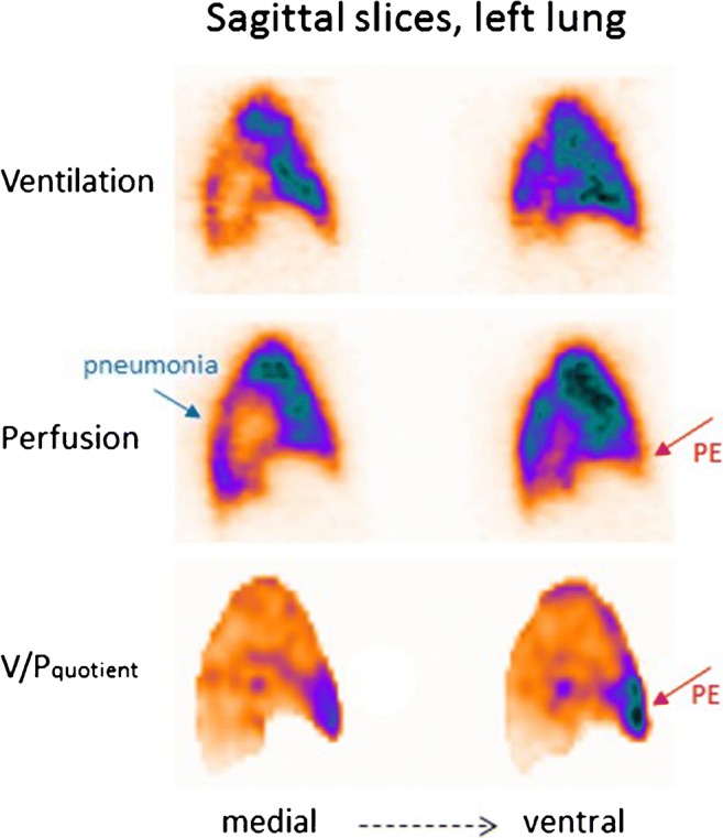
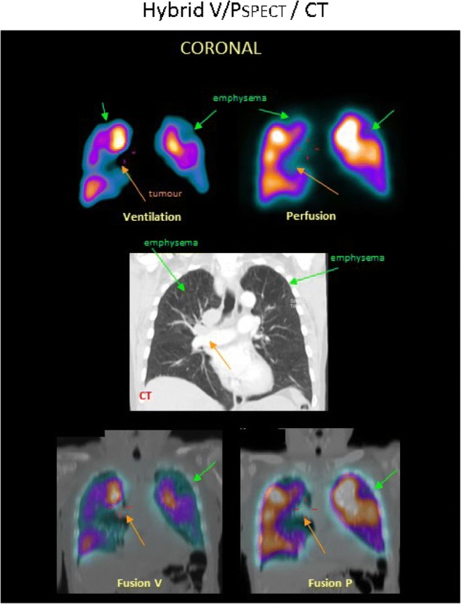
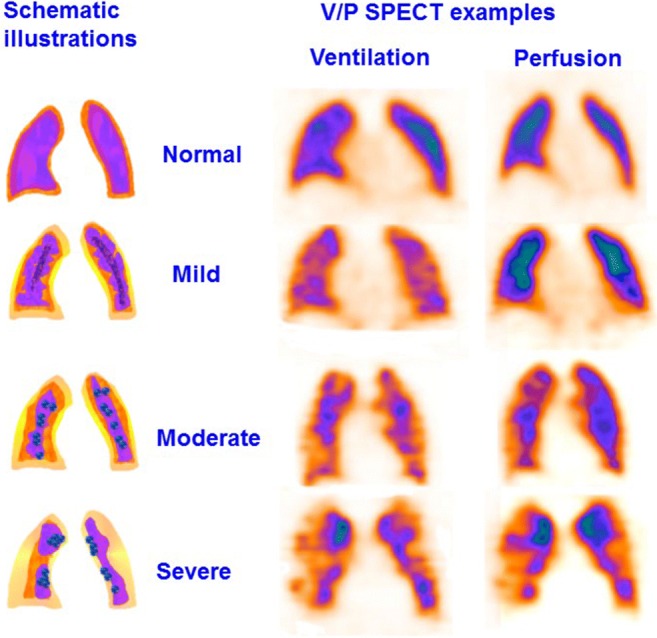
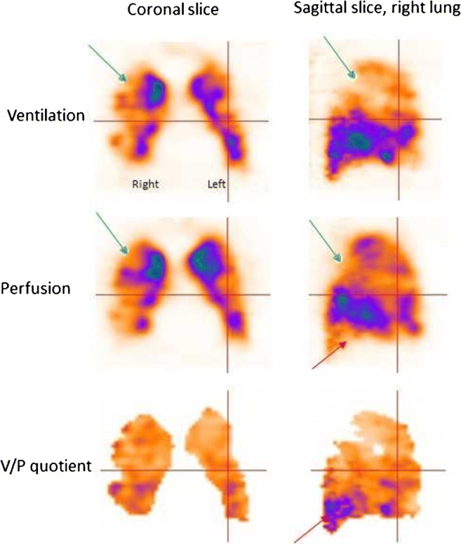
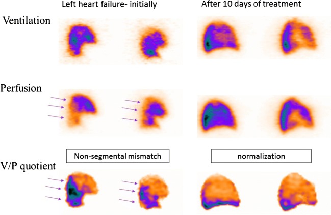
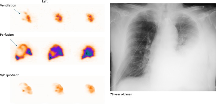
These guidelines update the previous EANM 2009 guidelines on the diagnosis of pulmonary embolism (PE). Relevant new aspects are related to (a) quantification of PE and other ventilation/perfusion defects; (b) follow-up of patients with PE; (c) chronic PE; and (d) description of additional pulmonary physiological changes leading to diagnoses of left ventricular heart failure (HF), chronic obstructive pulmonary disease (COPD) and pneumonia. The diagnosis of PE should be reported when a mismatch of one segment or two subsegments is found. For ventilation, Technegas or krypton gas is preferred over diethylene triamine pentaacetic acid (DTPA) in patients with COPD. Tomographic imaging with V/PSPECT has higher sensitivity and specificity for PE compared with planar imaging. Absence of contraindications makes V/PSPECT an essential method for the diagnosis of PE. When V/PSPECT is combined with a low-dose CT, the specificity of the test can be further improved, especially in patients with other lung diseases. Pitfalls in V/PSPECT interpretation are discussed. In conclusion, V/PSPECT is strongly recommended as it accurately establishes the diagnosis of PE even in the presence of diseases like COPD, HF and pneumonia and has no contraindications.

Keywords: COPD; CTPA; Chronic pulmonary embolism; Left heart failure; Pneumonia; Pulmonary embolism; Pulmonary hypertension; SPECT; V/P SPECT/CT; Ventilation-perfusion.


Conflict of interest statement

The authors declare that they have no conflict of interest.

Figures

Fig. 1
Fig. 1
Segmental map of the two lungs in four coronal slices and two sagittal slices for each lung. A bronchopulmonary segment is conical with its apex towards the hilum and its base projected onto the pleural surface. Thrombi occluding pulmonary arteries, therefore, produce characteristic lobar, segmental or subsegmental peripheral wedge-shaped defects with the base reaching the pleural surface
Fig. 2
Fig. 2
Coronal slices in a patient with PE. Multiple bilateral segmental perfusion defects (red arrows) in areas with normal ventilation. These are delineated on V/P quotient images which facilitate interpretation
Fig. 3
Fig. 3
Sagittal slices of both lungs in a patient with small PE (red arrows), initially (not treated) and 3 months later. On follow-up, the progression of perfusion defects as well as deterioration in the ventilation (blue arrow in the left lung) can be seen clearly. The patient also developed pneumonia
Fig. 4
Fig. 4
Coronal slices in a patient with recurrent PE (red arrows), initially referred after a diagnosis of pulmonary hypertension. PE was not identified on CT. The first follow-up scan shows new perfusion defects that occurred after stopping therapy, and the final follow-up identifies improving perfusion after 4 months of treatment
Fig. 5
Fig. 5
Sagittal slices of the left lung in a patient with PE in the medial lobe (red arrow) and pneumonia posteriorly (blue arrow). The VP mismatch may be highlighted in V/P quotient images
Fig. 6
Fig. 6
A patient with COPD, emphysema and tumour. Coronal slices display uneven distribution of ventilation with a pattern of deposition of 99mTc-Technegas® that is typical for COPD. Perfusion follows the ventilation pattern. Matched ventilation and perfusion defects are observed in both upper lobes (green arrows) and to the right of the mediastinum (orange arrows). In the medial row of the corresponding coronal CT slice, emphysema is seen in both upper lobes (green arrows), as is a tumour in the mediastinum (orange arrow). Fusion images of CT and ventilation SPECT and CT and perfusion SPECT are shown
Fig. 7
Fig. 7
Schematic presentation of the obstructive lung disease grading system and correlating representative V/PSPECT images that shows different degrees of airway obstruction on coronal slices. 0: normal, even distribution of Technegas® with good peripheral penetration and without accumulation in large or small airways. 1: mild airway obstruction, slightly uneven distribution with some deposition of aerosol in small and intermediate airways. Only minor areas with reduced peripheral penetration are observed. 2: moderate airway obstruction, deposition of Technegas® in intermediate and large airways, diminished peripheral penetration with maximum accumulation in the central half of the lung. 3: severe airway obstruction, central deposition in large airways with severely impaired penetration of Technegas® and major areas with reduced or abolished function
Fig. 8
Fig. 8
V/PSPECT images showing a severe degree of airway obstruction in coronal and sagittal projections in a patient with severe COPD, emphysema (green arrow) and PE (red arrow)
Fig. 9
Fig. 9
Sagittal slices from the lung show antigravitational redistribution of perfusion in left heart failure. Ventilation is less affected causing mismatch. Mind the pattern; it is not of segmental character!
Fig. 10
Fig. 10
Sagittal slices of the left lung in a patient with extensive pneumonia in whom the chest X-ray was interpreted as showing atelectasis (a). The left lung shows nearly absent ventilation in areas with much better perfusion. Arrow indicates stripe sign. V/P quotient highlight reverse mismatch
Fig. 11
Fig. 11
Patient with chronic PE. The perfusion is only maintained in the central part of the lung, leading to large nonsegmental perfusion defects (red arrows)

Comment in

  • Radioaerosols and the updated EANM guideline in ventilation/perfusion imaging.
    LaFrance N, Fournier F. LaFrance N, et al. Eur J Nucl Med Mol Imaging. 2020 Jul;47(7):1640-1642. doi: 10.1007/s00259-020-04793-z. Epub 2020 Apr 13. Eur J Nucl Med Mol Imaging. 2020. PMID: 32285154 Free PMC article. No abstract available.
  • Letter to editor.
    Bajc M, Jonson B. Bajc M, et al. Eur J Nucl Med Mol Imaging. 2020 Jul;47(7):1643-1644. doi: 10.1007/s00259-020-04813-y. Eur J Nucl Med Mol Imaging. 2020. PMID: 32303787 No abstract available.

References

    1. Bajc M, Neilly JB, Miniati M, Schuemichen C, Meignan M, Jonson B. EANM guidelines for ventilation/perfusion scintigraphy: Part 1. Pulmonary imaging with ventilation/perfusion single photon emission tomography. Eur J Nucl Med Mol Imaging. 2009;36:1356–1370. doi: 10.1007/s00259-009-1170-5. - DOI - PubMed
    1. Bajc M, Neilly JB, Miniati M, Schuemichen C, Meignan M, Jonson B. EANM guidelines for ventilation/perfusion scintigraphy: Part 2. Algorithms and clinical considerations for diagnosis of pulmonary emboli with V/P(SPECT) and MDCT. Eur J Nucl Med Mol Imaging. 2009;36:1528–1538. doi: 10.1007/s00259-009-1169-y. - DOI - PubMed
    1. Cohen AT, Agnelli G, Anderson FA, Arcelus JI, Bergqvist D, Brecht JG, et al. Venous thromboembolism (VTE) in Europe. The number of VTE events and associated morbidity and mortality. Thromb Haemost. 2007;98:756–764. doi: 10.1160/TH07-03-0212. - DOI - PubMed
    1. Kakkar VV, Howe CT, Flanc C, Clarke MB. Natural history of postoperative deep-vein thrombosis. Lancet (London, England) 1969;2:230–232. doi: 10.1016/S0140-6736(69)90002-6. - DOI - PubMed
    1. Moser KM, Fedullo PF, LitteJohn JK, Crawford R. Frequent asymptomatic pulmonary embolism in patients with deep venous thrombosis. Jama. 1994;271:223–225. doi: 10.1001/jama.1994.03510270069042. - DOI - PubMed

MeSH terms