. 2019 Aug 14;38(1):354.
doi: 10.1186/s13046-019-1359-9.

Estrogen receptor β inhibits breast cancer cells migration and invasion through CLDN6-mediated autophagy

Affiliations

Estrogen receptor β inhibits breast cancer cells migration and invasion through CLDN6-mediated autophagy

Peiye Song et al. J Exp Clin Cancer Res. .

Abstract

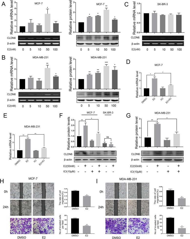
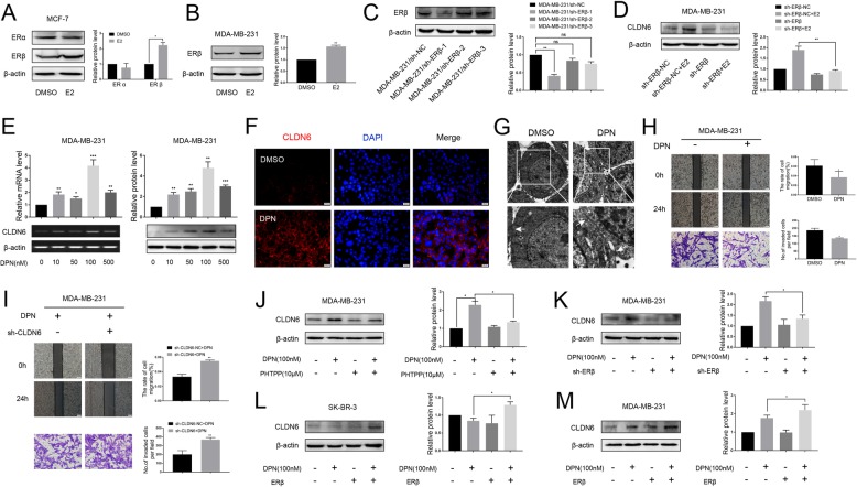
Background: Estrogen receptor β (ERβ) has been reported to play an anti-cancer role in breast cancer, but the regulatory mechanism by which ERβ exerts this effect is not clear. Claudin-6 (CLDN6), a tight junction protein, acts as a tumor suppressor gene in breast cancer. Our previous studies have found that 17β-estradiol (E2) induces CLDN6 expression and inhibits MCF-7 cell migration and invasion, but the underlying molecular mechanisms are still unclear. In this study, we aimed to investigate the role of ERβ in this process and the regulatory mechanisms involved.

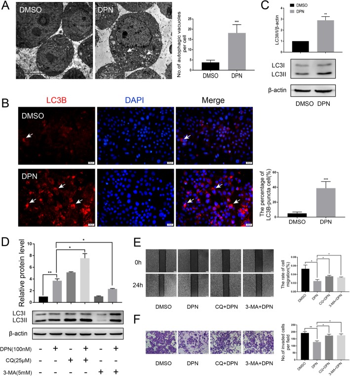
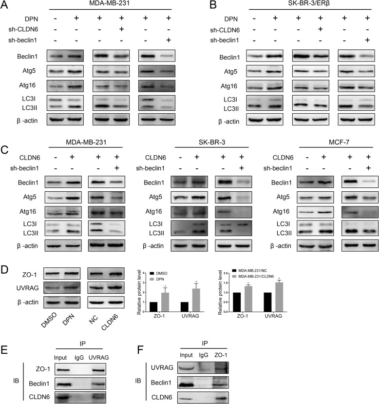
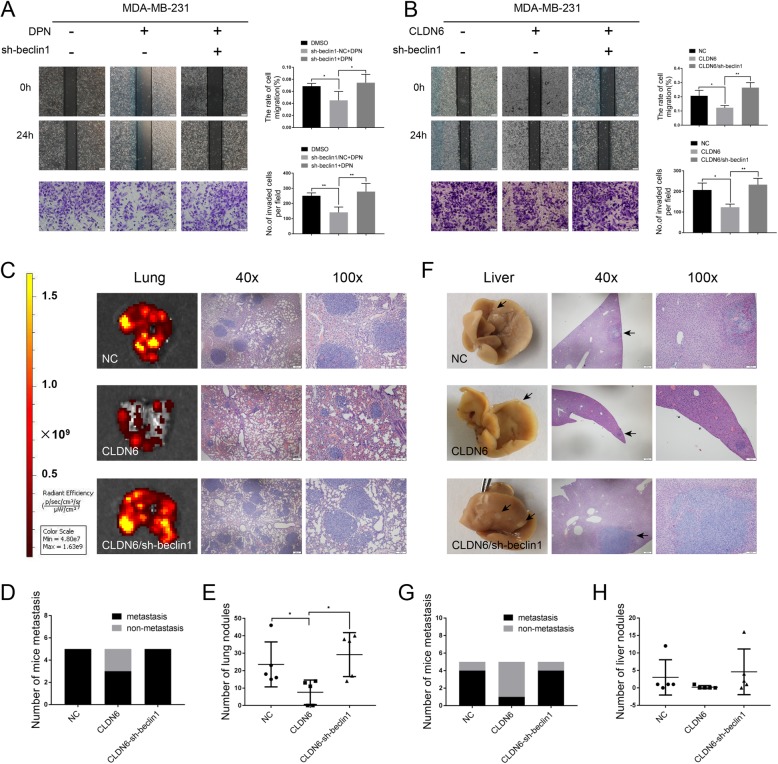
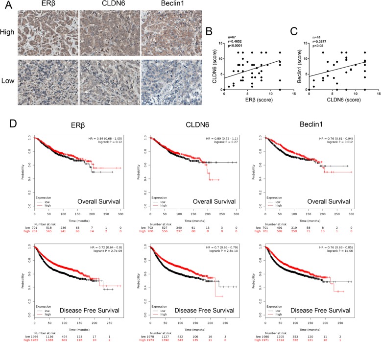
Methods: Polymerase chain reaction (PCR) and western blot were used to characterize the effect of E2 on the expression of CLDN6 in breast cancer cells. Chromatin immunoprecipitation (ChIP) assays were carried out to confirm the interaction between ERβ and CLDN6. Dual luciferase reporter assays were used to detect the regulatory role of ERβ on the promoter activity of CLDN6. Wound healing and Transwell assays were used to examine the migration and invasion of breast cancer cells. Western blot, immunofluorescence and transmission electron microscopy (TEM) were performed to detect autophagy. Xenograft mouse models were used to explore the regulatory effect of the CLDN6-beclin1 axis on breast cancer metastasis. Immunohistochemistry (IHC) was used to detect ERβ/CLDN6/beclin1 expression in breast cancer patient samples.

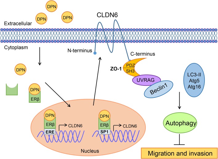
Results: Here, E2 upregulated the expression of CLDN6, which was mediated by ERβ. ERβ regulated CLDN6 expression at the transcriptional level. ERβ inhibited the migration and invasion of breast cancer cells through CLDN6. Interestingly, this effect was associated with CLDN6-induced autophagy. CLDN6 positively regulated the expression of beclin1, which is a key regulator of autophagy. Beclin1 knockdown reversed CLDN6-induced autophagy and the inhibitory effect of CLDN6 on breast cancer metastasis. Moreover, ERβ and CLDN6 were positively correlated, and the expression of CLDN6 was positively correlated with beclin1 in breast cancer tissues.

Conclusion: Overall, this is the first study to demonstrate that the inhibitory effect of ERβ on the migration and invasion of breast cancer cells was mediated by CLDN6, which induced the beclin1-dependent autophagic cascade.

Keywords: Autophagy; Breast cancer; CLDN6; Estrogen receptor β; Invasion; Migration.


Conflict of interest statement

The authors declare that they have no competing interests.

Figures

Fig. 1
Fig. 1
E2 upregulates the expression of CLDN6 in MCF-7 and MDA-MB-231 cells. MCF-7(a), MDA-MB-231(b) and SK-BR-3(c) cells were treated with DMSO or E2 (from 5 nM to 100 nM). Cells were harvested after 24 h treatment for analysis of gene and protein expression of CLDN6 using semiquantitative RT-PCR and western blot, respectively. Actin served as a loading control. Quantification of CLDN6 expression in MCF-7 (d) and MDA-MB-231 (e) cells cotreated with ICI by qRT-PCR. CLDN6 protein expression levels in MCF-7 (f) and MDA-MB-231 (g) cells after treatment with E2 or E2/ICI. CLDN6 protein expression in SK-BR-3 (f) cells after 50 nM E2 treatment. Wound healing and Transwell assays were used to detect the migration (scale bar, 200 μm) and invasion (scale bar, 50 μm) abilities of MCF-7 (h) and MDA-MB-231 (i) cells treated with E2. Data are presented as mean ± SD. The data shown are representative results of three independent experiments. *P < 0.05, **P < 0.01, ***P < 0.001
Fig. 2
Fig. 2
E2 regulates CLDN6 expression via ERβ. a Western blot analysis of ERα and ERβ expression in MCF-7 cells treated with E2. Actin served as a loading control. b Western blot analysis of ERβ in MDA-MB-231 cells treated with E2. c MDA-MB-231 cells were transfected with either negative control (sh-NC) or three different ERβ shRNAs for 48 h and were then subjected to western blot analysis to detect the protein abundance of ERβ. Actin was used as the loading control. d ERβ knockdown abolished the CLDN6 expression induced by E2. e MDA-MB-231 cells were incubated with DPN for 24 h at the indicated concentration. CLDN6 gene and protein expression levels were detected by using semiquantitative RT-PCR and western blot. f Immunofluorescence of CLDN6 (red) was prominent along the edges of the MDA-MB-231 cells upon DPN treatment. Nuclei were stained with 4, 6-diamino-2-phenylindole (DAPI) (blue) (scale bar, 20 μm). g Tight junctions (white arrowheads) between cells were prominent in MDA-MB-231 cells after DPN treatment as observed by TEM. h The migration (scale bar, 200 μm) and invasion (scale bar, 50 μm) abilities of MDA-MB-231 cells treated with DPN were decreased. i CLDN6 knockdown rescued the migration and invasion abilities of MDA-MB-231 cells after DPN treatment. The ERβ antagonist PHTPP (10 μM, 24 h) (j) and ERβ knockdown (k) abolished the DPN-induced CLDN6 expression. Overexpression of ERβ induced CLDN6 upregulation in SR-BR-3 (l) and MDA-MB-231 (m) cells after treatment with DPN. Data are presented as mean ± SD. The data shown are representative results of three independent experiments. *P < 0.05, **P < 0.01, ***P < 0.001
Fig. 3
Fig. 3
ERβ regulates CLDN6 expression at the transcriptional level. a ERβ protein expression in the nuclear and cytoplasmic fractions of DPN-treated MDA-MB-231 cells. PCNA served as a nuclear protein control, and β-tubulin was used as a cytoplasmic protein control. b Subcellular localization of ERβ. DPN promotes the transportation of ERβ from the cytoplasm to the nucleus (white arrowheads) in MDA-MB-231 cells. Nuclei were stained with DAPI (blue) (scale bar, 50 μm). c Schematic diagram of the sequence spanning from -2000 bp to + 250 bp relative to the translational start site (TSS) of the CLDN6 gene promoter containing the (half-ERE)-(N)x-(GC-box) motif. d ChIP assay. The ERβ binding sites (− 183/− 168 bp or + 155/+ 170 bp) of the CLDN6 promoter were detected by PCR in DPN-treated MDA-MB-231 cells. e ChIP assay. The Sp1 binding sites (− 60/− 47 bp or + 192/+ 203 bp) of the CLDN6 promoter were detected by PCR in DPN-treated MDA-MB-231 cells. f The luciferase activities of DPN-treated MDA-MB-231 cells transfected with pGL3-CLDN6 and renilla luciferase reporter (pRL-TK) plasmids were detected by dual-luciferase reporter assays. Data are presented as mean ± SD. The data shown are representative results of three independent experiments. *P < 0.05, **P < 0.01, ***P < 0.001
Fig. 4
Fig. 4
ERβ induces autophagy and suppresses the migration and invasion of breast cancer cells. a TEM analysis was performed on MDA-MB-231 cells treated or not treated for 24 h with 100 nM DPN. DPN-treated cells displayed several autophagic vacuoles with the characteristic double membrane (white arrowheads) which were not observed in the control (scale bar, 5 μm). b Immunofluorescence analysis showed that the numbers of LC3-II puncta (white arrowheads) were increased after DPN treatment. Nuclei were stained with DAPI (Scale bar, 20 μm). c The expression of LC3B was detected by western blot in DPN-treated or untreated MDA-MB-231 cells. Actin served as a loading control. d The expression of LC3B was detected by western blot in MDA-MB-231 cells cotreated with DPN and CQ (25 μM, 24 h) or DPN and 3-MA (5 mM, 24 h). CQ and 3-MA were pretreated for 2 h before treatment with DPN. Wound healing (e) and Transwell migration (f) assays showed that the DPN-inhibited migration (scale bar, 200 μm) and invasion (scale bar, 50 μm) abilities were attenuated by CQ and 3-MA. CQ and 3-MA were pretreated for 2 h before treatment with DPN. Data are presented as mean ± SD. The data shown are representative results of three independent experiments. *P < 0.05, **P < 0.01, ***P < 0.001
Fig. 5
Fig. 5
ERβ induces autophagy via CLDN6-mediated increase in beclin1. Expression of autophagy-related proteins was detected by using western blot in DPN-treated cells and in CLDN6-knockdown and beclin1-knockdown DPN-treated MDA-MB-231 (a) and SK-BR-3/ERβ (b) cells. Actin served as a loading control. c Expression of autophagy-related proteins was detected by using western blot in CLDN6-overexpressing MDA-MB-231, SK-BR-3 and MCF-7 cells and CLDN6-overexpressing beclin1-knockdown cells. d Western blot analysis of ZO-1 and UVRAG in DPN-treated and CLDN6-overexpressing MDA-MB-231 cells. e-f Co-IP assays were performed to detect the interaction between beclin1 and CLDN6 in MDA-MB-231 cells treated with DPN. Data are presented as mean ± SD. The data shown are representative results of three independent experiments. *P < 0.05
Fig. 6
Fig. 6
CLDN6 inhibits migration, invasion and metastasis of breast cancer through beclin1 in vitro and in vivo. Wound healing and Transwell migration assays showed that beclin1 knockdown drove the migration (scale bar, 200 μm) and invasion (scale bar, 50 μm) of DPN-treated (a) and CLDN6-overexpressing MDA-MB-231 cells (b). c IVIS imaging was performed to show lung metastatic sites. Representative H&E staining of lung sections from the three groups (n = 5). d The number of mice with lung metastasis was counted in each group. e The number of lung nodules in each group. f Representative images of liver metastasis and H&E staining in the three groups (n = 5). Black arrowheads indicate the metastatic sites. g The number of mice with liver metastasis was counted in each group. h The number of liver nodules in each group. Data are presented as mean ± SD. The data shown are representative results of three independent experiments. *P < 0.05, **P < 0.01
Fig. 7
Fig. 7
Clinical correlation analyses between ERβ, CLDN6 and beclin1 expression and prognosis in breast cancer patients. a Representative images of IHC analysis of ERβ, CLDN6 and beclin1 expression from tissue microarray (× 400). In breast cancer tissues and adjacent tissues, ERβ expression was observed in the nucleus and the cytoplasm, CLDN6 was expressed in the cytoplasm and the plasma membrane and beclin1 was mainly expressed in the cytoplasm. “Low” indicates proteins with low expression, and “high” indicates proteins with high expression. b Correlation between ERβ expression and CLDN6 expression in breast cancer tissue microarray. Pearson correlation test, n = 67, r = 0.4652, P < 0.0001. c Correlation between CLDN6 expression and beclin1 expression in breast cancer tissue microarray. Pearson correlation test, n = 44, r = 0.3677, P = 0.0141. d Kaplan-Meier analysis of the overall survival and disease-free survival of breast cancer patients with different ERβ, CLDN6 and beclin1 expression levels in the in Kaplan-Meier plotter database. Statistical differences were determined by log-rank test. Data are presented as mean ± SD
Fig. 8
Fig. 8
The proposed model for ERβ-induced autophagy inhibiting breast cancer cell migration and invasion. In this model, when ERβ bound with ligands (DPN), DPN-ERβ complexes directly bound to the ERE of the CLDN6 promoter and enhanced CLDN6 expression. In addition, activated ERβ can interact with Sp1 and bind the Sp1 transcriptional regulation domains of the CLDN6 promoter to induce CLDN6 expression. ZO-1 and UVRAG act as bridge molecules for the CLDN6-beclin1 interaction. CLDN6 and ZO-1/UVRAG/beclin1 form complexes and serve as a platform for recruiting other autophagy regulatory proteins (atg5, atg16 and LC3-II) and induce autophagy to suppress the migration and invasion of breast cancer cells

References

    1. Heldring N, Pike A, Andersson S, Matthews J, Cheng G, Hartman J, et al. Estrogen receptors: how do they signal and what are their targets. Physiol Rev. 2007;87(3):905–931. doi: 10.1152/physrev.00026.2006. - DOI - PubMed
    1. Thomas C, Gustafsson JA. The different roles of ER subtypes in cancer biology and therapy. Nat Rev Cancer. 2011;11(8):597–608. doi: 10.1038/nrc3093. - DOI - PubMed
    1. Huang B, Omoto Y, Iwase H, Yamashita H, Toyama T, Coombes RC, et al. Differential expression of estrogen receptor alpha, beta1, and beta2 in lobular and ductal breast cancer. Proc Natl Acad Sci U S A. 2014;111(5):1933–1938. doi: 10.1073/pnas.1323719111. - DOI - PMC - PubMed
    1. Clarke R, Tyson JJ, Dixon JM. Endocrine resistance in breast cancer--an overview and update. Mol Cell Endocrinol. 2015;418(Pt 3):220–234. doi: 10.1016/j.mce.2015.09.035. - DOI - PMC - PubMed
    1. Droog M, Beelen K, Linn S, Zwart W. Tamoxifen resistance: from bench to bedside. Eur J Pharmacol. 2013;717(1–3):47–57. doi: 10.1016/j.ejphar.2012.11.071. - DOI - PubMed

MeSH terms