. 2019 Aug 14;7(1):220.
doi: 10.1186/s40425-019-0702-1.

Indocyanine green and poly I:C containing thermo-responsive liposomes used in immune-photothermal therapy prevent cancer growth and metastasis

Affiliations

Indocyanine green and poly I:C containing thermo-responsive liposomes used in immune-photothermal therapy prevent cancer growth and metastasis

Li Xu et al. J Immunother Cancer. .

Abstract

Background: Efficient cancer therapy is sought not only for primary tumor treatment but also for the prevention of metastatic cancer growth. Immunotherapy has been shown to prevent cancer metastasis by inducing antigen-specific immune responses. Indocyanine green (ICG) has a peak spectral absorption at about 800 nm, which makes it a photothermal reagent for direct treatment of solid tumors by photothermal therapy (PTT). Since PTT alone cannot fully induce antigen-specific immune response for prevention of cancer metastasis, the combination of PTT and immunotherapy has been developed as a new strategy of cancer treatment.

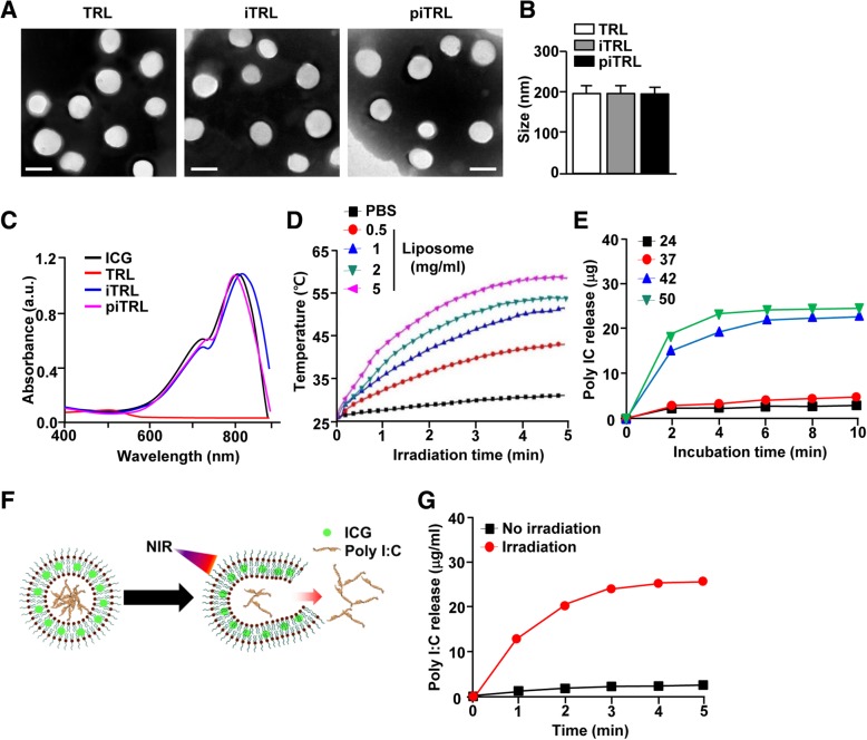
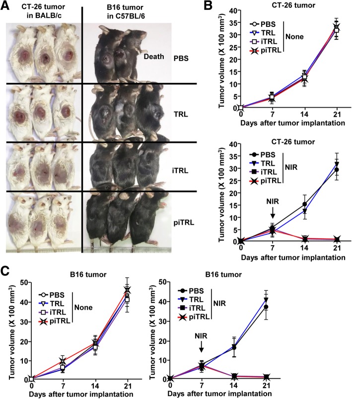
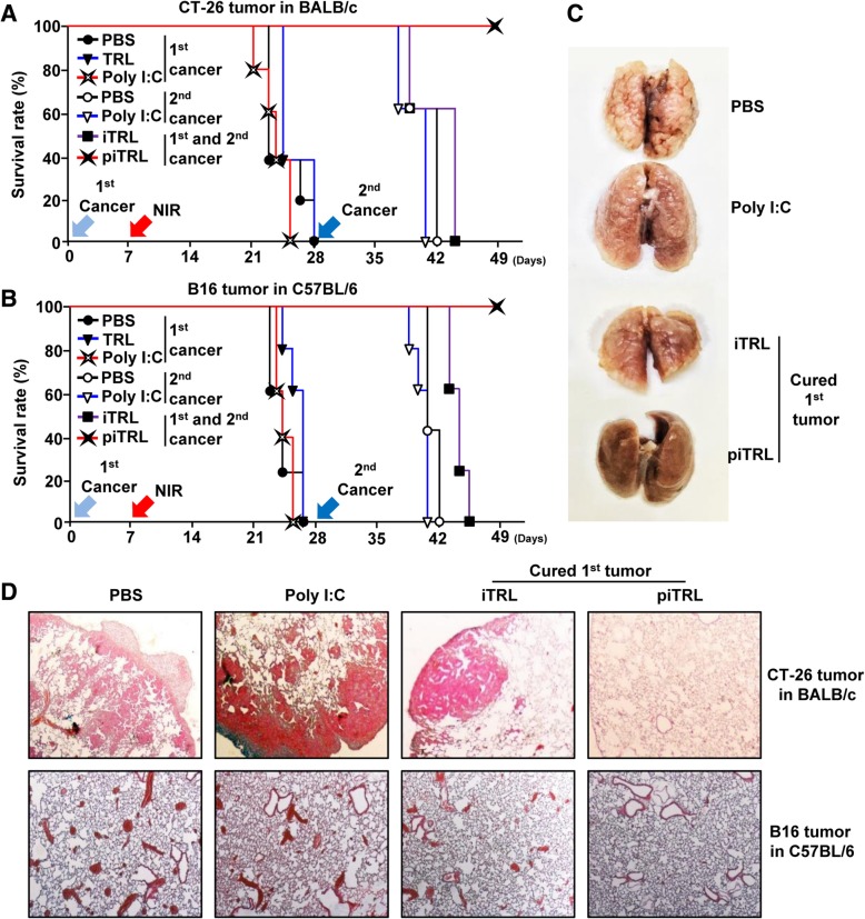
Methods: Thermal responsive liposomes (TRL) were synthesized by incorporating ICG into the lipid bilayer and encapsulating the water-soluble immune stimulatory molecule polyinosinic:polycytidylic acid (poly I:C) in the hydrophilic core. The poly I:C- and ICG-containing TRLs (piTRLs) were analyzed according to size, and their photothermal effect was evaluated following laser irradiation at 808 nm. Moreover, the temperature-dependent release of poly I:C was also measured. For cancer therapy, CT-26 (carcinoma) and B16 (melanoma) cells were subcutaneously inoculated to build the 1st transplanted tumor in BALB/c and C57BL/6 mice, respectively. These mice received a 2nd transplantation with the same cancer cells by intravenous inoculation, for evaluation of the anti-metastatic effects of the liposomes after PTT.

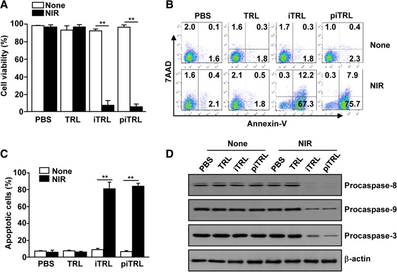
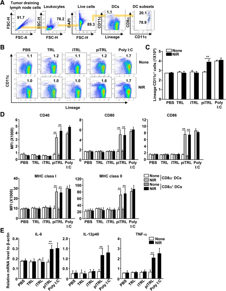
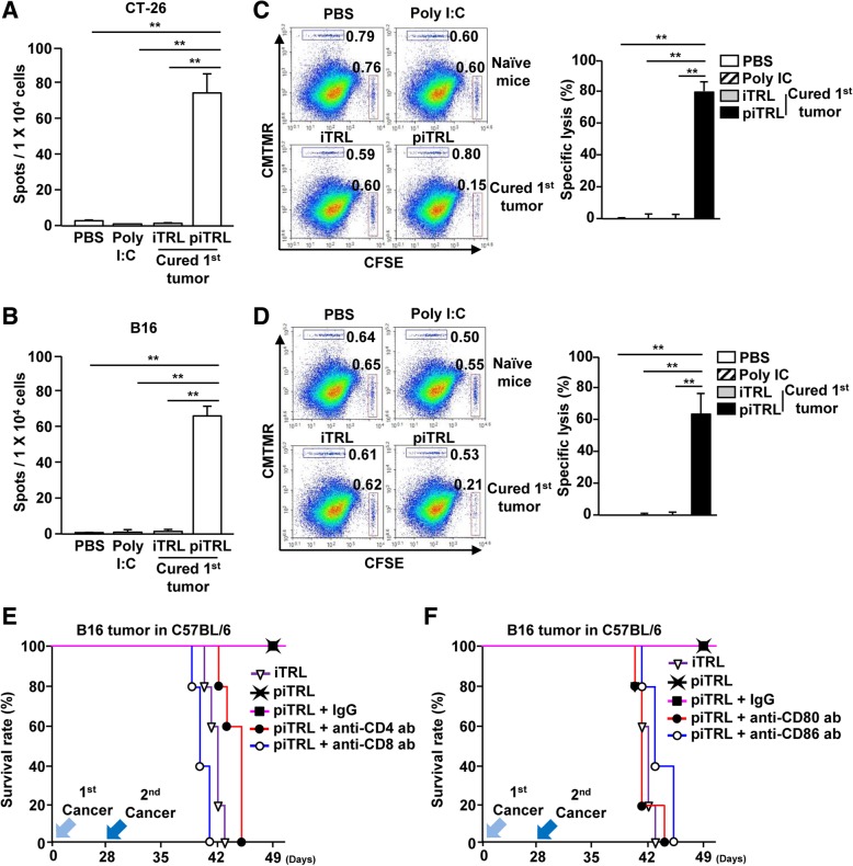
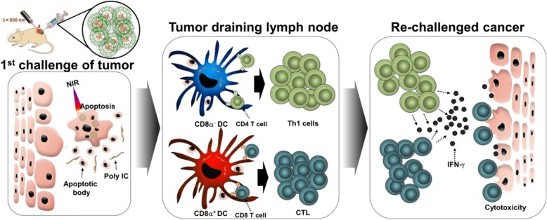
Results: Near-infrared (NIR) laser irradiation increased the temperature of piTRLs and effectively released poly I:C from the liposomes. The increased temperature induced a photothermal effect, which promoted cancer cell apoptosis and dissolution of the 1st transplanted tumor. Moreover, the released poly I:C from the piTRL induced activation of dendritic cells (DCs) in tumor draining lymph node (tdLN). Cancer cell apoptosis and DC-activation-mediated cancer antigen-specific immune responses further prevented growth of lung metastatic cancer developed following intravenous transplantation of cancer cells.

Conclusion: These results demonstrated the potential usage of a piTRL with laser irradiation for immuno-photothermal therapy against various types of cancer and their metastases.

Keywords: Anti-metastasis; Immunotherapy; Indocyanine green; Liposome; Photothermal therapy; Polyinosinic:polycytidylic acid.


Conflict of interest statement

The authors declare that they have no competing interests.

Figures

Fig. 1
Fig. 1
Characterization of liposomes. a FE-TEM images of TRL, iTRL, and piTRL. (Scale bars: 200 nm). b TEM corresponding size distribution of each liposome. c UV-vis absorption of liposomes is shown. d Photothermal heating curves of different concentrations of piTRL, irradiated for 5 min with an 808-nm laser at a power density of 1 W/cm2. e The cumulative release of poly I:C from piTRL at 24, 37, 42, and 50 °C. f Schematic diagram of poly I:C release from piTRL under NIR-laser irradiation. g The concentration of released poly I:C from piTRL under NIR-laser irradiation at a power intensity of 1 W/cm2
Fig. 2
Fig. 2
piTRL and laser irradiation promoted apoptosis of CT-26 carcinoma. CT-26 cells were incubated with PBS, TRL, iTRL or piTRL for 1 h, and the cells were treated with or without laser irradiation at 1 W/cm2 for 5 min and cultured for 24 h. a Cell viability of CT-26 was measured by MTT assay; ** p < 0.01. b Apoptosis was analyzed by annexin-V and 7AAD staining on flow cytometry. c Mean percentages of apoptotic cells, ** p < 0.01. d The expression levels of procaspase-8, − 9, and − 3 were assayed by western blotting analysis. β-actin was used as a loading control
Fig. 3
Fig. 3
Anti-cancer effect of piTRL treatment followed by laser irradiation against CT-26 carcinoma and B16 melanoma. BALB/c and C57BL/6 mice were subcutaneously (s.c.) inoculated with 1 × 106 CT-26 and B16 cells, respectively. The mice were intratumorally (i.t.) injected with PBS, TRL, iTRL, or piTRL on day 7 of tumor cell injection and were treated with or without laser irradiation at a power density of 1 W/cm2 for 5 min. a CT-26 (left panel) and B16 (right panel) tumor mass is shown on day 21 of tumor injection. b Tumor growth curves for CT-26 carcinoma with or without laser irradiation. c B16 melanoma tumor growth curves for the mice in the presence or absence of laser irradiation. Data are from the analyses of six individual mice (three mice per experiment for a total of two independent experiments)
Fig. 4
Fig. 4
piTRL treatment followed by laser irradiation promoted activation in tumor-draining lymph node (tdLN). CT-26 tumor-bearing mice were i.t. injected with PBS, TRL, iTRL, piTRL or poly I:C, and treated with or without laser irradiation for 5 min. tdLN were harvested 24 h after laser irradiation. a Definition of DC population in tdLN was shown. Lineage markers included CD3, Thy1.1, B220, Gr-1, CD49b, and TER-119. LineageCD11c+ DCs were further divided as CD8α+ and CD8α DCs. b Frequency of tdLN DCs is shown. c Mean absolute number of LineageCD11c+ cells in tdLN is shown, ** p < 0.01. d Mean fluorescence intensity (MFI) of co-stimulatory molecules and MHC class I and II in gated CD8α+ and CD8α DCs in tdLN was analyzed using flow cytometry. e Levels of IL-6, IL-12p40 and TNF-α mRNA in tdLN. All data are representative of the average of the analyses of six independent samples (i.e., three samples per experiment, two independent experiments)
Fig. 5
Fig. 5
Protective effect of piTRL treatment with laser irradiation against lung metastasis of cancer. On day 28 of the 1st transplanted tumor challenge, mice treated with iTRL or piTRL andlaser irradiation mice were further intravenously (i.v.) inoculated 2nd transplant of CT-26 and B16 cells, respectively. PBS- and poly I:C-treated mice were also i.v. injected with the cancer cells as a control. a The survival rate of CT-26 challenged BALB/c mice and b B16 challenged C57BL/6 mice were monitored, n = 5 for each group. c Representative images of CT-26 metastatic lung cancer. d H&E staining of lung on day 10 of 2nd transplant of CT-26 and B16 cell challenge. Data are representative of the analyses of six independent samples (i.e., three mice per experiment, two independent experiments)
Fig. 6
Fig. 6
Induction of cancer Ag-specific immune responses by piTRL. BALB/c and C57BL/6 mice were subcutaneously injected with cancer cells (1st transplanted tumor) and treated by the liposomes as shown in Fig. 5. a and b Spleens were harvested on day 10 of the 2nd transplantation of tumor. The splenocytes were stimulated with a CT-26 or b B16 self-Ag for 24 h and IFN-γ production was measured by ELISPOT. ** p < 0.01. c and d Specific lysis of cells was analyzed on day 10 of the 2nd transplantation of tumor in mice by transferring cancer Ag- or control peptide-coated splenocytes. ** p < 0.01. e and f B16 tumors in C57BL/6 mice were treated with piTRL and laser irradiation as indicated in Fig. 5. On day 25 of the 1st transplant of B16 cells, the mice received e depletion abs (anti-CD4 and anti-CD8 abs) or f blockade abs (anti-CD80 and anti-CD86 abs). The curves show survival rates of mice (n = 5 for each group)
Fig. 7
Fig. 7
Schematic illustration of poly I:C and ICG containing temperature-sensitive liposome (piTRL) induced immuno-photothermal therapy for treatment of 1st and 2nd transplanted cancers

References

    1. Ban Q, Bai T, Duan X, Kong J. Noninvasive photothermal cancer therapy nanoplatforms via integrating nanomaterials and functional polymers. Biomater Sci. 2017;5(2):190–210. doi: 10.1039/C6BM00600K. - DOI - PubMed
    1. Shanmugam V, Selvakumar S, Yeh CS. Near-infrared light-responsive nanomaterials in cancer therapeutics. Chem Soc Rev. 2014;43(17):6254–6287. doi: 10.1039/C4CS00011K. - DOI - PubMed
    1. Jang B, Moorthy MS, Manivasagan P, Xu L, Song K, Lee KD, Kwak M, Oh J, Jin JO. Fucoidan-coated CuS nanoparticles for chemo-and photothermal therapy against cancer. Oncotarget. 2018;9(16):12649–12661. doi: 10.18632/oncotarget.23898. - DOI - PMC - PubMed
    1. Jang B, Xu L, Moorthy MS, Zhang W, Zeng L, Kang M, Kwak M, Oh J, Jin JO. Lipopolysaccharide-coated CuS nanoparticles promoted anti-cancer and anti-metastatic effect by immuno-photothermal therapy. Oncotarget. 2017;8(62):105584–105595. doi: 10.18632/oncotarget.22331. - DOI - PMC - PubMed
    1. Mkandawire MM, Lakatos M, Springer A, Clemens A, Appelhans D, Krause-Buchholz U, Pompe W, Rodel G, Mkandawire M. Induction of apoptosis in human cancer cells by targeting mitochondria with gold nanoparticles. Nanoscale. 2015;7(24):10634–10640. doi: 10.1039/C5NR01483B. - DOI - PubMed

Publication types

LinkOut - more resources