. 2019 Oct 7;216(10):2302-2315.
doi: 10.1084/jem.20191061. Epub 2019 Aug 14.

Risk of Zika microcephaly correlates with features of maternal antibodies

Davide F Robbiani #  1 Priscilla C Olsen #  2   3 Federico Costa #  4   5 Qiao Wang #  6 Thiago Y Oliveira #  2 Nivison Nery Jr  7 Adeolu Aromolaran  4 Mateus S do Rosário  8 Gielson A Sacramento  7 Jaqueline S Cruz  7 Ricardo Khouri  7 Elsio A Wunder Jr  4 Adriana Mattos  8 Bruno de Paula Freitas  8   9 Manoel Sarno  5 Gracinda Archanjo  8 Dina Daltro  8 Gustavo B S Carvalho  8 Kleber Pimentel  8 Isadora C de Siqueira  7 João R M de Almeida  5 Daniele F Henriques  10 Juliana A Lima  10 Pedro F C Vasconcelos  10 Dennis Schaefer-Babajew  2 Stephanie A Azzopardi  11 Leonia Bozzacco  11 Anna Gazumyan  2 Rubens Belfort Jr  9 Ana P Alcântara  12 Gustavo Carvalho  12 Licia Moreira  12 Katiaci Araujo  13 Mitermayer G Reis  7   4   5 Rebekah I Keesler  14 Lark L Coffey  15 Jennifer Tisoncik-Go  16   17   18 Michael Gale Jr  16   17   18   19 Lakshmi Rajagopal  19   20   21 Kristina M Adams Waldorf  16   17   19   22 Dawn M Dudley  23 Heather A Simmons  24 Andres Mejia  24 David H O'Connor  23 Rosemary J Steinbach  25 Nicole Haese  26   27 Jessica Smith  27 Anne Lewis  28 Lois Colgin  26 Victoria Roberts  25 Antonio Frias  25   29 Meredith Kelleher  25 Alec Hirsch  26   27 Daniel N Streblow  26   27 Charles M Rice  11 Margaret R MacDonald  11 Antonio R P de Almeida  5 Koen K A Van Rompay #  14   15 Albert I Ko #  7   4 Michel C Nussenzweig #  30   31
Affiliations

Risk of Zika microcephaly correlates with features of maternal antibodies

Davide F Robbiani et al. J Exp Med. .

Abstract

Zika virus (ZIKV) infection during pregnancy causes congenital abnormalities, including microcephaly. However, rates vary widely, and the contributing risk factors remain unclear. We examined the serum antibody response to ZIKV and other flaviviruses in Brazilian women giving birth during the 2015-2016 outbreak. Infected pregnancies with intermediate or higher ZIKV antibody enhancement titers were at increased risk to give birth to microcephalic infants compared with those with lower titers (P < 0.0001). Similarly, analysis of ZIKV-infected pregnant macaques revealed that fetal brain damage was more frequent in mothers with higher enhancement titers. Thus, features of the maternal antibodies are associated with and may contribute to the genesis of ZIKV-associated microcephaly.


Figures

Figure 1.
Figure 1.
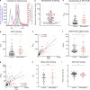
ZIKV neutralization capacity and ZEDIII binding are increased in mothers of microcephalic newborns. (a) The collection period of the maternal sera of this study (November 2015 to February 2016) is indicated in gray alongside the incidence of exanthematic diseases (blue, left y axis) and neonates born with microcephaly (red, right y axis) per epidemiological week in Bahia. Adapted from Ministry of Health of Brazil (2017). (b) 160 maternal sera (43 cases with microcephaly and 117 controls) were screened at 1:1,000 dilution for neutralization of ZIKV RVPs. Neutralization was expressed as the reciprocal of the luciferase activity normalized to no serum control. Samples below the dotted line (open symbols) were considered nonneutralizing. Each symbol represents the average of triplicate values for each donor. All triangles are maternal sera from microcephaly cases, and triangles pointing down represent ZIKV cases confirmed either by RT-PCR or IgM ELISA on cord blood. (c) ZIKV neutralization potency was determined using RVPs. The neutralization capacity was expressed as the reciprocal of the serum dilution resulting in 50% inhibition compared with RVPs alone (NT50). Each sample was evaluated in triplicate (n = 107, 40 cases with microcephaly and 67 controls). Three control samples that were identified as ZIKV neutralizers in the screening (panel b) were borderline in this assay and thus not plotted and omitted from further analysis. (d) IgG antibodies binding to ZEDIII were evaluated by ELISA. Binding was expressed as the reciprocal of the serum dilution resulting in 50% of maximal binding (BT50). Each value represents the average of two independent assays (n = 103, 40 cases with microcephaly and 63 controls). (e) Correlation between ZIKV neutralization potency, expressed as NT50, and ZEDIII binding, expressed as BT50. (f) Serum antibodies binding to the ZEDIII lateral ridge were determined by antigen competition ELISA and expressed as ΔBT50 (n = 103). (g) Correlation between ZIKV neutralization potency, expressed as NT50, and ZEDIII lateral ridge binding, expressed as ΔBT50. (h) IgG antibodies binding to UV-inactivated ZIKV were evaluated by ELISA. Optical densities were normalized by the control serum of a flavivirus naive individual vaccinated for YFV. Binding is expressed as the area under the curve (AUC) obtained in ELISA (n = 55, 27 microcephalies and 28 controls). (i) IgG antibodies binding to ZIKV NS1 protein were evaluated by ELISA. Optical densities were normalized as in panel h (n = 98, 39 cases with microcephaly and 59 controls). Each symbol represents an individual donor; black circles are from controls, and red triangles are from the microcephaly group. The P values in panels c, d, f, h, and i were determined with the Mann–Whitney test, and the mean and SD are shown. The P and ρ (rho) values for the correlation in panels e and g were determined with the Spearman test. *, P < 0.05; ***, P < 0.001. n.s., not significant.
Figure 2.
Figure 2.
Sera from mothers with microcephalic neonates have higher enhancing power and a higher peak enhancement titer. (a) Enhancement of infection (fitted curves) by ZIKV RVPs is presented as the average of the fold change in luciferase activity of each group compared with control antibody (see Materials and methods). The thick lines represent control (n = 64) and microcephaly (n = 40) groups with ZIKV neutralizing activity, and thin lines represent samples that lack ZIKV neutralizing activity (empty symbols in Fig. 1 b). Sera were serially diluted and the enhancement of infection at each dilution for each group is shown. Standard errors are indicated in gray. The profile of the individual samples is shown in Fig. S2 a (n = 160). (b) Evaluation of the serum enhancing power. The enhancing power is defined as the fold increase of infection at peak enhancement titer for each serum sample (Fig. S1 a; Halstead, 2003). (c) Evaluation of the peak enhancement titer. The peak enhancement titer is the serum dilution at which maximum infection occurs for any tested sample (Fig. S1 a; Halstead, 2003). (d) Correlation between enhancing power and neutralization capacity, expressed as NT50. (e) Correlation between peak enhancement titer and NT50. The P values in panels b and c were determined with the Mann–Whitney test, and the mean and SD are shown. Symbols represent individual donors (n = 104). The P and ρ (rho) values for the correlations in panels d and e were determined with the Spearman test. *, P < 0.05; **, P < 0.01; ***, P < 0.001.
Figure 3.
Figure 3.
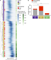
Clustering analysis identifies groups with different retrospective risks of microcephaly in humans. (a) Unsupervised hierarchical clustering of the log-normalized values for ZIKV RVP enhancement (ADE) combined with neutralization (NT50, n = 104). Clusters are indicated in the first column, and the presence or absence of neonatal microcephaly is indicated in the second column in red or gray, respectively. (b) Histogram with the number of microcephaly cases (red) and controls (gray) in the three clusters. Statistical analysis of the relative risks was performed using the Fisher’s exact test (***, P < 0.0001).
Figure 4.
Figure 4.
Clustering analysis identifies groups with different retrospective likelihood of fetal brain damage in macaques. (a) Unsupervised hierarchical clustering of the log-normalized values for ZIKV RVP enhancement (ADE) combined with neutralization (NT50, n = 32). Clusters are indicated in the first column and the degree of fetal brain damage is indicated in the second column in red or gray, respectively. (b) Histogram with the number of cases with moderate to severe fetal brain pathology or fetal loss (red) and controls with undetectable to mild lesion (gray) in the four clusters. The statistical analysis of the relative risks was performed using the Fisher’s exact test (*, P < 0.05). (c) Evaluation of gestational day (GD) of infection, duration of viremia, and last GD with detectable viremia. Duration of viremia is the difference between last detectable viremia and day of infection. The P values were not significant (P > 0.05 as determined with the Mann–Whitney test), and the mean and SD are shown. Symbols represent individual animals (n = 32, except in middle and bottom panel, where n = 31; see Table S2). n.s., not significant.

References

    1. Adams Waldorf K.M., Stencel-Baerenwald J.E., Kapur R.P., Studholme C., Boldenow E., Vornhagen J., Baldessari A., Dighe M.K., Thiel J., Merillat S., et al. 2016. Fetal brain lesions after subcutaneous inoculation of Zika virus in a pregnant nonhuman primate. Nat. Med. 22:1256–1259. 10.1038/nm.4193 - DOI - PMC - PubMed
    1. Adams Waldorf K.M., Nelson B.R., Stencel-Baerenwald J.E., Studholme C., Kapur R.P., Armistead B., Walker C.L., Merillat S., Vornhagen J., Tisoncik-Go J., et al. 2018. Congenital Zika virus infection as a silent pathology with loss of neurogenic output in the fetal brain. Nat. Med. 24:368–374. 10.1038/nm.4485 - DOI - PMC - PubMed
    1. Aldo P., You Y., Szigeti K., Horvath T.L., Lindenbach B., and Mor G.. 2016. HSV-2 enhances ZIKV infection of the placenta and induces apoptosis in first-trimester trophoblast cells. Am. J. Reprod. Immunol. 76:348–357. 10.1111/aji.12578 - DOI - PubMed
    1. Andrade D.V., and Harris E.. 2017. Recent advances in understanding the adaptive immune response to Zika virus and the effect of previous flavivirus exposure. Virus Res. 10.1016/j.virusres.2017.06.019 - DOI - PMC - PubMed
    1. Barba-Spaeth G., Dejnirattisai W., Rouvinski A., Vaney M.-C., Medits I., Sharma A., Simon-Lorière E., Sakuntabhai A., Cao-Lormeau V.-M., Haouz A., et al. 2016. Structural basis of potent Zika-dengue virus antibody cross-neutralization. Nature. 536:48–53. 10.1038/nature18938 - DOI - PubMed

Publication types

MeSH terms

Substances

Associated data