. 2019 Oct 15;93(21):e00699-19.
doi: 10.1128/JVI.00699-19. Print 2019 Nov 1.

Discovery of Novel Thrips Vector Proteins That Bind to the Viral Attachment Protein of the Plant Bunyavirus Tomato Spotted Wilt Virus

Affiliations

Discovery of Novel Thrips Vector Proteins That Bind to the Viral Attachment Protein of the Plant Bunyavirus Tomato Spotted Wilt Virus

Ismael E Badillo-Vargas et al. J Virol. .

Abstract

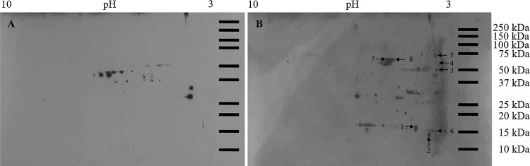
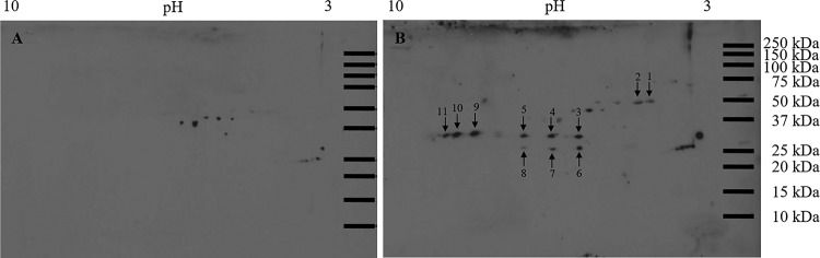
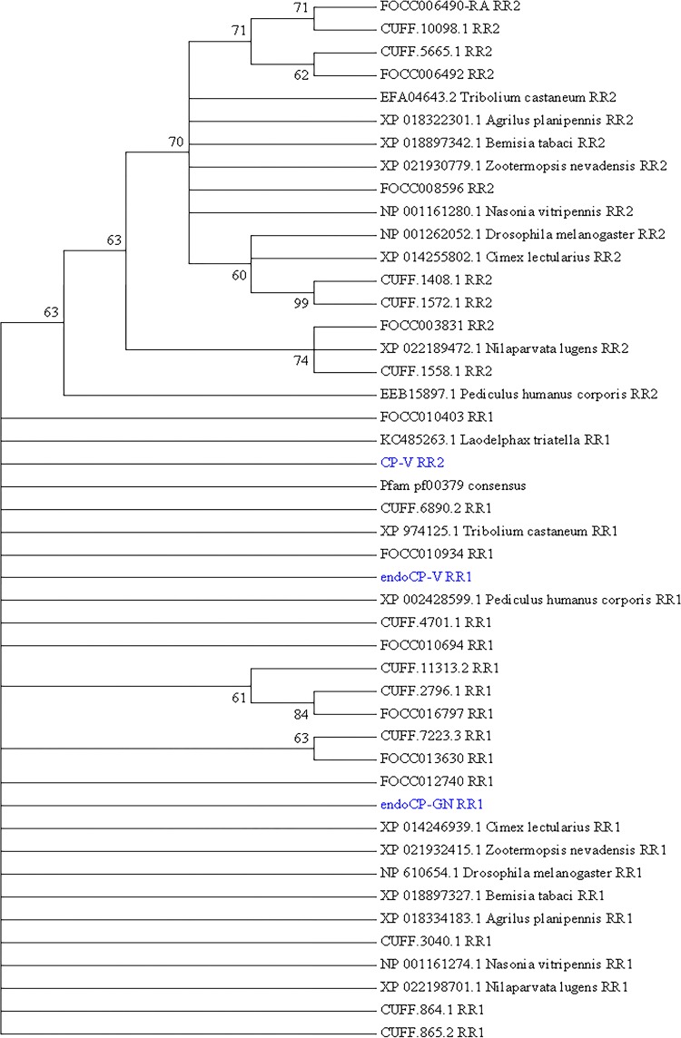
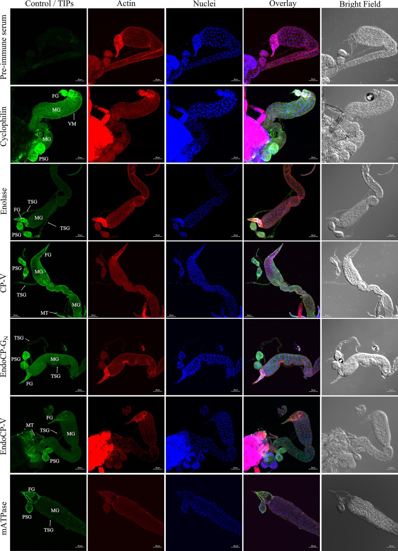
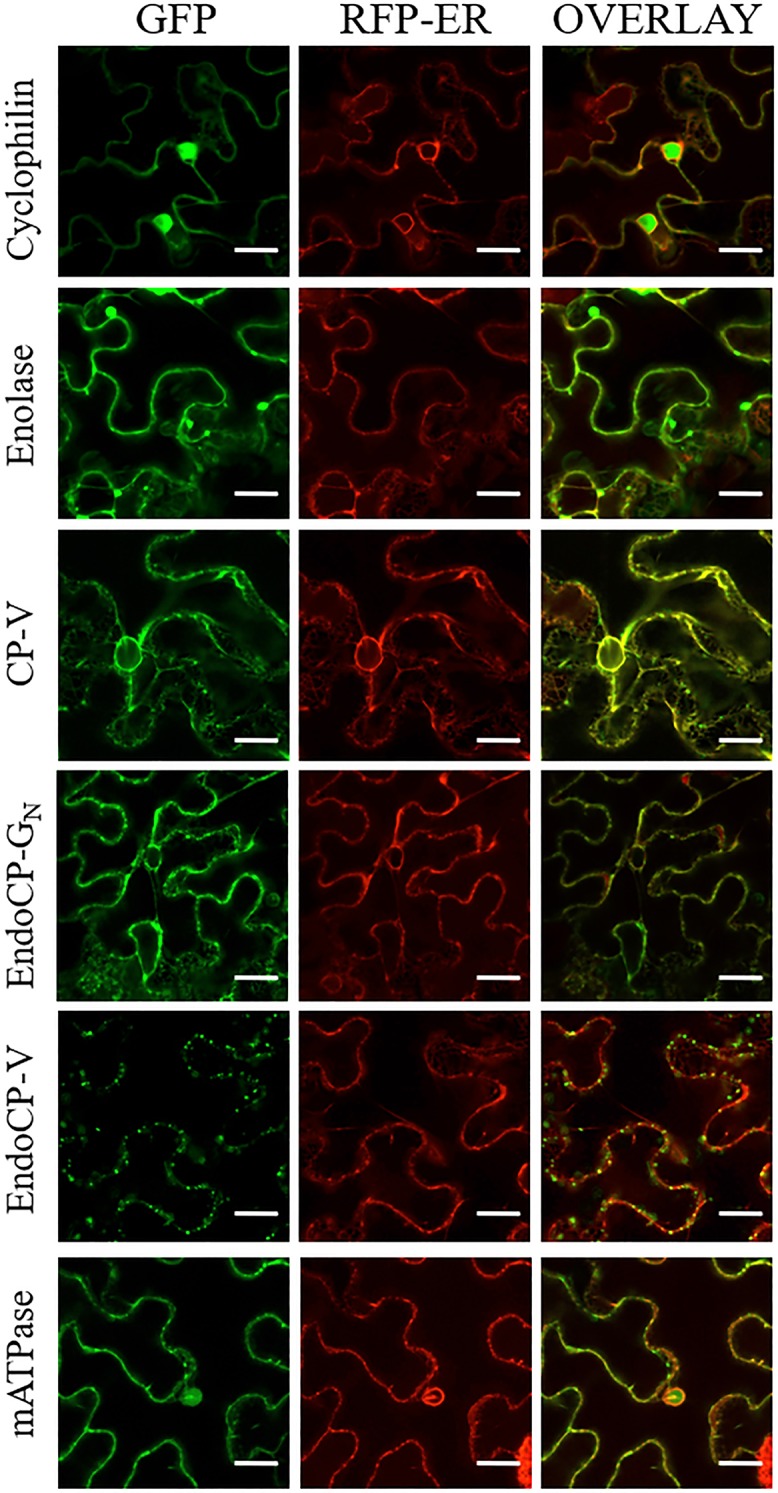
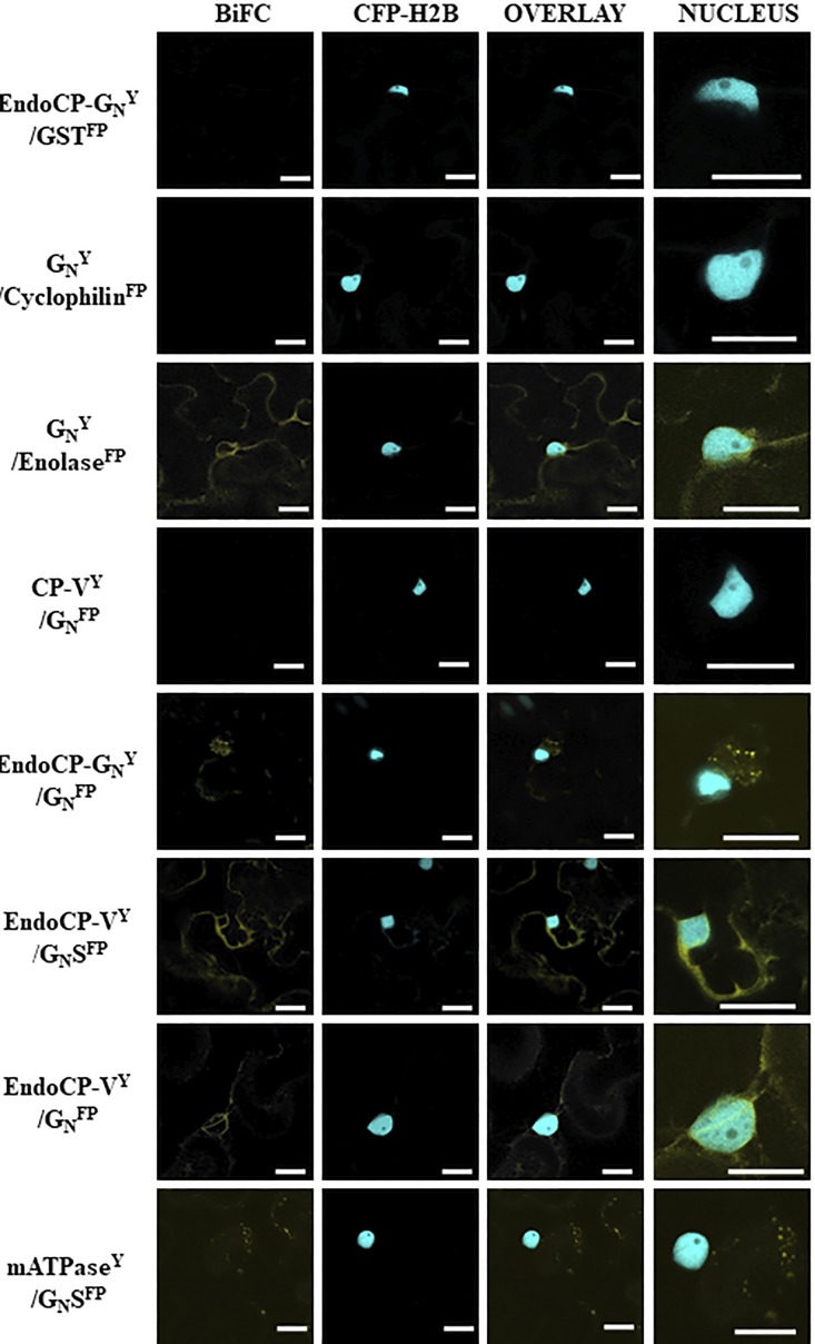
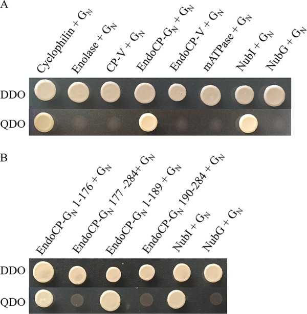
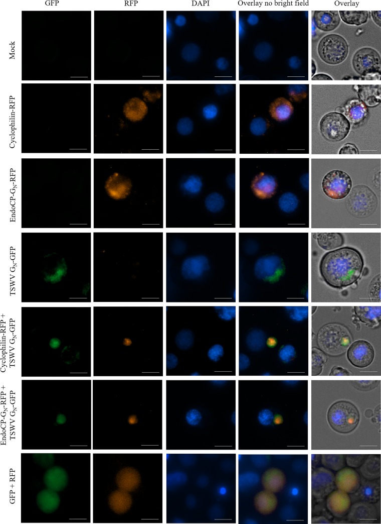
The plant-pathogenic virus tomato spotted wilt virus (TSWV) encodes a structural glycoprotein (GN) that, like with other bunyavirus/vector interactions, serves a role in viral attachment and possibly in entry into arthropod vector host cells. It is well documented that Frankliniella occidentalis is one of nine competent thrips vectors of TSWV transmission to plant hosts. However, the insect molecules that interact with viral proteins, such as GN, during infection and dissemination in thrips vector tissues are unknown. The goals of this project were to identify TSWV-interacting proteins (TIPs) that interact directly with TSWV GN and to localize the expression of these proteins in relation to virus in thrips tissues of principal importance along the route of dissemination. We report here the identification of six TIPs from first-instar larvae (L1), the most acquisition-efficient developmental stage of the thrips vector. Sequence analyses of these TIPs revealed homology to proteins associated with the infection cycle of other vector-borne viruses. Immunolocalization of the TIPs in L1 revealed robust expression in the midgut and salivary glands of F. occidentalis, the tissues most important during virus infection, replication, and plant inoculation. The TIPs and GN interactions were validated using protein-protein interaction assays. Two of the thrips proteins, endocuticle structural glycoprotein and cyclophilin, were found to be consistent interactors with GN These newly discovered thrips protein-GN interactions are important for a better understanding of the transmission mechanism of persistent propagative plant viruses by their vectors, as well as for developing new strategies of insect pest management and virus resistance in plants.IMPORTANCE Thrips-transmitted viruses cause devastating losses to numerous food crops worldwide. For negative-sense RNA viruses that infect plants, the arthropod serves as a host as well by supporting virus replication in specific tissues and organs of the vector. The goal of this work was to identify thrips proteins that bind directly to the viral attachment protein and thus may play a role in the infection cycle in the insect. Using the model plant bunyavirus tomato spotted wilt virus (TSWV), and the most efficient thrips vector, we identified and validated six TSWV-interacting proteins from Frankliniella occidentalis first-instar larvae. Two proteins, an endocuticle structural glycoprotein and cyclophilin, were able to interact directly with the TSWV attachment protein, GN, in insect cells. The TSWV GN-interacting proteins provide new targets for disrupting the viral disease cycle in the arthropod vector and could be putative determinants of vector competence.

Keywords: Bunyavirales; insect vector; orthotospovirus; plant virology; thrips; vector biology; virus-vector interactions.


Figures

FIG 1
FIG 1
Overlay assay using purified virions and F. occidentalis first-instar proteins resolved in two-dimensional gels. Total proteins (150 μg) extracted from pooled noninfected first-instar larvae (0 to 17 h old) of F. occidentalis were resolved by 2D gel electrophoresis and transferred to nitrocellulose membranes. After blocking, membranes were incubated overnight with blocking buffer (negative control) (A) or purified TSWV at 25 μg/ml (B), followed by incubation with polyclonal rabbit anti-TSWV GN antibodies. Only protein spots that consistently bound to purified TSWV in three (spots 1, 2, 4, 6, and 7) and four (spots 3, 5, and 8) biological replicates of the overlay assay were collected from three individual picking gels and subjected to ESI-mass spectrometry for protein identification. Protein spots observed in the no-overlay-control membrane represent nonspecific binding and were not collected for further analysis. Molecular mass (in kilodaltons) is shown on the y axis, and pI (as pH range) is shown on the x axis.
FIG 2
FIG 2
Overlay assay using recombinant GN and F. occidentalis first-instar proteins resolved in two-dimensional gels. Total proteins (150 μg) extracted from pooled healthy first-instar larvae (0 to 17 h old) of F. occidentalis were resolved by 2D gel electrophoresis and transferred to nitrocellulose membranes. The membranes were incubated overnight with blocking buffer (negative control) (A) or recombinant TSWV GN (3.5 μg/ml) (B). Using the polyclonal rabbit anti-TSWV GN, protein spots that consistently bound to the recombinant TSWV GN in two (spots 1 through 11) biological replicates of the overlay assay were collected from two individual picking gels and subjected to ESI-mass spectrometry for protein identification. Protein spots observed in the no-overlay-control membrane represent nonspecific binding and were not collected for further analysis. Molecular mass (in kilodaltons) is shown on the y axis, and pI (as pH range) is shown on the x axis.
FIG 3
FIG 3
Phylogenetic analysis of the extended R&R (Rebers and Riddiford, CPR-RR1 and CPR-RR2) consensus, a conserved chitin-binding motif (Chitin_bind_4 [CHB4]), of the three cuticle-associated TSWV-interacting proteins (TIPs) in first-instar larvae of Frankliniella occidentalis (cuticle protein-V [CP-V], endocuticle structural glycoprotein-V [endoCP-V], and endocuticle structural glycoprotein-GN [endoCP-GN]). The analysis involved 46 sequences, as follows: the three cuticle TIPs (in blue), the “gold standard” Pfam database extended R&R consensus sequence (pf00379), 19 insect orthologous sequences obtained from NCBI GenBank (accession numbers are present), and 23 structural CPs and endoCPs (translated transcripts, designated with FOCC or CUFF identifiers) previously reported to be differentially abundant in TSWV-infected first-instar larva of F. occidentalis (20). All positions with less than 30% site coverage in the multiple alignment were eliminated, resulting in 74 amino acid positions in the final data set. Evolutionary history was inferred by the maximum likelihood (ML) method based on the Jones-Taylor-Thornton (JTT) matrix-based model, and a discrete gamma distribution was used to model the variation among sites. The bootstrap consensus tree (500 replicates) was generated by the ML algorithm, and branches corresponding to partitions reproduced in less than 60% bootstrap replicates were collapsed. The numbers shown next to branches indicate the percentage of replicate trees in which the associated taxa (sequences) clustered together in the bootstrap test. The analysis was performed with MEGA7 (73).
FIG 4
FIG 4
Confirmation of binding specificity for antibodies produced against TSWV-interacting protein (TIP) (peptides) by dot blot analysis. Peptide antigens were diluted to 100 μg/ml (for cyclophilin, enolase, endoCP-GN, endoCP-V, and mATPase), and 2.5 mg/ml (for CP-V), and 2 μl of each peptide was used for each test. PBS buffer and preimmune serum (500,000 × dilution) were used as controls. All six diluted peptides and two controls were loaded onto six nitrocellulose membrane strips. Each strip was initially incubated with the homologous primary antibody (0.5 μg/ml, generated in mice), followed by incubation with anti-mouse-HRP (1:5,000 dilution). Each membrane strip was developed independently.
FIG 5
FIG 5
In situ detection of TSWV-interacting proteins (TIPs) in first-instar larvae of F. occidentalis. Synchronized first-instar larvae (0 to 17 h old) were kept on a 7% sucrose solution for 3 h to clean their guts from plant tissues. These larvae were then dissected and immunolabeled using specific antibodies against each TIP, as indicated. Thrips tissues incubated with preimmune mouse serum are depicted here. Confocal microscopy detection of green fluorescence (Alexa Fluor 488) represents the localization of each TIP, red represents Alexa Fluor 594-labeled actin, and blue represents DAPI-labeled nuclei. TIPs were mainly localized at the foregut (FG), midgut (MG), which includes epithelial cells and visceral muscle (VM), principal salivary glands (PSG), tubular salivary glands (TSG), and Malpighian tubules (MT). All scale bars = 50 μm.
FIG 6
FIG 6
Localization of TSWV-interacting proteins (TIPs) fused to green fluorescent protein (GFP) in Nicotiana benthamiana. Plants transgenic for an RFP-ER marker were infiltrated with Agrobacterium tumefaciens strain LBA 4404 suspensions of TIP constructs. Each row indicates the specific TIP-GFP fusion in relation to the RFP-ER marker. The columns are as follows, from left the right: GFP channel, RFP channel, and the overlay between the two channels. All scale bars = 20 μm.
FIG 7
FIG 7
Confirmation of interactions between TSWV proteins and TSWV-interacting proteins (TIPs) using bimolecular fluorescence complementation (BiFC) in Nicotiana benthamiana. Plants transgenic for a nuclear marker fused to cyan fluorescent protein (CFP-H2B) were infiltrated with suspensions of Agrobacterium tumefaciens transformed with plasmids encoding the GN protein (full-length or soluble form [GN-S]) and TIP proteins (endoCP-GN, cyclophilin, enolase, CP-V, endoCP-V, and mATPase) fused to either the amino or carboxy terminus of yellow fluorescent protein (YFP). The designation of Y indicates this is the N-terminal half of YFP, and FP represents the C-terminal half of YFP. The Y or FP position in the name indicates that all are carboxy-terminal fusions to the protein of interest. The positive interactors are seen by fluorescence of YFP in images shown in the BiFC column. The CFP-H2B column is indicated to give cellular reference, and the overlay between the two is also shown. The final column is the nucleus enlarged to show detail of the interacting TIPs within the cellular context. The first row is a representative negative control, including a TIP and glutathione S-transferase (all thrips and virus proteins were tested with the negative control to rule out nonspecific interactions). All scale bars = 20 μm.
FIG 8
FIG 8
Validation of interactions between GN and TSWV-interacting proteins (TIPs) and identification of the interacting domain of endoCP-GN using a split-ubiquitin membrane-based yeast two hybrid (MbY2H) assay. (A) Interactions between GN and six TIPs. GN was expressed as GN-Cub, and TIPs were expressed as NubG-TIPs using MbY2H vectors. (B) Interactions between TSWV GN and different regions of endoCP-GN. EndoCP-GN was expressed as either the N-terminal domain (amino acids 1 to 176 and 1 to 189) that includes the nonconserved region or the C-terminal region (amino acids 177 to 284 and 190 to 284) that includes the conserved Chitin_bind_4 motif (CHB4) of endoCP-GN. Interactions between GN-Cub and NubI and between GN-Cub and NubG were used as positive and negative controls, respectively, for all MbY2H assays. Cotransformation of pTSU2-APP and pNubG-Fe65 into NYM51 was used as another positive control (data not shown). DDO, yeast double-dropout (SD/−Leu/−Trp) medium; QDO, yeast quadruple-dropout (SD/−Ade/−His/−Leu/−Trp) medium.
FIG 9
FIG 9
Colocalizations of TSWV GN with endoCP-GN or cyclophilin in insect cells. Open reading frames of cyclophilin, endoCP-GN, and TSWV GN were cloned into Drosophila Gateway vectors (under the control of the Hsp70 promoter), and the resulting pHWR and pHWG expression plasmids were used for the following fusion proteins: cyclophilin-RFP, endoCP-GN-RFP, and TSWV GN-GFP. The recombinant plasmids, pHWR-cyclophilin, pHWR-endoCP-GN, and pHWG-TSWV-GN, were singly transfected or cotransfected into insect Sf9 cells. All transfection reactions were performed using the Cellfectin II reagent. Mock, no DNA treatment (top left panels) and cotransfection of pHRW and pHGW expression plasmids (bottom left panels) were used as controls. Cells were stained with DAPI 72 h posttransfection and then visualized using the Cytation 5 cell imaging multimode reader (BioTek, Winooski, VT) to detect red and green fluorescence. Exposure settings (LED intensity/integration time/camera gain) of the mock control were set as the baseline parameters for the analysis of all other treatments. Cells were visualized with a 40× objective. Scale bars = 10 μm.

References

    1. Gubler DJ. 1998. Resurgent vector-borne diseases as a global health problem. Emerg Infect Dis 4:442–450. doi:10.3201/eid0403.980326. - DOI - PMC - PubMed
    1. Gubler DJ. 2002. The global emergence/resurgence of arboviral diseases as public health problems. Arch Med Res 33:330–342. doi:10.1016/S0188-4409(02)00378-8. - DOI - PubMed
    1. Beaty BJ, Bishop DH. 1988. Bunyavirus-vector interactions. Virus Res 10:289–301. doi:10.1016/0168-1702(88)90071-8. - DOI - PubMed
    1. Elliott RM. 1997. Emerging viruses: the Bunyaviridae. Mol Med 3:572–577. doi:10.1007/BF03401814. - DOI - PMC - PubMed
    1. Horne KM, Vanlandingham DL. 2014. Bunyavirus-vector interactions. Viruses 6:4373–4397. doi:10.3390/v6114373. - DOI - PMC - PubMed

Publication types

MeSH terms

LinkOut - more resources