. 2019 Oct 15;93(21):e00996-19.
doi: 10.1128/JVI.00996-19. Print 2019 Nov 1.

Dengue Virus M Protein Promotes NLRP3 Inflammasome Activation To Induce Vascular Leakage in Mice

Affiliations

Dengue Virus M Protein Promotes NLRP3 Inflammasome Activation To Induce Vascular Leakage in Mice

Pan Pan et al. J Virol. .

Abstract

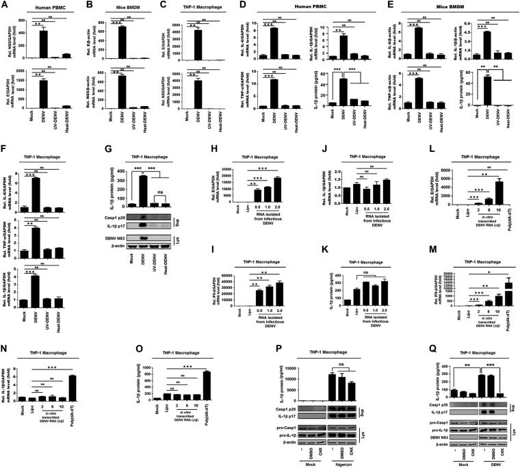
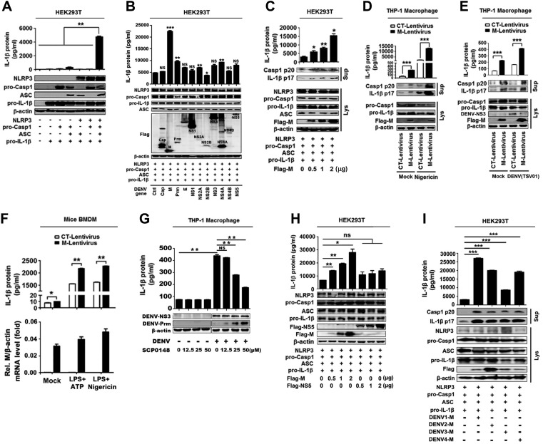
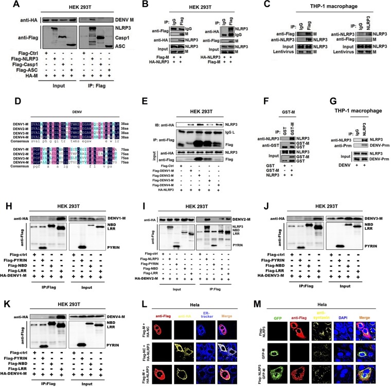
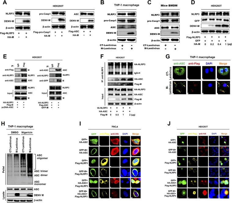
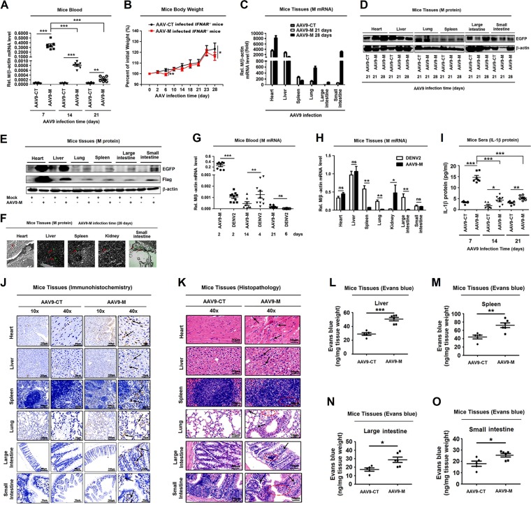
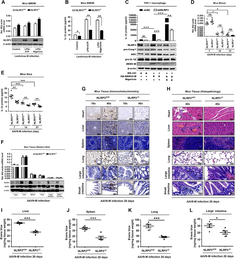
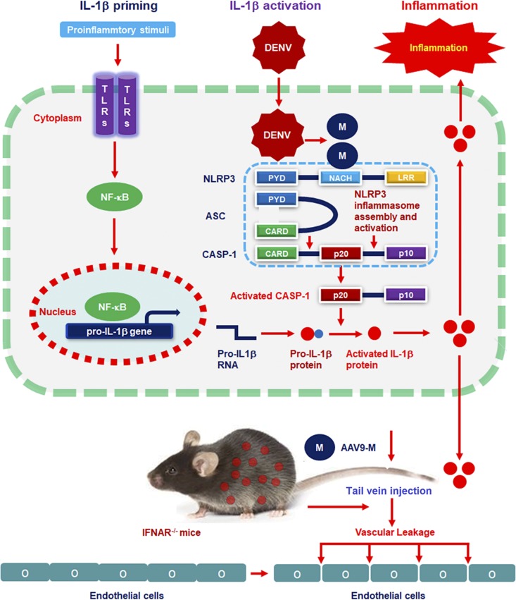
Dengue virus (DENV) infection causes serious clinical symptoms, including dengue hemorrhagic fever (DHF) and dengue shock syndrome (DSS). Vascular permeability change is the main feature of the diseases, and the abnormal expression of proinflammatory cytokines is the important cause of vascular permeability change. However, the mechanism underlying vascular permeability induced by DENV has not been fully elucidated. Here, we reveal a distinct mechanism by which DENV infection promotes NLRP3 inflammasome activation and interleukin-1 beta (IL-1β) release to induce endothelial permeability and vascular leakage in mice. DENV M protein interacts with NLRP3 to facilitate NLRP3 inflammasome assembly and activation, which induce proinflammatory cytokine IL-1β activation and release. Notably, M can induce vascular leakage in mouse tissues by activating the NLRP3 inflammasome and IL-1β. More importantly, inflammatory cell infiltration and tissue injuries are induced by M in wild-type (WT) mouse tissues, but they are not affected by M in NLRP3 knockout (NLRP3-/-) mouse tissues. Evans blue intensities in WT mouse tissues are significantly higher than in NLRP3-/- mouse tissues, demonstrating an essential role of NLRP3 in M-induced vascular leakages in mice. Therefore, we propose that upon DENV infection, M interacts with NLRP3 to facilitate inflammasome activation and IL-1β secretion, which lead to the induction of endothelial permeability and vascular leakage in mouse tissues. The important role of the DENV-M-NLRP3-IL-1β axis in the induction of vascular leakage provides new insights into the mechanisms underlying DENV pathogenesis and DENV-associated DHF and DSS development.IMPORTANCE Dengue virus (DENV) is a mosquito-borne pathogen, and infections by this virus are prevalent in over 100 tropical and subtropical countries or regions, with approximately 2.5 billion people at risk. DENV infection induces a spectrum of clinical symptoms, ranging from classical dengue fever (DF) to severe dengue hemorrhagic fever (DHF) and dengue shock syndrome (DSS). Therefore, it is important to understand the mechanisms underlying DENV pathogenesis. In this study, we reveal that the DENV membrane protein (M) interacts with the host NLRP3 protein to promote NLRP3 inflammasome activation, which leads to the activation and release of a proinflammatory cytokine, interleukin-1 beta (IL-1β). More importantly, we demonstrate that M protein can induce vascular permeability and vascular leakage and that NLRP3 is required for M-induced vascular leakage in mouse tissues. Collectively, this study reveals a distinct mechanism underlying DENV pathogeneses and provides new insights into the development of therapeutic agents for DENV-associated diseases.

Keywords: DENV; DENV membrane protein; IL-1β; M; NLRP3; NLRP3 inflammasome; Nod-like receptor (NLR) family pyrin domain containing 3; dengue virus; interleukin-1β; vascular leakage; vascular permeability.


Figures

FIG 1
FIG 1
The replication and protein synthesis of Dengue virus are essential for the activation of NLRP3 inflammasome. Human PBMCs (A, D), GM-CSF differentiated mouse BMDMs (B, E), and PMA-differentiated THP-1 macrophages (C, F, and G) were treated with infectious, UV-, or heat-inactivated DENV2(NGC) at an MOI of 5 for 24 h. The DENV E gene mRNA and NS5 gene mRNA were quantified by RT-PCR (A to C). IL-1β RNA, IL-6 mRNA, and TNF-α expression was determined by qRT-PCR, and IL-1β was measured by ELISA (D to F). IL-1β in cell supernatants was measured by ELISA and proteins in cell extracts (Lys) were analyzed by Western blotting (WB) (G). THP-1 macrophages were transfected with RNA extracted from infectious DENV2(NGC) at 0.5, 1, and 2 μg (H to K) or with in vitro-transcribed DENV2(TSV01) RNA at 2, 6, and 10 μg or treated with 10 μg/ml poly(dA·dT) (L to O). The DENV E gene mRNA (H, L), IFN-β RNA (I, M), or IL-1β RNA (J, N) was determined by RT-PCR, and IL-1β was measured by ELISA (K, O). (P, Q) PMA-differentiated THP-1 macrophages were pretreated with 100 μM CHX for 2 h and stimulated with nigericin (2 μM) for 2 h (P) or infected with DENV at an MOI of 5 for 48 h (Q). Supernatants were analyzed (top) by ELISA for IL-1β. Cell lysates were analyzed (bottom) by immunoblotting. Data are representative of three independent experiments. Mock, supernatant of C6/36 cells without DENV2 infection (A to G, P, and Q) or unprocessed cells (H to O); Lipo, transfection with Lipofectamine 2000 reagent. Values are means ± SEMs. ns, not significant; *, P ≤ 0.05; **, P ≤ 0.01; ***, P ≤ 0.001.
FIG 2
FIG 2
M protein promotes the activation of the NLRP3 inflammasome. (A) HEK293T cells were transfected with the expression plasmid pro-IL-1β, pro-IL-1β, and pro-caspase-1, any three of the four plasmids, and four plasmids (pro-IL-1β, pro-caspase-1, ASC, and NLRP3) for 48 h. (Top) Supernatants were analyzed by ELISA for IL-1β. (Bottom) Cell lysates were analyzed by immunoblotting. (B) HEK293T cells were cotransfected with plasmids encoding NLRP3, ASC, pro-caspase-1 (pro-Casp1), or pro-IL-1β and with plasmids encoding Cap, M, Prm, E, NS1, NS2A, NS2B, NS3, NS4A, NS4B, or NS5 for 48 h. (Top) Supernatants were analyzed by ELISA for IL-1β. (Bottom) Cell lysates were analyzed by immunoblotting. (C) HEK293T cells were cotransfected with plasmids encoding NLRP3, ASC, pro-Casp1, and pro-IL-1β and with different concentrations of plasmid encoding DENV2 M for 48 h. (Top) Supernatants were analyzed by ELISA for IL-1β. (Bottom) Cell lysates were analyzed by immunoblotting. (D) THP-1 cells were stably infected with lentivirus-CT or lentivirus-M, differentiated into macrophages, and stimulated with 2 μM nigericin. IL-1β in cell supernatants was measured by ELISA and proteins in Lys were analyzed by WB. (E) THP-1 cells were stably infected with lentivirus-CT or lentivirus-M, differentiated into macrophages, and stimulated with DENV2(TSV01) at an MOI of 5 for 48 h. IL-1β in cell supernatants was measured by ELISA and proteins in Lys were analyzed by WB. (F) Mouse BMDMs were infected with lentivirus-CT or lentivirus-M and stimulated with LPS (1 μg/ml) plus ATP (2.5 mM) or LPS (1 μg/ml) plus nigericin (2 μM). IL-1β in supernatants was measured by ELISA and M gene mRNA was quantified by RT-PCR. (G) PMA-differentiated THP-1 macrophages were infected with DENV2(NGC) at an MOI of 5 for 12 h, and then Furin protease inhibitor (SCP0148) was added at different concentrations (12.5, 25, 50 μM) and the infection allowed to continue for 12 h. IL-1β in cell supernatants was measured by ELISA and proteins in Lys were analyzed by WB. (H) HEK293T cells were cotransfected with plasmids encoding NLRP3, ASC, pro-Casp1, and pro-IL-1β and with different concentrations of plasmid encoding DENV2 M or DENV2 NS5 (0, 0.5, 1, and 2 μg) for 48 h. (Top) Supernatants were analyzed by ELISA for IL-1β. (Bottom) Cell lysates were analyzed by immunoblotting. (I) HEK293T cells were cotransfected with plasmids encoding NLRP3, ASC, pro-Casp1, and pro-IL-1β, and transfected with plasmid encoding DENV1-M, DENV2-M, DENV3-M, and DENV4-M (2 μg) for 48 h. (Top) Supernatants were analyzed by ELISA for IL-1β. (Bottom) Cell lysates were analyzed by immunoblotting. Ctrl, empty plasmid (B); Mock, unprocessed cells (D, E, and F). Data are representative of three independent experiments. Values are means ± SEMs. ns, not significant; *, P ≤ 0.05; **, P ≤ 0.01; ***, P ≤ 0.001.
FIG 3
FIG 3
Dengue virus M only interacts with NLRP3 protein. (A) HEK293T cells were cotransfected with HA-M and Flag-NLRP3, Flag-pro-caspase-1, or Flag-ASC. Cell lysates were immunoprecipitated using anti-Flag antibody and analyzed using anti-Flag and anti-HA antibodies. Cell lysates (40 μg) were used as input. (B) HEK293T cells were cotransfected with HA-NLRP3 and Flag-M. Cell lysates were immunoprecipitated using anti-Flag antibody or anti-HA antibody and analyzed using anti-Flag and anti-HA antibodies. Cell lysates (40 μg) were used as input. (C) THP-1 macrophages were stably infected with lentivirus-M. Cell lysates were immunoprecipitated using anti-Flag antibody or anti-NLRP3 antibody and analyzed using anti-Flag and anti-NLRP3 antibodies. Cell lysates (50 μg) were used as input. (D) Sequence alignment of dengue virus M protein with different serotypes. (E) HEK293T cells were cotransfected with HA-NLRP3 and Flag-DENV1-M, Flag-DENV2-M, Flag-DENV3-M, or Flag-DENV4-M. Cell lysates were immunoprecipitated using anti-Flag antibody and analyzed using anti-Flag and anti-HA antibodies. Cell lysates (40 μg) were used as input. (F) Purified GST (10 μg) or GST-M (10 μg) was incubated with cell lysates of Flag-NLRP3-transfected HEK293T cells. (G) THP-1 macrophages were infected with DENV2(NGC) for 48 h. Cell lysates were immunoprecipitated using anti-NLRP3 antibody and analyzed using anti-DENV2-Prm antibody. Cell lysates (40 μg) were used as input. (H to K) HEK293T cells were cotransfected with HA-DENV1-M, HA-DENV2-M, HA-DENV3-M, HA-DENV4-M, and Flag-NLRP3, Flag-PYRIN, Flag-NBD, or Flag-LRR. Cell lysates were immunoprecipitated using anti-Flag antibody and analyzed using anti-Flag and anti-HA antibodies. Cell lysates (40 μg) were used as input. (L) HeLa cells were transfected with Flag-M, HA-NLRP3, or Flag-M/HA-NLRP3 for 24 h. Endoplasmic reticulum marker ER-Tracker (blue), Flag-M (red), and HA-NLRP3 (yellow) were then visualized with confocal microscopy. (M) HeLa cells were transfected with Flag-NLRP3, GFP-M, or GFP-M/Flag-NLRP3 for 24 h. Golgi apparatus marker antibodies against syntaxin 6 (yellow), Flag-tagged NLRP3 (red), and GFP-tagged M (green) and nucleus marker DAPI (blue) were then visualized with confocal microscopy. Flag-Ctrl, Flag-NC, or HA-NC indicates pcDNA3.1(+)-3×Flag or pCAggs-HA empty plasmid. Data are representative of three independent experiments.
FIG 4
FIG 4
Dengue virus M facilitates the assembly of NLRP3 inflammasome by interacting with NLRP3. (A) HEK293T cells were cotransfected with HA-CT or HA-M plus Flag-NLRP3, Flag-pro-Casp1, or Flag-ASC for 36 h. The indicated proteins in cell extracts were analyzed by WB. (B) THP-1 cells were stably infected with lentivirus-CT or lentivirus-M and differentiated into macrophages, and the indicated proteins in cell extracts were analyzed by WB. (C) Mouse BMDMs were infected with lentivirus-CT or lentivirus-M for 48 h, and the indicated proteins in cell extracts were analyzed by WB. (D) HEK293T cells were cotransfected with different concentrations of HA-M plus Flag-NLRP3 and GFP for 36 h. The indicated proteins in cell extracts were analyzed by WB. (E) HEK293T cells were cotransfected with Flag-M, HA-NLRP3, and pcDNA3.1(+)-ASC for 24 h. Cell lysates were immunoprecipitated using anti-Flag antibody and analyzed using anti-NLRP3, anti-ASC, and anti-Flag antibodies. HEK293T cells were cotransfected with GFP-M, HA-NLRP3, and Flag-ASC. Cell lysates were immunoprecipitated using anti-Flag antibody and analyzed using anti-NLRP3, anti-ASC, and anti-GFP antibodies. Cell lysates (40 μg) were used as inputs. (F) HEK293T cells were cotransfected with different concentrations of Flag-M plus HA-NLRP3 and HA-ASC for 24 h. Cell lysates were immunoprecipitated using anti-NLRP3 antibody and analyzed using anti-HA and anti-Flag antibodies. (G) THP-1 macrophages were stably infected with lentivirus-CT or lentivirus-M. Subcellular locations of ASC, Flag-M, and DAPI were visualized under confocal microscopy. (H) THP-1 macrophages were stably infected with lentivirus-CT or lentivirus-M and stimulated by 2 μM nigericin. ASC oligomerization was analyzed by immunoblotting. HeLa cells (I) and HEK293T cells (J) were cotransfected with GFP/HA-ASC, GFP-M/HA-ASC, GFP/Flag-NLRP3, GFP-M/Flag-NLRP3, GFP/HA-ASC/Flag-NLRP3, or GFP-M/HA-ASC/Flag-NLRP3 for 24 h. The subcellular locations of HA-tagged ASC (red), Flag-tagged NLRP3 (yellow), GFP-tagged M (green), and nucleus marker DAPI (blue) were visualized with confocal microscopy. The data are representative of three independent experiments.
FIG 5
FIG 5
DENV M protein induces IL-1β activation and vascular leakage in mice. IFNAR−/− C57BL/6 genetic background mice received tail vein injections of 300 μl containing 5 × 1011 vg of AAV9-EGFP or AAV9-EGFP-M. Serum was collected every 7 days for AAV9 groups from the orbits. (A) Total RNA was extracted from the plasma from mice. The RNA level of M protein was quantified by qRT-PCR. (B) Mice (n = 8 per group) were weighed every 2 days, and weight was expressed as the percentage of the initial weight. (C) The RNA levels of M protein in heart, liver, spleen, lung, large intestine, and small intestine were quantified by qRT-PCR. (D) Western blot analysis of EGFP expression in heart, liver, spleen, lung, large intestine, and small intestine after AAV9-EGFP and AAV9-EGFP-M infection. (E) Western blot analysis of EGFP and Flag-M expression in heart, liver, spleen, lung, large intestine, and small intestine in mock- and AAV9-EGFP-M-infected mice. (F) Immunofluorescence analysis of EGFP expression in heart, liver, spleen, kidney, and small intestine after AAV9-EGFP-M infection. (G) Total RNAs was extracted from blood samples from DENV2(NGC)-infected IFNAR−/− mice (n = 10) at 2, 4, and 6 days postinfection or AAV9-EGFP-M-infected IFNAR−/− mice (n = 8) at 2, 14, and 21 days postinfection. The DENV M gene mRNA was quantified by RT-PCR. (H) Total RNA was extracted from tissues, including heart, liver, spleen, lung, kidney, large intestine, and small intestine, from DENV2(NGC)-infected mice after 7 days or AAV9-EGFP-M-infected mice after 28 days. The DENV M gene mRNA was quantified by RT-PCR. (I) IL-1β in sera was measured by ELISA. Points represent the IL-1β values from each serum sample. At 28 days postinfection, mice were euthanized, and tissues were collected. (J) Immunohistochemistry analysis of IL-1β in the tissues, including heart, liver, spleen, lung, large intestine, and small intestine after AAV9-EGFP or AAV9-EGFP-M infection. Black arrows indicated the immunostaining of IL-1β. (K) Histopathology analysis of tissues, including heart, liver, spleen, lung, large intestine, and small intestine, after AAV9-EGFP or AAV9-EGFP-M infection. Black arrows indicated the infiltrated inflammatory cells; red circles indicate the aberrant cells. Evans blue dye was intravenously injected into mice 28 days after infection with AAV9-EGFP-M (n = 6) and AAV9-EGFP control (n = 5). The dye circulated for 2 h before mice were euthanatized, tissues including liver (L), spleen (M), large intestine (N), and small intestine (O) were collected, and the value of Evans blue was measured at OD610. Data are representative of two independent experiments. Values are means ± SEMs. ns, not significant; *, P ≤ 0.05; **, P ≤ 0.01; ***, P ≤ 0.001.
FIG 6
FIG 6
NLRP3 is crucial for M-induced IL-1β activation and vascular leakage in mice. (A and B) GM-CSF differentiated NLRP3+/+ mouse BMDM cells and NLRP3−/− mouse BMDM cells were infected with M-encoding lentivirus for 48 h and then stimulated with LPS (1 μg/ml) for 6 h and ATP (2.5 mM) for 20 min or nigericin (2 μM) for 2 h. (A) The relative M RNA level was measured by RT-PCR, and NLRP3 and β-actin proteins were determined by Western blotting. (B) IL-1β secreted in cell supernatants was analyzed by ELISA. PMA-differentiated THP-1 macrophages stably expressing short hairpin RNAs (sh-RNAs), sh-NLRP3, and transfected with HA-ctrl or HA-DENV2-M plasmid for 48 h were stimulated with nigericin (2 μM) for 2 h. (C) IL-1β in cell supernatants was measured by ELISA (top) and proteins in cell extract (Lys) were analyzed by WB (bottom). (D and E) NLRP3+/+ C57BL/6 mice (n = 6) or NLRP3−/− C57BL/6 mice (n = 6) received tail vein injections of 300 μl of M-encoding AAV9 (5 × 1011 vg) (AAV9-EGFP-M). Serum was collected every 7 days for AAV9 groups from the orbits. Total RNA was extracted from plasma samples from mice. (D) The RNA level of M protein was quantified by qRT-PCR. (E) IL-1β in the sera was measured by ELISA. Points represent the IL-1β values from each serum sample. At 28 days postinfection, mice were euthanized, and tissues were collected. (F) The levels of M RNA in heart, liver, spleen, lung, large intestine, and small intestine were quantified by qRT-PCR analyses (top), and the levels of NLRP3 protein and EGFP were determined by Western blot analyses (bottom). (G) Immunohistochemistry analysis of IL-1β in the tissues, including heart, liver, spleen, lung, large intestine, and small intestine, after AAV9-EGFP-M infection. Black arrows indicated the immunostaining of IL-1β. (H) Histopathology analysis of tissues, including heart, liver, spleen, lung, large intestine, and small intestine, after AAV9-EGFP-M infection. Black arrows indicate the infiltrated inflammatory cells. At 28 days postinfection, mice were intravenously injected with Evans blue dye. The dye was circulated for 2 h before mice were euthanized, and tissues including liver (I), spleen (J), lung (K), and large intestine (L) were collected. The value of Evans blue was measured at OD610. Data are representative of two independent experiments. Values are means ± SEMs. *, P ≤ 0.05; **, P ≤ 0.01; ***, P ≤ 0.001.
FIG 7
FIG 7
A proposed model in which DENV M induces vascular leakage by activating the NLRP3 inflammasome. Upon DENV infection, the Prm protein is cleaved by Furin protease in the Golgi apparatus to form M protein. M protein then interacts with NLRP3 to facilitate NLRP3 inflammasome assembly. Once the NLRP3 inflammasome is activated, a large proportion of IL-1β is produced and released. Mature proinflammatory cytokine IL-1β then causes changes in endothelial cell permeability, including in the liver, spleen, large intestine, and small intestine.

References

    1. Wilder-Smith A, Ooi EE, Horstick O, Wills B. 2019. Dengue. Lancet 393:350–363. doi:10.1016/S0140-6736(18)32560-1. - DOI - PubMed
    1. Guzman MG, Gubler DJ, Izquierdo A, Martinez E, Halstead SB. 2016. Dengue infection. Nat Rev Dis Primers 2:16055. doi:10.1038/nrdp.2016.55. - DOI - PubMed
    1. Bhatt S, Gething PW, Brady OJ, Messina JP, Farlow AW, Moyes CL, Drake JM, Brownstein JS, Hoen AG, Sankoh O, Myers MF, George DB, Jaenisch T, Wint GR, Simmons CP, Scott TW, Farrar JJ, Hay SI. 2013. The global distribution and burden of dengue. Nature 496:504–507. doi:10.1038/nature12060. - DOI - PMC - PubMed
    1. Martina BE, Koraka P, Osterhaus AD. 2009. Dengue virus pathogenesis: an integrated view. Clin Microbiol Rev 22:564–581. doi:10.1128/CMR.00035-09. - DOI - PMC - PubMed
    1. Guzman MG, Halstead SB, Artsob H, Buchy P, Farrar J, Gubler DJ, Hunsperger E, Kroeger A, Margolis HS, Martinez E, Nathan MB, Pelegrino JL, Simmons C, Yoksan S, Peeling RW. 2010. Dengue: a continuing global threat. Nat Rev Microbiol 8:S7–S16. doi:10.1038/nrmicro2460. - DOI - PMC - PubMed

Publication types

MeSH terms

LinkOut - more resources