. 2019 Oct 15;93(21):e00929-19.
doi: 10.1128/JVI.00929-19. Print 2019 Nov 1.

Phylogeography of Lassa Virus in Nigeria

Affiliations

Phylogeography of Lassa Virus in Nigeria

Deborah U Ehichioya et al. J Virol. .

Abstract

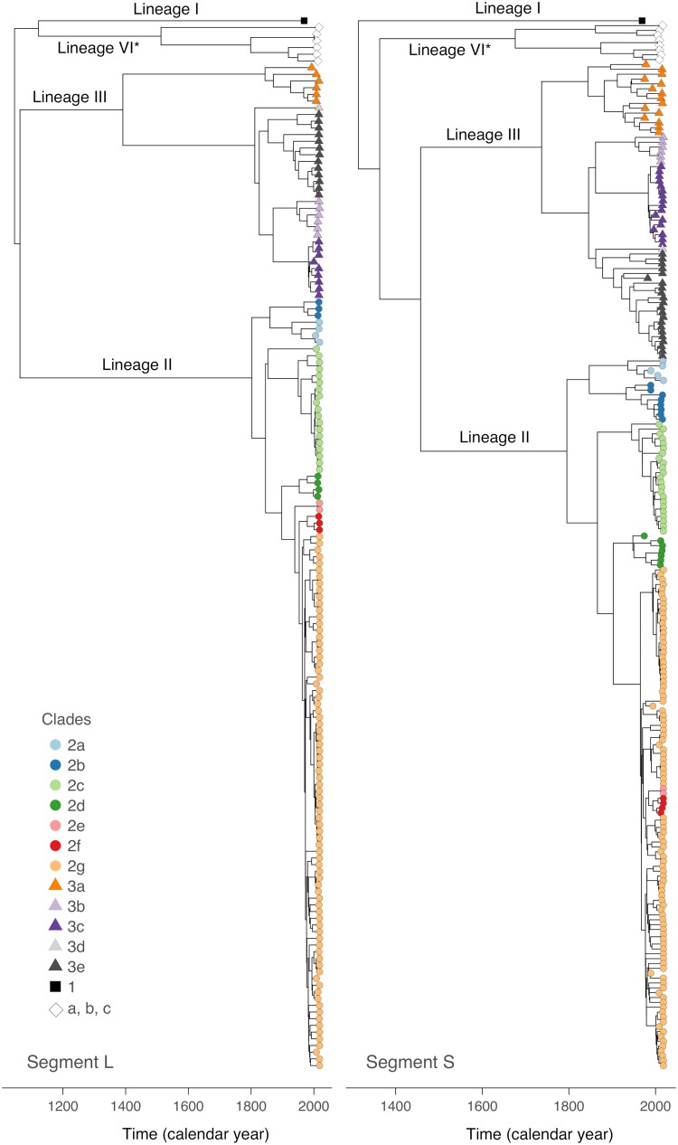
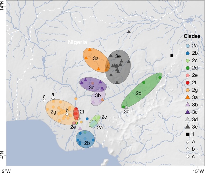
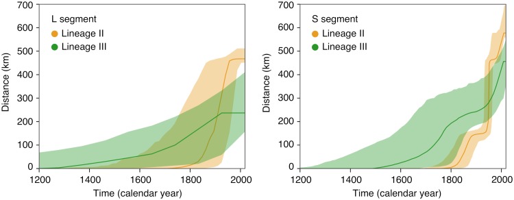
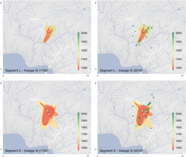
Lassa virus is genetically diverse with several lineages circulating in West Africa. This study aimed at describing the sequence variability of Lassa virus across Nigeria and inferring its spatiotemporal evolution. We sequenced and isolated 77 Lassa virus strains from 16 Nigerian states. The final data set, including previous works, comprised metadata and sequences of 219 unique strains sampled between 1969 and 2018 in 22 states. Most of this data originated from Lassa fever patients diagnosed at Irrua Specialist Teaching Hospital, Edo State, Nigeria. The majority of sequences clustered with the main Nigerian lineages II and III, while a few sequences formed a new cluster related to Lassa virus strains from Hylomyscus pamfi Within lineages II and III, seven and five sublineages, respectively, were distinguishable. Phylogeographic analysis suggests an origin of lineage II in the southeastern part of the country around Ebonyi State and a main vector of dispersal toward the west across the Niger River, through Anambra, Kogi, Delta, and Edo into Ondo State. The frontline of virus dispersal appears to be in Ondo. Minor vectors are directed northeast toward Taraba and Adamawa and south toward Imo and Rivers. Lineage III might have spread from northern Plateau State into Kaduna, Nasarawa, Federal Capital Territory, and Bauchi. One sublineage moved south and crossed the Benue River into Benue State. This study provides a geographic mapping of lineages and phylogenetic clusters in Nigeria at a higher resolution. In addition, we estimated the direction and time frame of virus dispersal in the country.IMPORTANCE Lassa virus is the causative agent of Lassa fever, a viral hemorrhagic fever with a case fatality rate of approximately 30% in Africa. Previous studies disclosed a geographical pattern in the distribution of Lassa virus strains and a westward movement of the virus across West Africa during evolution. Our study provides a deeper understanding of the geography of genetic lineages and sublineages of the virus in Nigeria. In addition, we modeled how the virus spread in the country. This knowledge allows us to predict into which geographical areas the virus might spread in the future and prioritize areas for Lassa fever surveillance. Our study not only aimed to generate Lassa virus sequences from across Nigeria but also to isolate and conserve the respective viruses for future research. Both isolates and sequences are important for the development and evaluation of medical countermeasures to treat and prevent Lassa fever, such as diagnostics, therapeutics, and vaccines.

Keywords: Lassa virus; Nigeria; phylogeny.


Figures

FIG 1
FIG 1
Time-scaled maximum clade credibility trees for S and L segments. The evolutionary relationships among the Lassa virus lineages in Nigeria were investigated by BEAST analysis using a simple constant population size model. Detailed phylogenies of lineages II and III based on a flexible skygrid coalescent model are shown in Fig. S3 to S6 in the supplemental material. The sublineages (clades 2a through 2g and 3a through 3e for lineages II and III, respectively) have been defined according to the phylogenies inferred separately for lineages II and III and geographic location of the strains (see Fig. S3 to S8 in the supplemental material). Detailed phylogenies of lineage I (clade 1) and the cluster including Lassa virus strains from H. pamfi (lineage VI, clade c) and related human sequences (clades a and b) are shown in Fig. S1 and S2 in the supplemental material. The molecular clock rate estimated for the L and S tree was 7.8 × 10−4 substitutions/(site × year) (95% highest posterior density interval [5.4, 10.2]) and 7.8 × 10−4 substitutions/(site × year) (95% highest posterior density interval [6.3, 9.1]), respectively. *, tentative lineage designation.
FIG 2
FIG 2
Map of Lassa virus lineages and sublineages in Nigeria. The putative core areas for circulation of sublineages, i.e., areas with the highest density of strains belonging to a certain sublineage, are encircled and colored according to the clade. Clades 2a through 2g and 3a through 3e represent the sublineages of lineages II and III, respectively (see Fig. S3 to S6 in the supplemental material). Clade 1 and clades a, b, and c represent lineage I and the cluster including Lassa virus strains from H. pamfi and related human sequences, respectively (see Fig. S1 and S2 in the supplemental material). Each symbol on the map marks a single strain or several strains with the same coordinates. Separate maps for each sublineage within lineages II and III are shown in Fig. S7 and S8 in the supplemental material; for coordinates of each strain see Table S1 in the supplemental material. Map made with Natural Earth.
FIG 3
FIG 3
Evolution of maximal wavefront distance estimated for Lassa virus lineages II and III based on the continuous phylogeographic analysis of segments L and S. The plots display the temporal evolution of the maximal wavefront distance, i.e., the spatial distance between the furthest extent of the wavefront and the position of the most ancestral node.
FIG 4
FIG 4
Snapshots of the spatiotemporal diffusion of Lassa virus lineage II estimated from continuous phylogeographic reconstructions based on segments L and S. The plots show temporal snapshots of the mapped maximum clade credibility (MCC) trees and 95% highest posterior density (HPD) regions based on 1,000 trees subsampled from the post burn-in posterior distribution of trees. Nodes of the MCC tree (dots) are colored according to a color scale ranging from red (the time to the most recent common ancestor) to green (most recent sampling time). The 95% HPD regions were computed for successive time layers and then superimposed using the same color scale reflecting time. Lines connecting the dots depict the direction of virus spread. Lines pointing to outlier sampling sites likely to be artifacts due to patient movement or sample mix-up are identified by a “?”; the respective sequences are marked with an asterisk in Fig. S5 in the supplemental material. The spatiotemporal diffusion is shown for S and L segment trees for the years 1850 and 2018. International borders and rivers are represented by white dashed lines and blue lines, respectively. Maps made with Natural Earth.
FIG 5
FIG 5
Snapshots of the spatiotemporal diffusion of Lassa virus lineage III estimated from continuous phylogeographic reconstructions based on segments L and S. The plots show temporal snapshots of the mapped maximum clade credibility (MCC) trees and 95% highest posterior density (HPD) regions based on 1,000 trees subsampled from the post burn-in posterior distribution of trees. Nodes of the MCC tree (dots) are colored according to a color scale ranging from red (the time to the most recent common ancestor) to green (most recent sampling time). The 95% HPD regions were computed for successive time layers and then superimposed using the same color scale reflecting time. Lines connecting the dots depict the direction of virus spread. Lines pointing to outlier sampling sites likely to be artifacts due to patient movement or sample mix-up are identified by a “?”; the respective sequences are marked with an asterisk in Fig. S3 in the supplemental material. The spatiotemporal diffusion is shown for S and L segment trees for the years 1750 and 2018. International borders and rivers are represented by white dashed lines and blue lines, respectively. Maps made with Natural Earth.

References

    1. Okokhere P, Colubri A, Azubike C, Iruolagbe C, Osazuwa O, Tabrizi S, Chin E, Asad S, Ediale E, Rafiu M, Adomeh D, Odia I, Atafo R, Aire C, Okogbenin S, Pahlman M, Becker-Ziaja B, Asogun D, Fradet T, Fry B, Schaffner SF, Happi C, Akpede G, Günther S, Sabeti PC. 2018. Clinical and laboratory predictors of Lassa fever outcome in a dedicated treatment facility in Nigeria: a retrospective, observational cohort study. Lancet Infect Dis 18:684–695. doi:10.1016/S1473-3099(18)30121-X. - DOI - PMC - PubMed
    1. Asogun DA, Adomeh DI, Ehimuan J, Odia I, Hass M, Gabriel M, Olschläger S, Becker-Ziaja B, Folarin O, Phelan E, Ehiane PE, Ifeh VE, Uyigue EA, Oladapo YT, Muoebonam EB, Osunde O, Dongo A, Okokhere PO, Okogbenin SA, Momoh M, Alikah SO, Akhuemokhan OC, Imomeh P, Odike MAC, Gire S, Andersen K, Sabeti PC, Happi CT, Akpede GO, Günther S. 2012. Molecular diagnostics for Lassa fever at Irrua specialist teaching hospital, Nigeria: lessons learnt from two years of laboratory operation. PLoS Negl Trop Dis 6:e1839. doi:10.1371/journal.pntd.0001839. - DOI - PMC - PubMed
    1. Shaffer JG, Grant DS, Schieffelin JS, Boisen ML, Goba A, Hartnett JN, Levy DC, Yenni RE, Moses LM, Fullah M, Momoh M, Fonnie M, Fonnie R, Kanneh L, Koroma VJ, Kargbo K, Ottomassathien D, Muncy IJ, Jones AB, Illick MM, Kulakosky PC, Haislip AM, Bishop CM, Elliot DH, Brown BL, Zhu H, Hastie KM, Andersen KG, Gire SK, Tabrizi S, Tariyal R, Stremlau M, Matschiner A, Sampey DB, Spence JS, Cross RW, Geisbert JB, Folarin OA, Happi CT, Pitts KR, Geske FJ, Geisbert TW, Saphire EO, Robinson JE, Wilson RB, Sabeti PC, Henderson LA, Khan SH, Bausch DG, Branco LM, Garry RF, Viral Hemorrhagic Fever Consortium. 2014. Lassa fever in post-conflict Sierra Leone. PLoS Negl Trop Dis 8:e2748. doi:10.1371/journal.pntd.0002748. - DOI - PMC - PubMed
    1. Bowen MD, Rollin PE, Ksiazek TG, Hustad HL, Bausch DG, Demby AH, Bajani MD, Peters CJ, Nichol ST. 2000. Genetic diversity among Lassa virus strains. J Virol 74:6992–7004. doi:10.1128/jvi.74.15.6992-7004.2000. - DOI - PMC - PubMed
    1. Safronetz D, Lopez JE, Sogoba N, Traore SF, Raffel SJ, Fischer ER, Ebihara H, Branco L, Garry RF, Schwan TG, Feldmann H. 2010. Detection of Lassa virus, Mali. Emerg Infect Dis 16:1123–1126. doi:10.3201/eid1607.100146. - DOI - PMC - PubMed

Publication types