Clinical Trial
. 2019 Aug 14;11(505):eaaw5680.
doi: 10.1126/scitranslmed.aaw5680.

Regulatable interleukin-12 gene therapy in patients with recurrent high-grade glioma: Results of a phase 1 trial

Affiliations
Clinical Trial

Regulatable interleukin-12 gene therapy in patients with recurrent high-grade glioma: Results of a phase 1 trial

E Antonio Chiocca et al. Sci Transl Med. .

Abstract

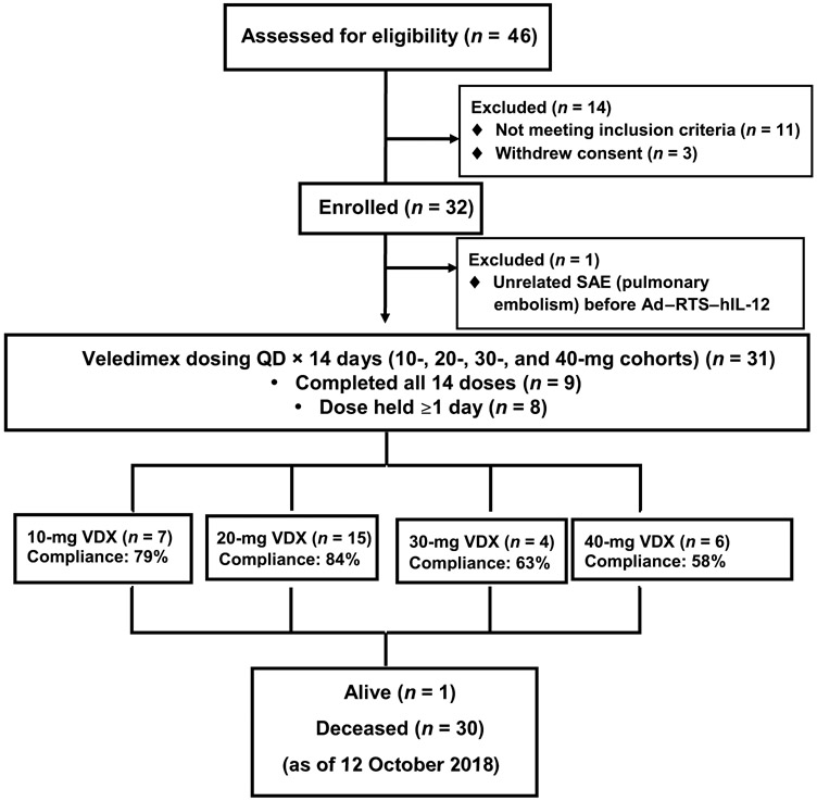
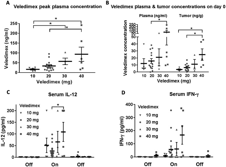
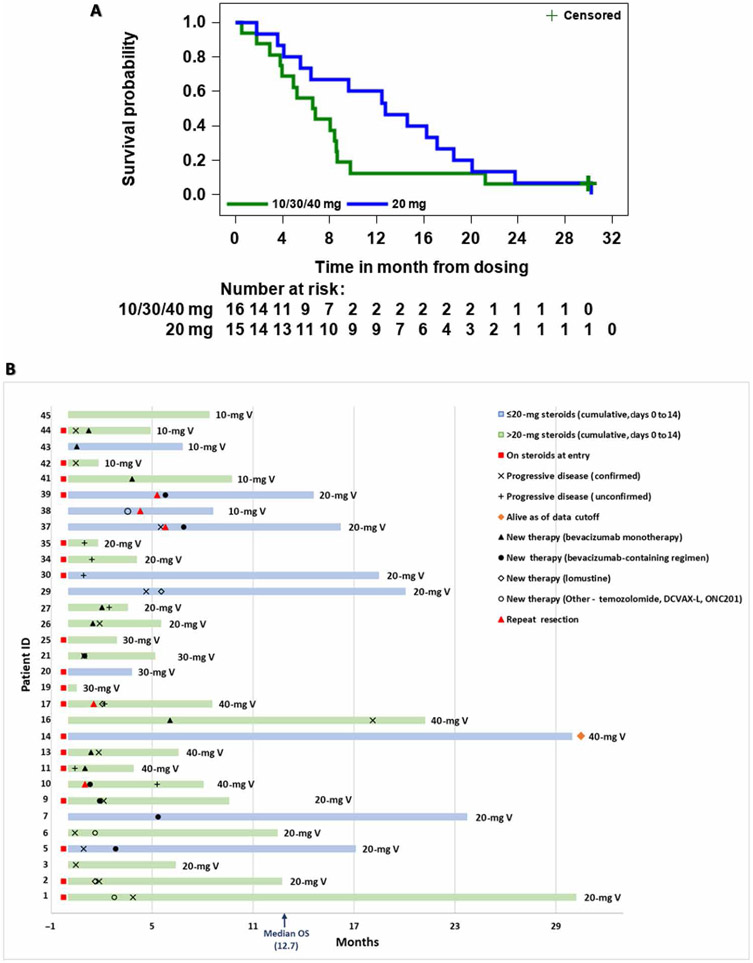
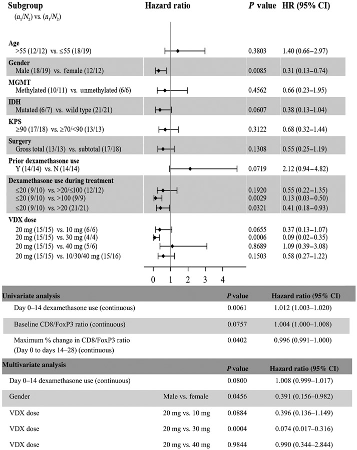
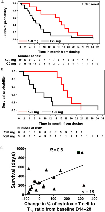
Human interleukin-12 (hIL-12) is a cytokine with anticancer activity, but its systemic application is limited by toxic inflammatory responses. We assessed the safety and biological effects of an hIL-12 gene, transcriptionally regulated by an oral activator. A multicenter phase 1 dose-escalation trial (NCT02026271) treated 31 patients undergoing resection of recurrent high-grade glioma. Resection cavity walls were injected (day 0) with a fixed dose of the hIL-12 vector (Ad-RTS-hIL-12). The oral activator for hIL-12, veledimex (VDX), was administered preoperatively (assaying blood-brain barrier penetration) and postoperatively (measuring hIL-12 transcriptional regulation). Cohorts received 10 to 40 mg of VDX before and after Ad-RTS-hIL-12. Dose-related increases in VDX, IL-12, and interferon-γ (IFN-γ) were observed in peripheral blood, with about 40% VDX tumor penetration. Frequency and severity of adverse events, including cytokine release syndrome, correlated with VDX dose, reversing promptly upon discontinuation. VDX (20 mg) had superior drug compliance and 12.7 months median overall survival (mOS) at mean follow-up of 13.1 months. Concurrent corticosteroids negatively affected survival: In patients cumulatively receiving >20 mg versus ≤20 mg of dexamethasone (days 0 to 14), mOS was 6.4 and 16.7 months, respectively, in all patients and 6.4 and 17.8 months, respectively, in the 20-mg VDX cohort. Re-resection in five of five patients with suspected recurrence after Ad-RTS-hIL-12 revealed mostly pseudoprogression with increased tumor-infiltrating lymphocytes producing IFN-γ and programmed cell death protein 1 (PD-1). These inflammatory infiltrates support an immunological antitumor effect of hIL-12. This phase 1 trial showed acceptable tolerability of regulated hIL-12 with encouraging preliminary results.


Figures

Fig. 1.
Fig. 1.. Study CONSORT diagram.
Patient accrual at four institutions shown as the number of patients treated at doses of 10, 20, 30, and 40 mg of VDX. The percentage of compliance represents the number of days that the VDX was orally administered versus the total number of days that each patient was expected to take the drug over 14 days. SAE, serious adverse event; QD, once a day.
Fig. 2.
Fig. 2.. VDX, IL-12, and IFN-γ concentrations upon VDX treatment.
(A) Peak plasma concentrations of VDX at each drug dosage. Each symbol represents plasma from a single patient. (B) VDX in plasma and intratumorally at the time of surgical resection. VDX was administered to each patient about 3 hours before the start of the craniotomy. Serum and tumor obtained at the time of resection were assayed for VDX concentrations. (C) IL-12 in peripheral blood before, during, and after VDX dosing. (D) IFN-γ in peripheral blood before, during, and after VDX dosing. *P < 0.05.
Fig. 3.
Fig. 3.. Radiologic and immunologic analyses of tumors after treatment.
(A) Three patients with suspected progression after treatment underwent re-resection of contrast-enhancing suspected tumor. The MRI images shown are from one patient who had a right occipital recurrent GBM resected. The MRI scans from 1 day after surgery (baseline) and from weeks 4, 8, and 24 are shown. The injections were given in an area of the occipital lobe and one area more superior toward the parietal lobe. Red and yellow arrows show areas with changes in enhancement in the occipital and parietal needle tracks. (B) Left panels: GBM from the patient shown in (A) at the time of resection before injection of Ad–RTS–hIL-12 [shown in the top panel at 20× magnification (scale bar, 100 μm) and in the bottom panel at 100× magnification (scale bar, 50 μm)]. Right panels: GBM from the same patient 175 days after treatment (at the time of suspected pseudoprogression). Resected material from the occipital lesion was analyzed by immunofluorescence histochemistry for expression of CD3+ (yellow), CD8+ (red), CD3+CD8+ (orange), PD-1+ (green), PD-L1+ (cyan), and GFAP (white) [shown in the top panel at 20× magnification (scale bar, 100 μm) and in the bottom panel at 100× magnification (scale bar, 50 μm)]. (C and D) Quantitative analyses of baseline and posttreatment expression of immunologic markers in tumors for the three patients undergoing re-resection after injection. (C) Counts of CD3+-, CD3+CD8+-, PD-1+-, CD3+CD4+FoxP3+-, CD56+-, and PD-L1+–expressing cells per square millimeter of tumor. (D) IFN-γ in the three GBMs before and after treatment.
Fig. 4.
Fig. 4.. Analyses of treatment efficacy.
(A) Kaplan-Meier analysis of overall survival (OS) for the 20-mg (blue line) versus combined 10-, 30-, and 40-mg cohorts (green line). The + censored label refers to the single patient alive at time of data cutoff. (B) Survival swimmer plot. The x axis lists survival time in months, with each patient number on the y axis. Blue and green colors represent patients who received 20 mg or less or more than 20 mg of cumulative dexamethasone, respectively, during days 0 to 14 of VDX treatment. The 10-, 20-, 30-, and 40-mg V designations at the end of each bar represent the dose of VDX that each patient received. Patients on steroids at entry, timing of progressive disease, and other therapy events are listed. The median OS was 12.7 months.
Fig. 5.
Fig. 5.. Forest plots of prognostic factors of subgroups examined for OS.
HR, hazard ratio.
Fig. 6.
Fig. 6.. Additional efficacy analyses.
(A and B) Kaplan-Meier survival curves based on cumulative dexamethasone dosage (red line, ≤20 mg of dexamethasone; black line, >20 mg of dexamethasone) for patients treated with (A) 10, 20, 30, or 40 mg of VDX (days 0 to 14) or (B) 20 mg of VDX (days 0 to 14). (C) Correlation of survival with peripheral blood CD8+ (cytotoxic T cell)/FOXP3+ [regulatory T cell (Treg)] ratio at 14 to 28 days after viral injection. Triangles represent deceased patients, and square represents an alive patient (P = 0.0071, R = 0.6).

References

    1. Kamiya-Matsuoka C, Gilbert MR, Treating recurrent glioblastoma: An update. CNS Oncol. 4, 91–104 (2015). - PMC - PubMed
    1. Mrugala MM, Advances and challenges in the treatment of glioblastoma: A clinician’s perspective. Discov. Med 15, 221–230 (2013). - PubMed
    1. Reardon DA, Wen PY, Wucherpfennig KW, Sampson JH, Immunomodulation for glioblastoma. Curr. Opin. Neurol 30, 361–369 (2017). - PubMed
    1. Ricklefs FL, Alayo Q, Krenzlin H, Mahmoud AB, Speranza MC, Nakashima H, Hayes JL, Lee K, Balaj L, Passaro C, Rooj AK, Krasemann S, Carter BS, Chen CC, Steed T, Treiber J, Rodig S, Yang K, Nakano I, Lee H, Weissleder R, Breakefield XO, Godlewski J, Westphal M, Lamszus K, Freeman GJ, Bronisz A, Lawler SE, Chiocca EA, Immune evasion mediated by PD-L1 on glioblastoma-derived extracellular vesicles. Sci. Adv 4, eaar2766 (2018). - PMC - PubMed
    1. Da Ros M, De Gregorio V, Iorio AL, Giunti L, Guidi M, de Martino M, Genitori L, Sardi I, Glioblastoma chemoresistance: The double play by microenvironment and blood-brain barrier. Int. J. Mol. Sci 19, E2879 (2018). - PMC - PubMed

Publication types

MeSH terms

Associated data