. 2019 Aug 14;9(1):11829.
doi: 10.1038/s41598-019-47352-9.

The PINK1 kinase-driven ubiquitin ligase Parkin promotes mitochondrial protein import through the presequence pathway in living cells

Affiliations

The PINK1 kinase-driven ubiquitin ligase Parkin promotes mitochondrial protein import through the presequence pathway in living cells

M Jacoupy et al. Sci Rep. .

Abstract

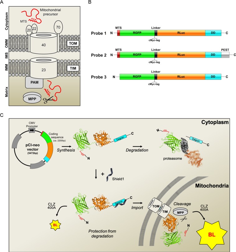
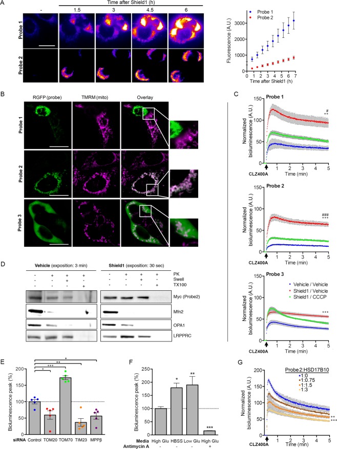
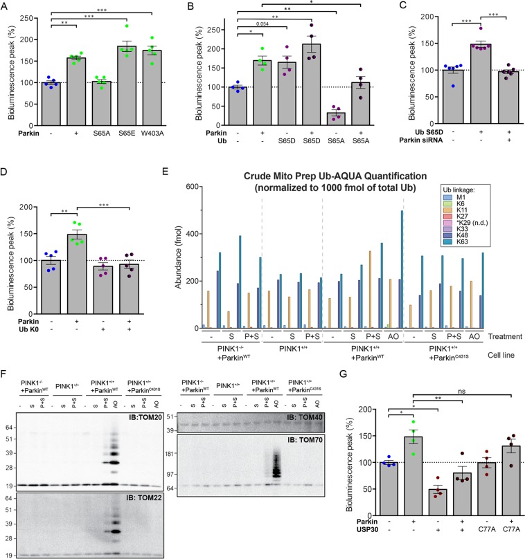
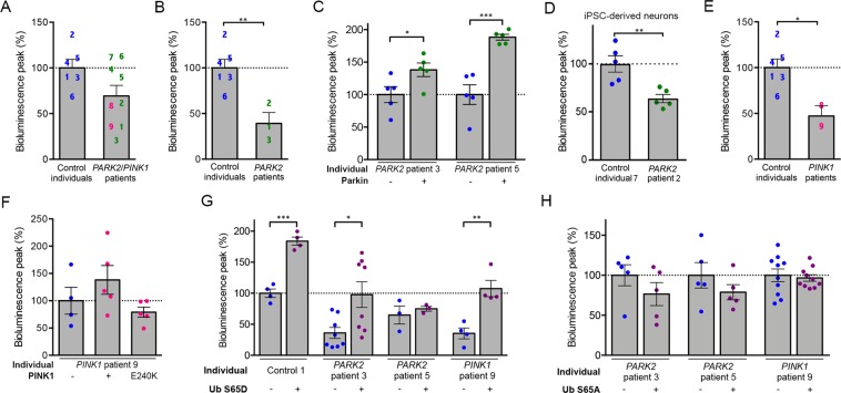
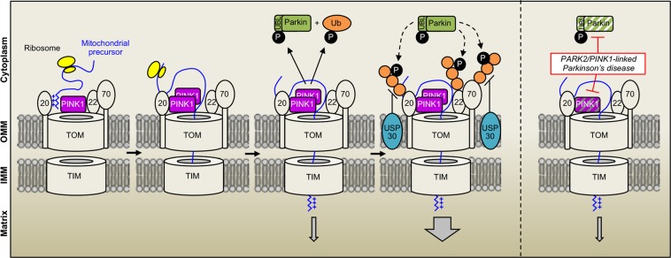
Most of over a thousand mitochondrial proteins are encoded by nuclear genes and must be imported from the cytosol. Little is known about the cytosolic events regulating mitochondrial protein import, partly due to the lack of appropriate tools for its assessment in living cells. We engineered an inducible biosensor for monitoring the main presequence-mediated import pathway with a quantitative, luminescence-based readout. This tool was used to explore the regulation of mitochondrial import by the PINK1 kinase-driven Parkin ubiquitin ligase, which is dysfunctional in autosomal recessive Parkinson's disease. We show that mitochondrial import was stimulated by Parkin, but not by disease-causing Parkin variants. This effect was dependent on Parkin activation by PINK1 and accompanied by an increase in the abundance of K11 ubiquitin chains on mitochondria and by ubiquitylation of subunits of the translocase of outer mitochondrial membrane. Mitochondrial import efficiency was abnormally low in cells from patients with PINK1- and PARK2-linked Parkinson's disease and was restored by phosphomimetic ubiquitin in cells with residual Parkin activity. Altogether, these findings uncover a role of ubiquitylation in mitochondrial import regulation and suggest that loss of this regulatory loop may underlie the pathophysiology of Parkinson's disease, providing novel opportunities for therapeutic intervention.


Conflict of interest statement

The authors declare no competing interests.

Figures

Figure 1
Figure 1
Genetically encoded reporters for the assessment of protein import through the TOM complex. (A) Molecular components of the presequence import pathway: translocase of the outer mitochondrial membrane (TOM) with its receptor subunits TOM20, TOM22 and the channel subunit TOM40; translocase of inner mitochondrial membrane (TIM23); presequence translocase-associated motor (PAM); mitochondrial processing peptidase (MPP). OMM, outer mitochondrial membrane; IMM inner mitochondrial membrane; IMS, intermembrane space. The TOM70 receptor recognizes proteins with internal mitochondrial targeting information. (B) Probes designed to monitor import through the presequence pathway. MTS, mitochondrial targeting signal; RGFP, green fluorescent protein from Renilla reniformis; RLuc, luciferase from Renilla reniformis; DD, destabilizing domain of FK506 binding protein (FKBP); PEST, sequence rich in proline (P), glutamic acid (E), serine (S), and threonine (T) associated with short-lived proteins; the D433A and D434A PEST mutations accelerate degradation. (C) Bioluminescence (BL)-based assay for mitochondrial import through the presequence pathway. In the absence of the small molecule Shield1, Probes 1 and 2 are rapidly degraded by the proteasome. Shield1 stabilizes the probes, which are then imported into the mitochondrial matrix through the TOM and TIM complexes. Light emission in the cytoplasm is weak, due to the presence of the MTS, blocking the N-terminus of RGFP. Cleavage of the MTS by MPP allows interaction between the RGFP and RLuc modules, leading to a characteristic light emission due to resonance energy transfer and quantum yield enhancement in the presence of the Rluc substrate coelenterazine 400A (CLZ400A). The RGFP and Rluc protein modules (PDB ID: 2RH7 and PDB ID: 2PSD) were obtained from the Protein Data Bank at the following URL: http://www.rcsb.org.
Figure 2
Figure 2
Validation of the probes for the assessment of the presequence import pathway in living cells. (A) Representative fluorescence images and the corresponding quantitative analysis (graph), illustrating the expression of Probes 1 and 2 (RGFP signal) at different time points after the addition of Shield1 to the culture medium of HEK293T cells. n = 15 (B) Representative fluorescence images and higher magnifications (framed regions in the overlay) showing the colocalization of each probe (RGFP signal) with TMRM in cells treated with Shield1 (24 h). Note the loss of TMRM staining in a cell with high levels of Probe 1, and the expected cytosolic localization of Probe 3. (C) Quantitative analysis of the bioluminescent signals emitted by Probes 1-3 in HEK293T cells with and without Shield1 and/or CCCP treatment (24 h), following the addition of CLZ400A. n = 6 wells from one of three independent experiments. (D) Western blot analysis of mitochondrial subfractions obtained by subjecting mitochondrion-enriched fractions to proteinase K (PK) treatment, combined or not with osmotic swelling (Swell) or solubilization with Triton X‐100 (TX100). The subfractions were probed for Mfn2 (OMM), OPA1 (IMS/IMM), LRPPRC (matrix). Note the presence of Probe2 in the mitochondrial matrix. (EG) Changes to bioluminescent signal emitted by Probe 2 in HEK293T cells following (E) siRNA-mediated silencing of key components of the presequence pathway (n = 5 independent experiments), (F) switch to nutrient-free (HBSS) or low glucose medium (1 g/l), or treatment with antimycin A (n = 3 independent experiments), or (G) cotransfection with various amounts of plasmid encoding HSD17B10 (V5 epitope-tagged; n = 5 wells from one of at least three independent experiments). Results are expressed as means ± SEM. *p < 0.05, **p < 0.01, ***p < 0.001 versus vehicle/vehicle (C; two-way repeated-measures ANOVA with Tukey’s posthoc test), Control (E; one-way ANOVA with Dunnett’s posthoc test), High Glu (E; one-way ANOVA with Dunnett’s posthoc test) or 1:0 ratio of Probe 2 to HSD17B10-V5 (F, two-way repeated-measures ANOVA with Dunnett’s posthoc test). #p < 0.05, ###p < 0.001 versus Shield1/CCCP. Scale bar: 10 µm.
Figure 3
Figure 3
Parkin modulates the mitochondrial presequence import pathway independently of its role in mitophagy. Quantitative analyses of the relative bioluminescent signals for Probe 2 in HEK293T cells (A) overproducing Parkin after 3, 8 or 24 h of treatment with Shield1, (B) silenced for Parkin or PINK1 or (C) overproducing pathogenic Parkin variants, showing the enhancement of import by normal Parkin but not by PD-linked or artificial (ΔUbl) variants. (D) Inhibition of autophagy (3-Methyladenine, ATG5 silencing), lysosomal (E-64d, Bafilomycin A1) or proteasomal (Epoxomicin) functions, or (E) siRNA-mediated silencing of PGC-1-α siRNA do not affect facilitation of mitochondrial import by Parkin. (F) Overproduction of Parkin is sufficient for rescuing the mitochondrial import decrease of the probe following HSD17B10 overproduction. Results are expressed as means ± SEM. n = 4 (A) to 6 (B) independent experiments. Data were analyzed by student t-test (A,D) one-way (C) or two-way ANOVA (B,E,F) with Dunnett’s or Holm-Sidak’s posthoc tests. *p < 0.05, **p < 0.01, ***p < 0.001. The schematic diagram illustrates Parkin with its main domains and the location of the PD-linked (yellow circles) or artificial (blue circles) variants examined.
Figure 4
Figure 4
Parkin modulates mitochondrial import through its E3 ubiquitin-protein ligase activity induced by PINK1. Effect of relief of Parkin autoinhibition by (A) the activating W403A substitution, or by mimicking the PINK1-mediated phosphorylation of Parkin UBL (S65E) or (B) Ub (S65D), showing exacerbation of the Parkin-dependent enhancement of the bioluminescent signal of Probe 2 in HEK293T cells. By contrast, non-phosphorylatable S65A Parkin or Ub variants abolish the effect of Parkin. (C) In these cells, Ub S65D mimics the effect of Parkin overproduction in a manner dependent on endogenous Parkin (Parkin siRNA). (D) Effect of the lysine-less Ub K0 variant on the intensity of the bioluminescent signal of Probe 2 in HEK293T, indicating that the effect of Parkin is mediated by polyubiquitylation. (E) Quantification by UB-AQUA proteomics of individual Ub chain linkage types associated with mitochondria in HeLa Flp-In T-REx ParkinWT or ParkinC431S cells depleted or not of PINK1 and expressing Probe 2 in the presence of Shield1 (P + S), or treated with S or AO. (F) Immunoblot analysis of ubiquitylated proteins pulled down with TUBEs from mitochondrion-enriched fractions analyzed in (E) shows ubiquitylation of the TOM subunits TOM20, TOM22 and TOM70 in cells expressing Probe 2 (P + S). (G) Analysis of the impact of the mitochondrial ubiquitin-specific protease USP30 or its inactive C77A variant on the signal of Probe in the presence or absence of overexpressed Parkin. n = 4 (A,B,G), 5 (D) or 6 (C) independent experiments. Results are expressed as means ± SEM. Data were analyzed by one-way (A) or two-way ANOVA (B,C,G) with Dunnett’s or Holm-Sidak’s posthoc tests. *p < 0.05, **p < 0.01, ***p < 0.001. ns: non-significant.
Figure 5
Figure 5
Defects in the mitochondrial presequence import pathway in fibroblasts from PD patients with PARK2 and PINK1 mutations are rescued by phosphomimetic ubiquitin. (AF) Mean bioluminescent signal for Probe 1 in primary fibroblasts (A–C,EH) or iPSC-derived neurons (D) from control individuals and PARK2/PINK1 patients (A), a subset of or single PARK2 patients (BD,G,H), or PINK1 patients (EH), showing a weaker signal in PINK1 patients and PARK2 Patients 1-3, associated with presence of the partially functional Parkin R42P substitution, and rescue effects of Parkin, PINK1, PINK1 E240K or Ub S65D/A overexpression. The numbers on the graphs indicate the mean signals for individual patients, as presented in Supplementary Table 1, with n = 4–6 independent experiments (A,B,E), n = 4–5 wells from one experiment representative of three (C,F), or n = 4–10 wells from one of 2–5 independent experiments (D,G,H). Note that the overproduction of Parkin, PINK1 or phosphomimetic Ub S65D partially normalizes the signal in fibroblasts from individual PARK2 or PINK1 patients, whereas PINK1 E240K and Ub S65A do not have any effect. Results are expressed as means ± SEM. *p < 0.05, **p < 0.01, ***p < 0.001 versus control individuals (A,B,D,E, Student’s t-test) or the conditions indicated (C,FH, one-way ANOVA with Dunnett’s (C,F) or Holm-Sidak’s (G,H) posthoc tests).
Figure 6
Figure 6
Model illustrating facilitation of mitochondrial protein import by the PINK1 kinase-driven E3 Ub ligase Parkin and lack thereof in PARK2/PINK1-linked Parkinson’s disease. Under physiological reductions in mitochondrial protein import efficiency, the PINK1 kinase associates with the TOM complex and activates Parkin by phosphorylating S65 of Parkin and ubiquitin. By ubiquitylating receptor subunits of the TOM complex, Parkin facilitates the import of proteins targeted to mitochondria by the presequence pathway. The mitochondrial ubiquitylase USP30 antagonizes these effects. Mutations in PINK1 or PARK2 impair the system and its regulatory effect on mitochondrial protein import, contributing to the pathophysiology of autosomal recessive PD.

Similar articles

Cited by

References

    1. Chacinska A, Koehler CM, Milenkovic D, Lithgow T, Pfanner N. Importing mitochondrial proteins: machineries and mechanisms. Cell. 2009;138:628–644. doi: 10.1016/j.cell.2009.08.005. - DOI - PMC - PubMed
    1. Devi L, Prabhu BM, Galati DF, Avadhani NG, Anandatheerthavarada HK. Accumulation of amyloid precursor protein in the mitochondrial import channels of human Alzheimer’s disease brain is associated with mitochondrial dysfunction. J Neurosci. 2006;26:9057–9068. doi: 10.1523/JNEUROSCI.1469-06.2006. - DOI - PMC - PubMed
    1. Li Q, et al. ALS-linked mutant superoxide dismutase 1 (SOD1) alters mitochondrial protein composition and decreases protein import. Proc Natl Acad Sci USA. 2010;107:21146–21151. doi: 10.1073/pnas.1014862107. - DOI - PMC - PubMed
    1. Bender A, et al. TOM40 mediates mitochondrial dysfunction induced by alpha-synuclein accumulation in Parkinson’s disease. PLoS One. 2013;8:e62277. doi: 10.1371/journal.pone.0062277. - DOI - PMC - PubMed
    1. Gottschalk, W. K. et al. The Broad Impact of TOM40 on Neurodegenerative Diseases in Aging. J Parkinsons Dis Alzheimers Dis 1, 10.13188/2376-922X.1000003 (2014). - PMC - PubMed

Publication types