. 2019 Aug 14;10(8):621.
doi: 10.1038/s41419-019-1848-y.

The POU4F2/Brn-3b transcription factor is required for the hypertrophic response to angiotensin II in the heart

Affiliations

The POU4F2/Brn-3b transcription factor is required for the hypertrophic response to angiotensin II in the heart

Laura Mele et al. Cell Death Dis. .

Abstract

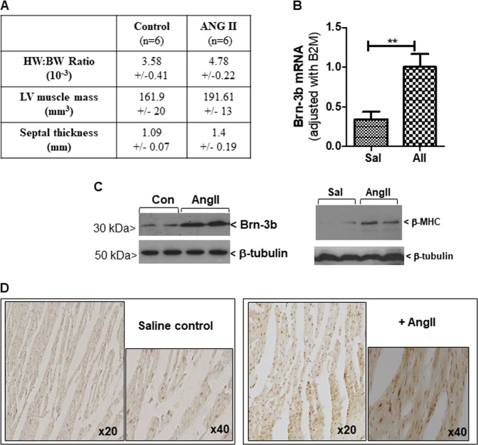
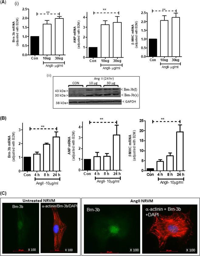
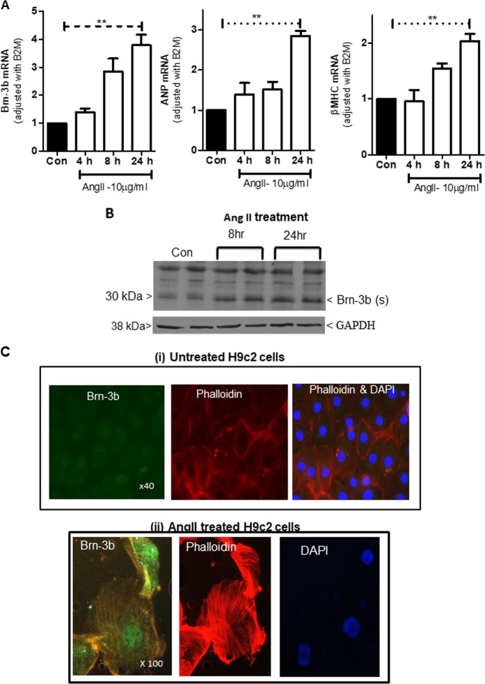
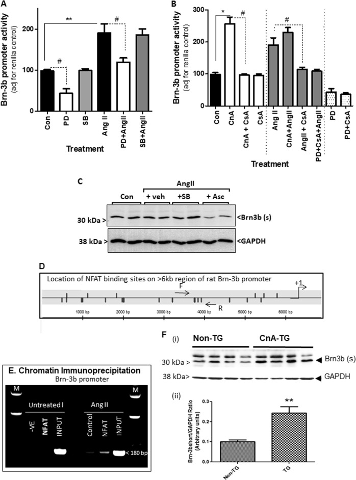
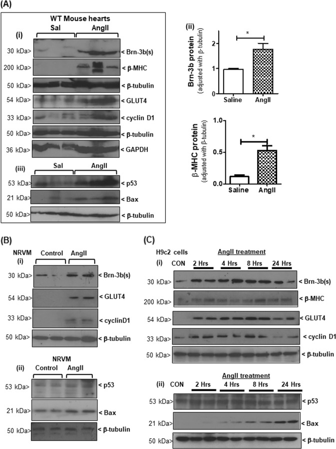
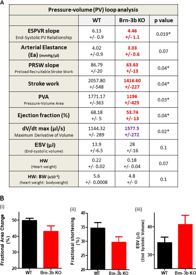
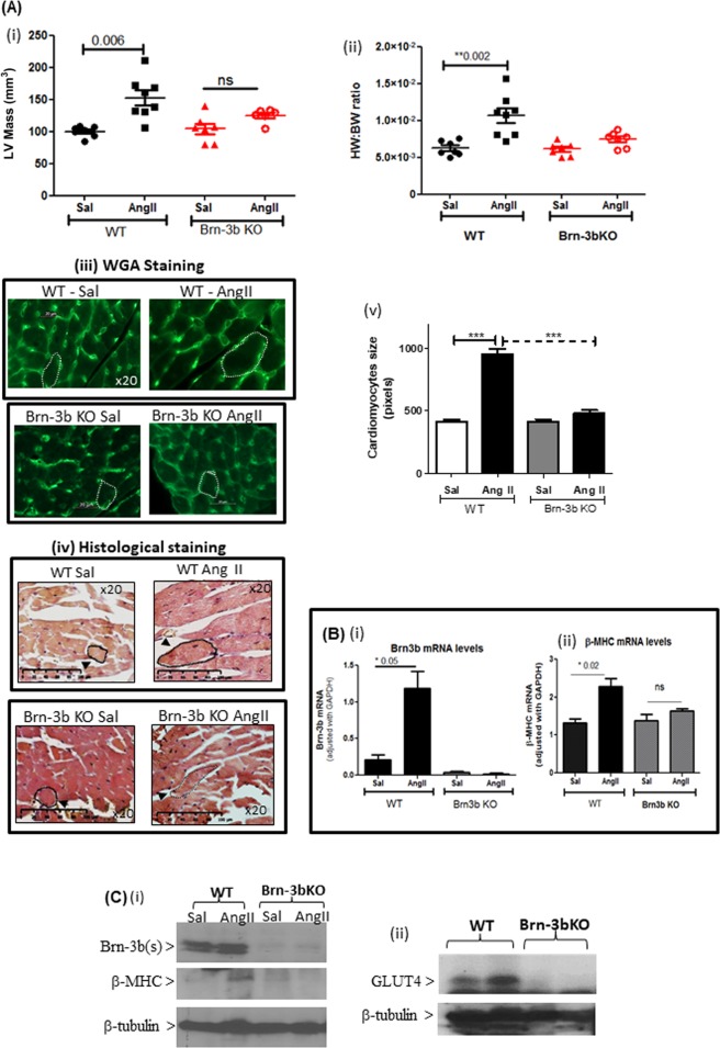
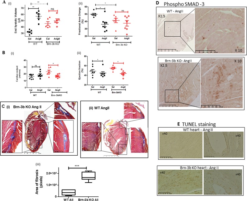
Adult hearts respond to increased workload such as prolonged stress or injury, by undergoing hypertrophic growth. During this process, the early adaptive responses are important for maintaining cardiac output whereas at later stages, pathological responses such as cardiomyocyte apoptosis and fibrosis cause adverse remodelling, that can progress to heart failure. Yet the factors that control transition from adaptive responses to pathological remodelling in the heart are not well understood. Here we describe the POU4F2/Brn-3b transcription factor (TF) as a novel regulator of adaptive hypertrophic responses in adult hearts since Brn-3b mRNA and protein are increased in angiotensin-II (AngII) treated mouse hearts with concomitant hypertrophic changes [increased heart weight:body weight (HW:BW) ratio]. These effects occur specifically in cardiomyocytes because Brn-3b expression is increased in AngII-treated primary cultures of neonatal rat ventricular myocytes (NRVM) or foetal heart-derived H9c2 cells, which undergo characteristic sarcomeric re-organisation seen in hypertrophic myocytes and express hypertrophic markers, ANP/βMHC. The Brn-3b promoter is activated by known hypertrophic signalling pathways e.g. p42/p44 mitogen-activated protein kinase (MAPK/ERK1/2) or calcineurin (via NFAT). Brn-3b target genes, e.g. cyclin D1, GLUT4 and Bax, are increased at different stages following AngII treatment, supporting distinct roles in cardiac responses to stress. Furthermore, hearts from male Brn-3b KO mutant mice display contractile dysfunction at baseline but also attenuated hypertrophic responses to AngII treatment. Hearts from AngII-treated male Brn-3b KO mice develop further contractile dysfunction linked to extensive fibrosis/remodelling. Moreover, known Brn-3b target genes, e.g. GLUT4, are reduced in AngII-treated Brn-3b KO hearts, suggesting that Brn-3b and its target genes are important in driving adaptive hypertrophic responses in stressed heart.


Conflict of interest statement

The authors declare that they have no conflict of interest.

Figures

Fig. 1
Fig. 1. Altered Brn-3b expression mRNA in mouse hearts treated with AngII.
a Assessment of hypertrophic responses in hearts taken from AngII-treated wild-type C57BL/6 mice and compared with saline-treated control mouse hearts. Measurements of heart weight:body weight ratio (HW:BW); LV mass and septal thickness is shown as mean and standard error from different experimental groups (n = 6). b Results of quantitative RT-PCR (qRT-PCR) showing increased Brn-3b mRNA in hearts taken from AngII-treated mice compared with saline controls. Statistical significance between groups (≥5 mice) (**p < 0.01) was analysed using students t test. c Representative western blot showing increased (i) Brn-3b or (ii) beta MHC proteins in mouse hearts either treated with AngII for up to 4 weeks, compared with saline control (Con). d Representative DAB immunostaining showing increased Brn-3b protein expression in AngII-treated mouse hearts. Images were taken using the Hamamatsu Nanozoomer scanner and shown at ×20 and ×40, as indicated
Fig. 2
Fig. 2. Brn-3b induction by AngII in neonatal rat ventricular myocytes (NRVM) cultures.
(a) (i) qRT-PCR data showing changes in mRNA encoding Brn-3b or hypertrophic markers ANF and β-MHC in NRVM treated with different concentrations (10 or 30 µg/ml) of AngII for 24 h, when compared with untreated control cells (Con). Variations in RNA from different samples were standardised using the housekeeping gene, β2-micro-globulin (B2M) mRNA expression and values were expressed relative to untreated control cells set at 1. Graphs represent the mean values ± SD from independent NRVM samples (n = 6). *p < 0.05. (ii) Representative western blot analysis showing induction of Brn-3b proteins following treatment with 10 µg/ml AngII for 24 h. Different Brn-3b isoforms (~43 and ~32 kDa) are indicated and the invariant GAPDH protein indicated any variation in protein loading. (b) Quantification of mRNA encoding Brn-3b or hypertrophic markers, ANP and β-MHC in NRVM cultures treated with 10/ml AngII for different times (4, 8, 24 h). Variations in mRNA between samples were standardised using B2M and values expressed relative to untreated control cells set at 1. Graphs represent the mean values ± SD from six independent experiments (n = 6). **p < 0.01 compared with untreated controls (Con). (c) Representative immunofluorescent images showing localisation of Brn-3b protein (Alexa Fluor 488–green) in AngII-treated NRVM co-stained with phalloidin (red) to demonstrate actin cytoskeleton reorganisation and DAPI staining (blue) showing cell nuclei. Images shown were taken at ×100 magnification (oil immersion lens) using the Zeiss Axioskop 2 microscope
Fig. 3
Fig. 3. Brn-3b induction by AngII in H9c2 cell cultures.
a qRT-PCR data showing changes in mRNA encoding Brn-3b, ANP and β-MHC in H9c2 cells treated with 10 µg/ml of AngII for 4, 8 or 2 h when compared with untreated controls (Con). Variation between RNA samples was standardised using B2M, and values are expressed relative to the untreated control cells set at 1. Data represent values from six independent experiments and **p < 0.01, where (n = 6). b Representative western blot analysis showing induction of Brn-3b(s) protein following treatment with 10 µg/ml AngII for 24 h and the invariant GAPDH protein indicates any variability in protein loading. c Representative immunofluorescent images showing Brn-3b protein localisation (green) in AngII-treated H9c2 cells stained with phalloidin (red) to show cytoskeletal re-modelling. DAPI staining (blue) shows the cell nuclei. Untreated control cells are shown at ×40 to demonstrate little protein expression in a larger field of view, whereas ×100 magnification of AngII-treated cells highlights nuclear Brn-3b expression in cells that display extensive cytoskeletal remodelling
Fig. 4
Fig. 4. Signalling pathways involved in AngII activation of Brn-3b.
a Results of reporter assays carried out using extracts from H9c2 cells transfected with Brn-3b promoter either alone (Con) or following treatment as specified i.e. angiotensin II (AngII) (10 µg/ml) ±MEK1/2 inhibitor (PD) at 20 µM (or ±p38 MAPK inhibitor (SB) at  5 µM. Promoter activity was expressed as % untreated control cells (transfected with reporter construct or empty expression vector), set arbitrarily at 100%. Data from six independent samples are shown and statistical significance (*p < 0.05 increase, #p < 0.05 reduction) was determined using two-way ANOVA followed by post-hoc Bonferroni test. b Results of reporter gene assays showing changes in Brn-3b promoter activity in cells that co-express constitutively active CnA either alone (CnA) or with the calcineurin inhibitor, CsA (CnA + CsA), when compared with CsA alone (1 µM). Promoter activity are also shown under different combinations of treatment i.e. CnA + AngII, in the absence or presence of different inhibitors, CsA and/or PD. The data represent the results of independent experiments (n = 6) and are expressed relative to levels found in untreated control cells (set at 100). Significant increases: **p < 0.01, *p < 0.05; ##significant reduction following treatment with inhibitor CsA (1 µM). c Western blot analysis showing Brn-3b expression in H9c2 cells either untreated (Con) or pretreated as indicated with either MAPK/ERK inhibitor, SB203580 (SB:5 µM) or calcineurin inhibitor, ascomycin (Asc:1uM) for 30 min prior to AngII treatment (10 µg/ml). d Schematic diagram showing the location of multiple NFAT binding sites on 6 kb region of the rat Brn-3b promoter following bioinformatic analysis using MatInspector™. +1 indicates the approximate position of the putative start site. PCR primers were designed to flank putative NFAT binding sites in the Brn-3b promoter. Horizontal arrows indicate positions of forward (F) and reverse (R) primers which were well conserved between rat and human sequence and which were used for ChIP assay. e PCR products obtained using the indicated primers set flanking NFAT sites, indicated by arrows (above) input DNA (chromatin DNA prior to immunoprecipitation) or ChIP DNA obtained after immunoprecipitation with NFAT Abs in either untreated or AngII-treated H9c2 cells. This was compared with negative ChIP DNA control (incubated with second Ab) or PCR negative (without DNA). The marker lane (M) shows the DNA ladder used to identify fragment size in the gel. f (i) Representative western blot showing Brn-3b(s) protein in hearts isolated from 4-week transgenic (TG) mice with cardiac-specific CnA overexpression or non-transgenic (non TG) littermates. The invariant protein GAPDH was used to control for protein loading. (ii) Quantification of Brn-3b(s) proteins in WT or CnA-TG mouse hearts, following normalisation with control GAPDH protein. **p < 0.01
Fig. 5
Fig. 5. Brn-3b target genes are regulated following AngII treatment.
a (i) Representative western blots showing changes in different proteins including Brn-3b(s) and hypertrophic marker β-MHC as well as known Brn-3b target genes GLUT4 and cyclin D1, in hearts taken from wild-type mice treated with AngII and compared with control hearts taken from age-matched control mice infused with saline (Sal). The approximate molecular weight (kD) of each protein is indicated. β-tubulin or GADPH are included to show variation in protein loading between samples. (ii) Graphical representation showing changes in Brn-3b and β-MHC proteins in AngII-treated WT hearts compared with saline controls (n ≥ 7). *p < 0.05, evaluated with students’ t test. (iii) Representative blots showing Bax and p53 proteins quantified in cellular extracts (as above). β-tubulin indicates variation in proteins loading from different samples. b Representative western blot showing changes in Brn-3b(s), GLUT4 and cyclin D1 protein levels in NRVM following treatment with AngII, compared with untreated controls while β-tubulin indicates variation in proteins loading from different samples. c Representative western blots showing Brn-3b, βMHC and Brn-3b target genes, GLUT4 and cyclin D1 in H9c2 cells, at different times after AngII treatment. (ii) Western blots showing increased pro-apoptotic Bax protein at later stages after AngII treatment
Fig. 6
Fig. 6. Baseline differences in Brn-3b KO heart function.
a Summary of results from pressure–volume measurements in mouse hearts collected using the Scisense ADVantageTM Admittance PV loop system, analysed with PowerLab software and statistical significance determined using students t test in Excel and Prism. Data represents mean and standard error (±) from six mice within each group (11 months old). *p ≤ 0.05. ESPVR = end systolic pressure–volume relationship; Ea arterial elastance; PRSW preload recruitable stroke work; PVA pressure–volume area; dV/dtMax maximum conductance velocity; Ves end systolic volume; HW heart weight; HW:BW ratio heart weight:body weight ratio. b Echocardiography data showing parameters that are altered in Brn-3b KO mouse hearts when compared with age-matched WT control hearts. Data represent mean and standard error of multiple hearts (n ≥ 5)
Fig. 7
Fig. 7. Attenuated hypertrophic responses to AngII in Brn-3b KO hearts.
Echocardiography data showing changes in (i) LV mass or (ii) HW:BW ratio, in hearts from male Brn-3b KO (KO) mice and age-matched, WT control mice following AngII or saline infusion for 4 weeks. Groups of 6–8 mice were used and data represent the mean and standard error.**p < 0.01 using either students t test or two-way ANOVA and post-hoc Bonferroni analyses. (iii) Representative images showing changes in cardiomyocyte size in hearts taken from WT or Brn-3b KO mice, infused with either saline (Sal) or AngII and stained with wheat germ agglutinin (WGA) or (iv) histochemical staining, e.g Masson’s trichrome. Images shown at ×20 magnification. Dotted lines show representative cell surface area used for analysing differences in cardiomyocyte size in wild-type or Brn-3b KO hearts following AngII-treated and compared with appropriate saline controls (v) The mean ± SEM of cell surface area measurements taken from multiple hearts (n ≥ 3 independent hearts) with >30 cells analysed from each heart section. b Data from qRT-PCR to analyse changes in (i) Brn-3b or (ii) β−MHC mRNA in hearts taken from AngII or saline-treated WT male mice or Brn-3b KO mutants. c Representative western blots showing changes in protein expression, as indicated, in hearts taken from WT and Brn-3b KO mutants, treated with AngII or saline controls (Sal). β-tubulin blots show difference in total protein between samples
Fig. 8
Fig. 8. Reduced cardiac function in male Brn-3b KO hearts following AngII treatment.
a. Echocardiography data showing changes in (i) end systolic volumes or (ii) fractional area change in Brn-3b KO hearts compared with WT control either in control saline (Sal) hearts or hearts infused with AngII (AII) for 4 weeks. Significant differences between groups are indicated by (**), as determined either using Students t test or two-way ANOVA and Bonferroni post-hoc test. b Changes in cardiac output (i) or ejection fraction (ii) in untreated control (Sal) WT or Brn-3b KO mice or treated with AngII for up to 4 weeks. Data represents the mean and standard error from 6–8 mice per group. Statistical significance between groups was determined using Students t test or two-way ANOVA and Bonferroni post-hoc test (*<0.05; **<0.01). c Representative images showing Masson’s trichrome staining of hearts sections prepared from (i) Brn-3b KO mice treated with AngII, or (ii) WT control mice treated with AngII. Images were captured using Hammamatsu Nanozoomer imaging system and shown at ×1.5–40 magnification. Red/Pink staining represent cytoplasm staining in muscle cells, dark purple/black indicates cell nuclei and blue staining indicates deposition of extracellular matrix proteins e.g. collagen. (iii) Graph representing differences in the areas with signs of fibrosis (increased ECM deposition) in WT or Brn-3b heart following AngII treatment. Areas of fibrosis were measured using image J in ≥5 independent heart sections, shown as ± SEM and significance determined using students t test (***p < 0.001). d Representative images showing anti-phospho-SMAD 3 antibody DAB immunostaining in sections of hearts taken from AngII-treated mice as indicated [WT mice (top panel) or Brn-3b KO mice (lower panel)]. Positive cells are stained brown in images shown either at lower magnification (×2.5) to highlight the extent of increased SMAD protein expression in AngII-treated Brn-3b KO hearts or at higher magnification (×10) displays the intensity of staining. e Representative TUNEL staining in heart sections taken from AngII-treated WT mice (top panel) or Brn-3b KO mice (lower panel). TUNEL positive cells appear brown in the images (shown at ×40 magnification)

References

    1. Frey N, Olson EN. Cardiac hypertrophy: the good, the bad, and the ugly. Annu. Rev. Physiol. 2003;65:45–79. doi: 10.1146/annurev.physiol.65.092101.142243. - DOI - PubMed
    1. Komuro I. Molecular mechanism of cardiac hypertrophy and development. Jpn. Circ. J. 2001;65:353–358. doi: 10.1253/jcj.65.353. - DOI - PubMed
    1. Richey PA, Brown SP. Pathological versus physiological left ventricular hypertrophy: a review. J. Sports Sci. 1998;16:129–141. doi: 10.1080/026404198366849. - DOI - PubMed
    1. Yazaki Y, Tsuchimochi H, Kurabayashi M, Komuro I. Molecular adaptation to pressure overload in human and rat hearts. J. Mol. Cell Cardiol. 1989;21(Suppl 5):91–101. doi: 10.1016/0022-2828(89)90775-X. - DOI - PubMed
    1. Anversa P, Kajstura J, Olivetti G. Myocyte death in heart failure. Curr. Opin. Cardiol. 1996;11:245–251. doi: 10.1097/00001573-199605000-00004. - DOI - PubMed

Publication types

MeSH terms