. 2019 Aug;572(7770):533-537.
doi: 10.1038/s41586-019-1482-y. Epub 2019 Aug 15.

Insights into ubiquitin chain architecture using Ub-clipping

Affiliations

Insights into ubiquitin chain architecture using Ub-clipping

Kirby N Swatek et al. Nature. 2019 Aug.

Abstract

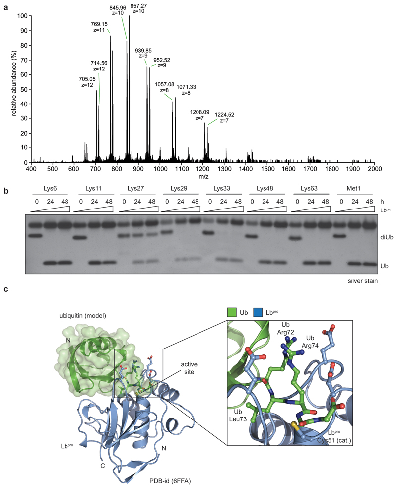
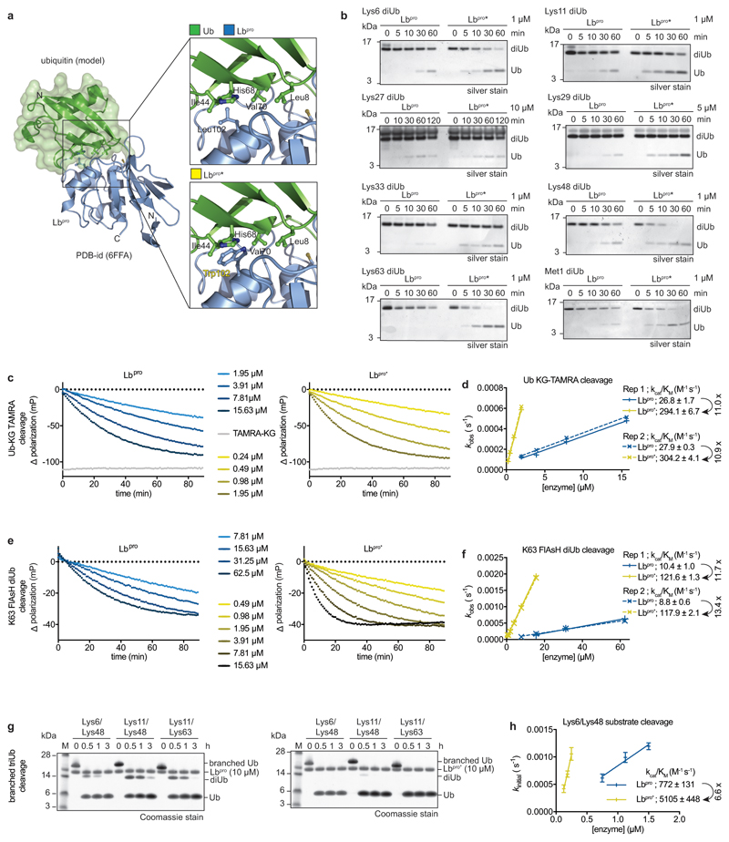
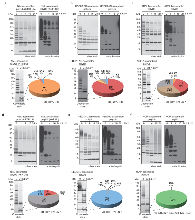
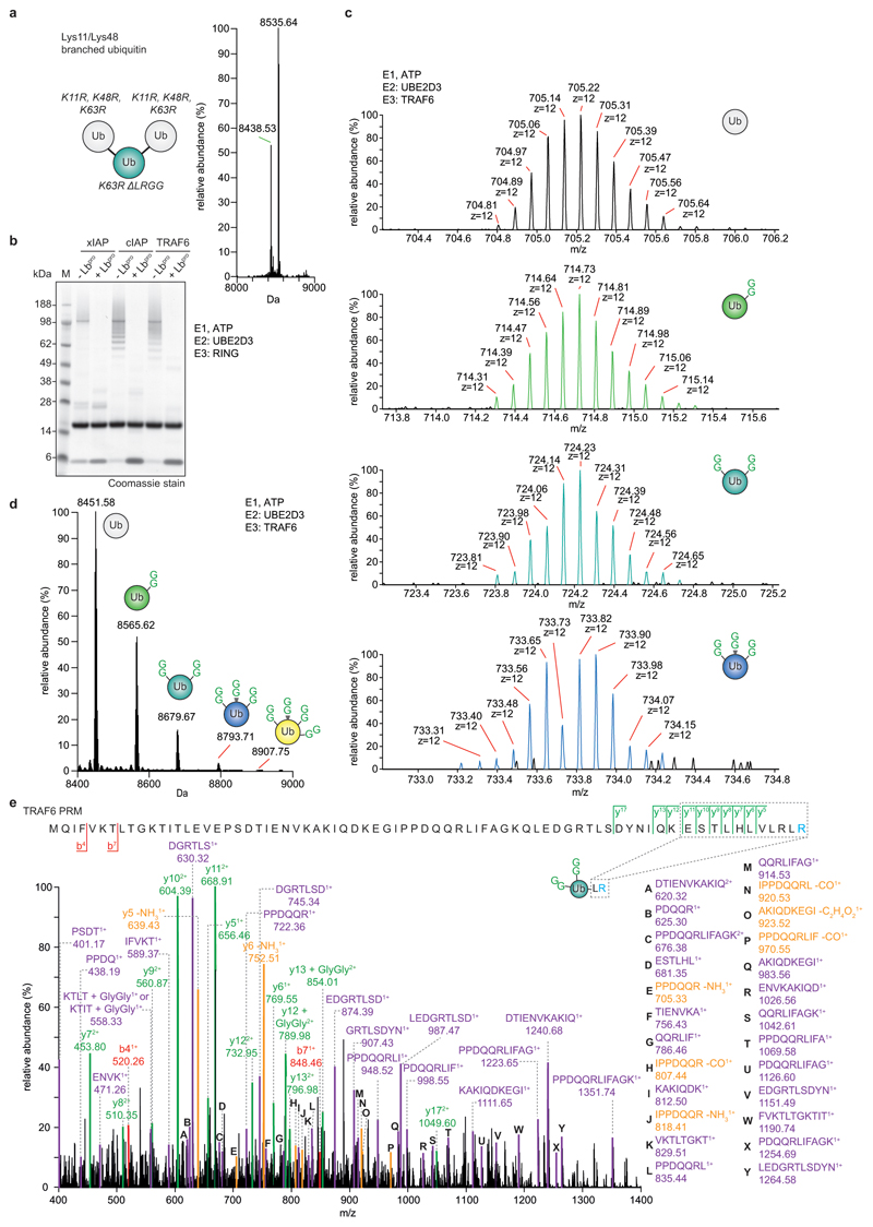
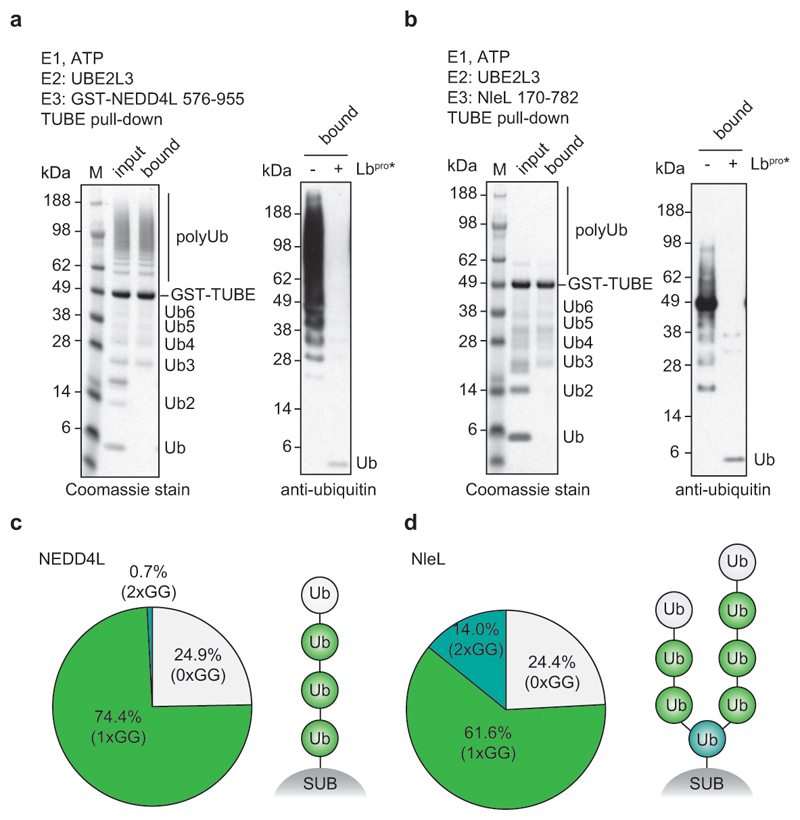
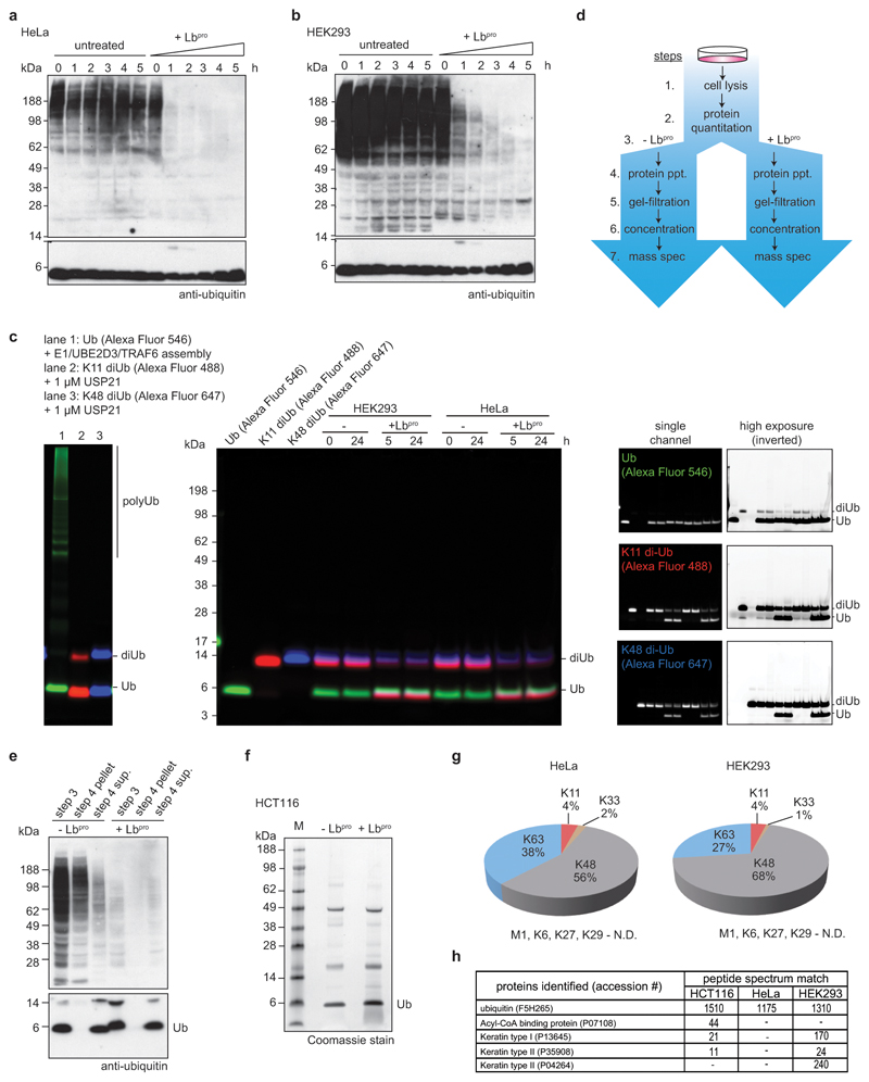
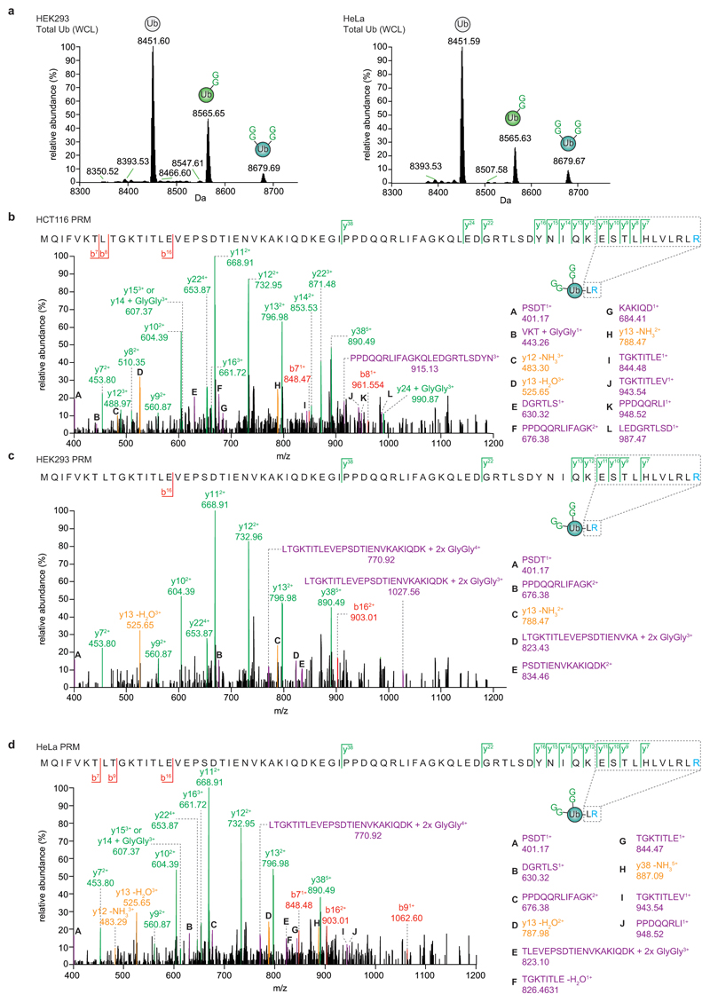
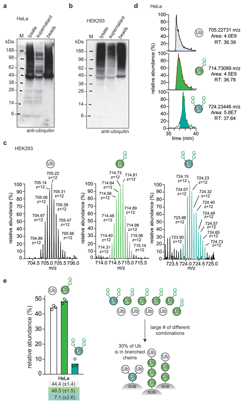
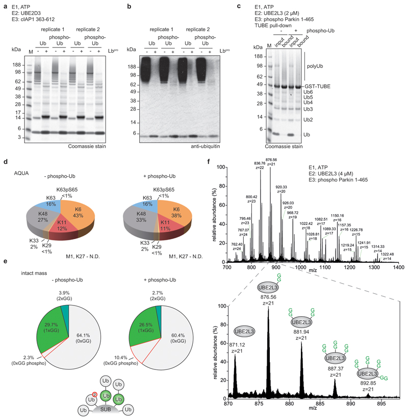
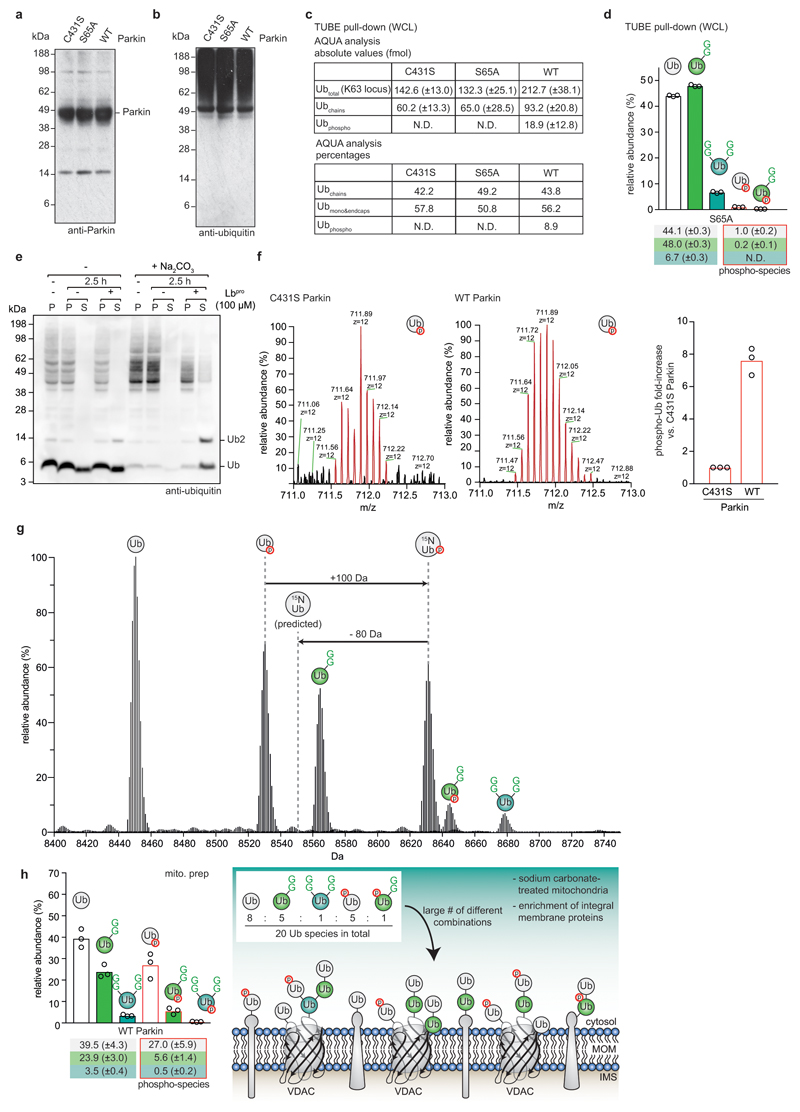
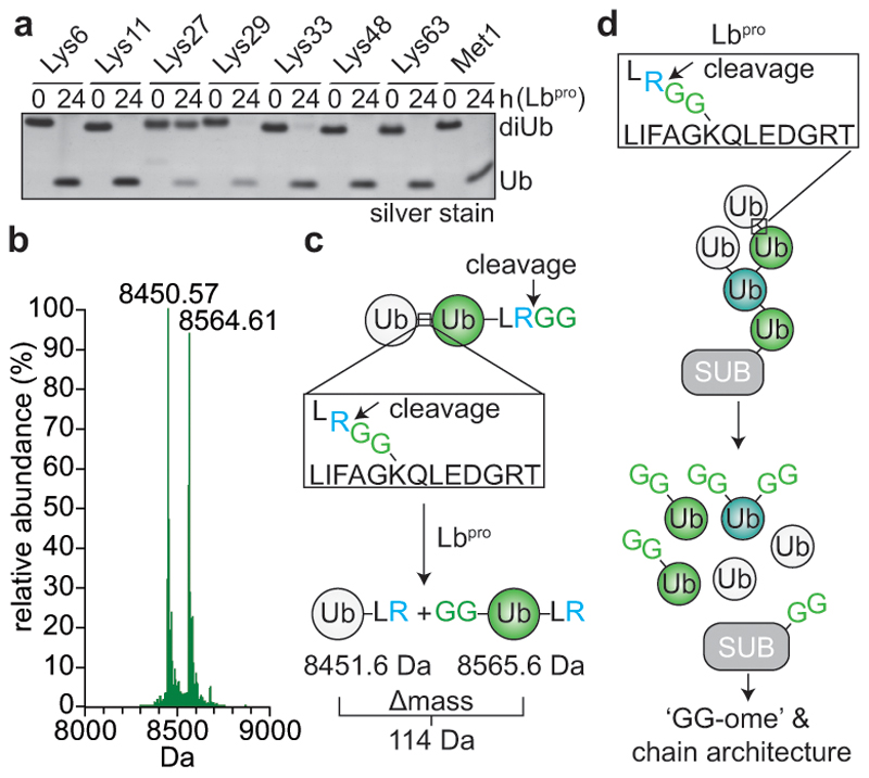
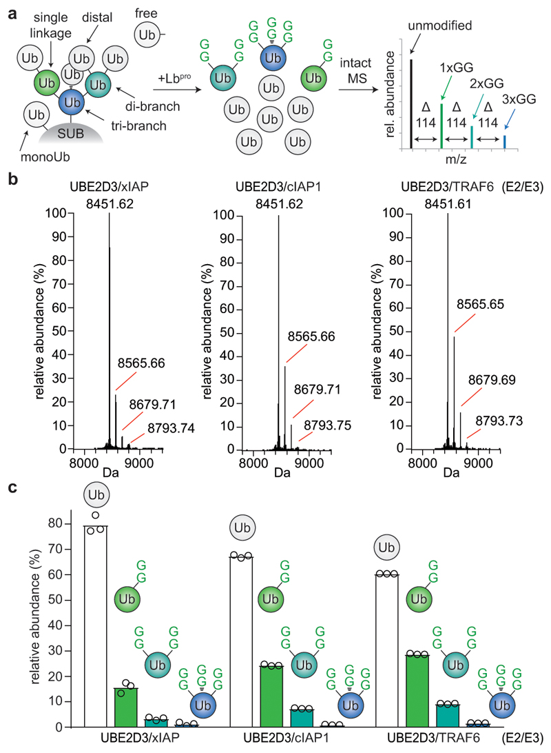
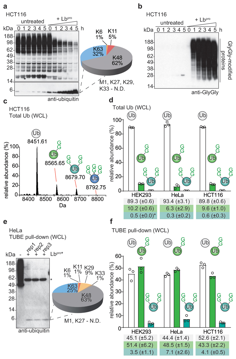
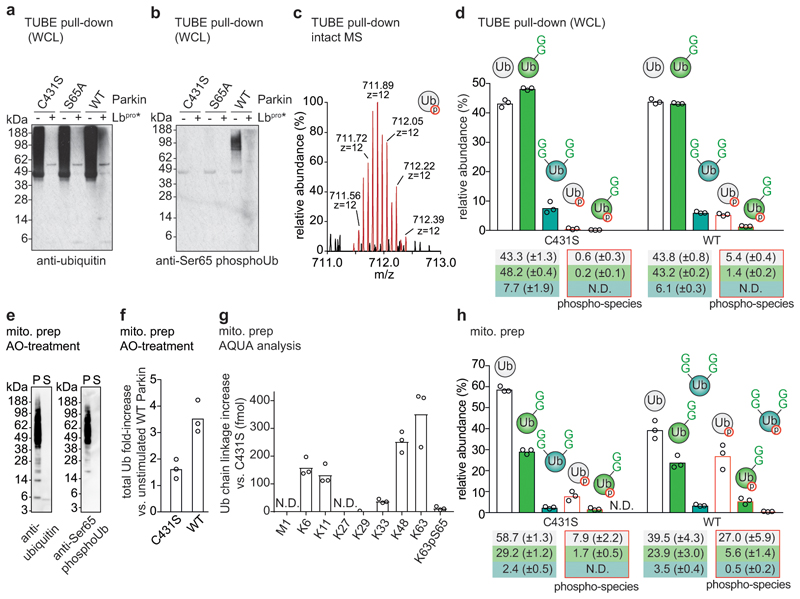
Protein ubiquitination is a multi-functional post-translational modification that affects all cellular processes. Its versatility arises from architecturally complex polyubiquitin chains, in which individual ubiquitin moieties may be ubiquitinated on one or multiple residues, and/or modified by phosphorylation and acetylation1-3. Advances in mass spectrometry have enabled the mapping of individual ubiquitin modifications that generate the ubiquitin code; however, the architecture of polyubiquitin signals has remained largely inaccessible. Here we introduce Ub-clipping as a methodology by which to understand polyubiquitin signals and architectures. Ub-clipping uses an engineered viral protease, Lbpro∗, to incompletely remove ubiquitin from substrates and leave the signature C-terminal GlyGly dipeptide attached to the modified residue; this simplifies the direct assessment of protein ubiquitination on substrates and within polyubiquitin. Monoubiquitin generated by Lbpro∗ retains GlyGly-modified residues, enabling the quantification of multiply GlyGly-modified branch-point ubiquitin. Notably, we find that a large amount (10-20%) of ubiquitin in polymers seems to exist as branched chains. Moreover, Ub-clipping enables the assessment of co-existing ubiquitin modifications. The analysis of depolarized mitochondria reveals that PINK1/parkin-mediated mitophagy predominantly exploits mono- and short-chain polyubiquitin, in which phosphorylated ubiquitin moieties are not further modified. Ub-clipping can therefore provide insight into the combinatorial complexity and architecture of the ubiquitin code.


Conflict of interest statement

Conflict of Interest Statement

The authors declare no competing interests.

Figures

Extended Data Figure 1
Extended Data Figure 1. Characterisation of Lbpro ubiquitin cleavage.
a, Representative raw spectrum of Lbpro-treated Lys48-linked diubiquitin (diUb) analysed by electrospray ionization MS. Two species arise due to internal cleavage of ubiquitin after Arg74. One scan is shown from analysis performed in triplicate. b, After 24 h of Lbprotreatment, diubiquitin was further supplemented with fresh Lbpro and incubated for an additional 24 h. There are no changes in ubiquitin band intensities, suggesting that Lbpro products are stable. Lys27 diubiquitin in this panel and in Fig. 1a was generated chemically from synthetically produced ubiquitin and was refolded; this generates a variable fraction of substrate that cannot be hydrolysed by DUBs, leading to apparent lower activity due to incomplete hydrolysis. Diubiquitin cleavage assays were performed in duplicate. c, Model of ubiquitin cleavage by Lbpro. Ubiquitin (green) was modelled based on the crystal structure of Lbpro (blue) bound to the C-terminal domain of ISG15 (PDB-id 6FFA, ). A close-up view shows the C-terminus of ubiquitin placed across the active site, enabling cleavage between Arg74 and Gly75.
Extended Data Figure 2
Extended Data Figure 2. Engineered Lbpro has enhanced activity against ubiquitin.
a, Left, structural model of ubiquitin-bound Lbpro as in Extended Data Fig. 1c with ubiquitin under a green surface. Top right, close-up view of the ubiquitin Ile44 patch (Leu8, Ile44, His68, Val70) and its predicted interactions with Lbpro. Differences in the equivalent surface in ISG15 explain its higher affinity . Bottom right, modeling of an improved hydrophobic contact between Lbpro and ubiquitin. The corresponding L102W Lbpro mutant is denoted as Lbpro*. b, Diubiquitin cleavage assays, as in Fig. 1a. The cleavage of each diubiquitin (diUb) linkage type was compared for Lbproand Lbpro*. Assays were performed in duplicate. c, Example ubiquitin-KG-TAMRA cleavage assays comparing Lbpro (left) and Lbpro*(right), the difference in polarization relative to a substrate-only negative control is shown. The average trace of assays performed in technical triplicate is shown. TAMRA-KG represents a cleaved product positive control. d, Catalytic efficiencies derived from two independent sets of ubiquitin-KG-TAMRA cleavage measurements as in c. Slope and errors values derived from linear regression are reported for each replicate individually. Fold improvement of Lbpro* over Lbpro in catalytic efficiency towards this substrate is indicated. e, Example diubiquitin K63-FlAsH cleavage assays as in c. f, Catalytic efficiencies derived from two independent sets of diubiquitin K63-FlAsH cleavage measurements as in e. Slope and errors values derived from linear regression are reported for each replicate individually. Fold improvement of Lbpro* over Lbpro in catalytic efficiency towards this substrate is indicated. The REP1 [Lbpro] = 7.81μM data point has been excluded, as reliable exponential decay parameters could not be fitted for this point. g, Branched ubiquitin cleavage assays. Three different branched ubiquitin chains (Lys6/Lys48; Lys11/Lys48; Lys11/Lys63, described in ) were used in Lbproand Lbpro* cleavage assays. Assays were performed in triplicate. h, Catalytic efficiencies derived from gel-based analysis of three independent Lys6/Lys48 branched triubiquitin cleavage assays performed at three different enzyme concentrations. Fold improvement of Lbpro* over Lbpro in catalytic efficiency towards this substrate is indicated. Centred values correspond to the mean of independent experiments performed in triplicate. Error values represent s.d. from the mean. Slope and errors values derived from linear regression based on the mean values are reported. For gel source data, see Supplementary Fig. 1.
Extended Data Figure 3
Extended Data Figure 3. Lbpro cleavage of ubiquitin chains from in vitro assembly reactions.
To test the activity of Lbpro on different chain topologies, ubiquitin chain assembly systems that produce a variety of chain types in vitro were analysed using Ub-clipping. Cleavage of polyubiquitin by Lbpro was followed by SDS-PAGE with silver staining and anti-ubiquitin Western blotting (top), and the produced monoubiquitin band was excised from a Coomassie stained gel and analysed by trypsin digest and AQUA MS (bottom). Gel-based assays were performed in duplicate and AQUA MS was performed in technical duplicate. N.D., not detected. a, The bacterial HECT-like E3 ligase NleL was used with UBE2L3 and ubiquitin K48R to produce Lys6-linked chains. b, The E2 enzyme UBE2S lacking the C-terminal Lys-rich sequence (UBE2S ΔC) assembles predominantly Lys11 linkages. c, The HECT E3 ligase AREL1 with UBE2L3 assembles Lys11-, Lys33- and Lys48-linked polyubiquitin. d, Reaction with NleL/UBE2L3 as in a but using a ubiquitin K6R mutant. e, The HECT E3 ligase NEDD4L assembles Lys63-linked polyubiquitin. Also see Extended Data Fig. 5a. f, The RBR E3 ligase HOIP assembles Met1-linked polyubiquitin.
Extended Data Figure 4
Extended Data Figure 4. Characterisation of branched ubiquitin chains with Ub-clipping.
a, Quantitative intact mass analysis for a branched triubiquitin, in which one ubiquitin moiety is modified on both Lys11 and Lys48 (ref. ). Lbpro cleavage generates the expected ratio of 1:2 for double-GlyGly and unmodified ubiquitin species. Intact MS analysis was performed in triplicate. b, In vitro assembly reactions from Fig. 2b and 2c. Experiments were performed in triplicate. c, Isotopic distribution of unmodified and different GlyGly-modified ubiquitin species from a Lbpro-treated TRAF6 assembly reaction with UBE2D3 (see b and Fig. 2b). Select spectra are shown from experiments performed in triplicate. d, Intact MS analysis of in vitro TRAF6 assembly reactions from b. Samples were separated by liquid chromatography (LC) prior to MS analysis and spectra deconvolution. LC-MS allowed for the detection of a ubiquitin species with four GlyGly modifications. These experiments were performed three independent times with similar results. e, LC-MS parallel reaction monitoring (PRM) analysis of double-GlyGly-modified ubiquitin as produced by TRAF6 in b, to confirm complete clipping of the C-terminal GlyGly by Lbpro. Green, y ions; red, b ions; purple, internal ions; yellow, neutral loss ions. Experiments were performed in triplicate.
Extended Data Figure 5
Extended Data Figure 5. TUBE-purified ubiquitin from in vitro assembly reactions.
a, NEDD4L-assembled polyubiquitin chains (see Extended Data Fig. 3e) were purified by GST-TUBE pull-downs (left) and treated with Lbpro(right). TUBE pull-down enriches chains and removes mono- and short polyubiquitin from the reaction (left, bound), and Lbprocleaves TUBE-bound ubiquitin as monitored by anti-ubiquitin Western blots. Experiments were performed in triplicate. b, As in a, using NleL for an E3 ligase known to make branched polyubiquitin. Coomassie-stained assays were performed in triplicate and Western blots in duplicate. c, Intact MS analysis of samples from a shows relative amounts of each identified ubiquitin species. A ratio of 3:1 for GlyGly-modified vs. unmodified ubiquitin from the NEDD4L-assembled polyubiquitin smear suggests that the average chain length in the reaction is four ubiquitin molecules. d, Samples from b were analysed as in c. A significant fraction of branched ubiquitin suggests that a large percentage of all chains in the reaction are branched. Relative quantities of roughly 2:5:1 for unmodified vs. GlyGly-modified vs. double-GlyGly modified ubiquitin lead to potential ubiquitinated species including the one depicted schematically. The caveat with this model is that chains in the reaction account for a range of lengths. Assays from c and d were performed in duplicate.
Extended Data Figure 6
Extended Data Figure 6. Characterisation of Ub-clipping in cells.
a, b Whole cell lysates of (a) HeLa or (b) HEK293 cells were treated with Lbproand analysed by anti-ubiquitin Western blots (compare Fig. 3a). Experiments were performed in triplicate. c, Assessment of endogenous ubiquitin ligase or deubiquitinase activity during Lbpro treatment. HEK293 and HeLa lysates were incubated with fluorescently labelled monoubiquitin, Lys11-linked diubiquitin, and Lys48-linked diubiquitin for the indicated time points. Despite being competent for ligation and susceptible to DUB cleavage (left panel), after 24 h incubation there was no visible DUB or ligase activity, even at high exposures (right panels). This experiment was performed in triplicate. d, Workflow of Lbpro ubiquitin purifications from whole cell lysates. e, Recovery of monoubiquitin after precipitation (dialysis in water) as shown in workflow from d. Western blots were performed on one of three experiments. f, Representative purification of ubiquitin from HCT116 cells. Samples were analysed by intact MS enabled by lack of background protein. These experiments were performed three independent times with similar results. g, Ubiquitin purified from HeLa and HEK293 cells in this manner (see c-e) was analysed by AQUA MS. A representative example of experiments performed in triplicate is shown. Relative amounts of ubiquitin chain linkages are very similar to whole-cell lysate tryptic digests. N.D., not detected. h, Lbpro-generated monoubiquitin bands purified as in e were excised and analysed using a shotgun proteomics approach to identify contaminant proteins. Experiments were performed in triplicate.
Extended Data Figure 7
Extended Data Figure 7. Intact mass spectrometry and PRM analysis of Lbpro-treated whole cell lysates.
a, Deconvolution of intact MS spectra for ubiquitin from HEK293 (left) and HeLa (right) cells, as in Fig. 3c. b-d, Parallel reaction monitoring (PRM) assay of branched ubiquitin isolated from HCT116 (b), HEK293 (c), and HeLa (d) cells. The mass corresponding to a +12 charge state of branched species (2xGG) was isolated and fragmented. Products ions are assigned to the amino acid sequence of ubiquitin, and coloured as in Extended Data Fig. 4e. This control shows that the double-GlyGly-modified ubiquitin originates from a branched species and not from single-GlyGly-modified ubiquitin with an intact C-terminus. All experiments were performed three times independently with similar results.
Extended Data Figure 8
Extended Data Figure 8. Characterisation of TUBE pull-downs from cells.
a, b Efficiency of polyubiquitin depletion by TUBE pull-down from (a) HeLa and (b) HEK293 cells. Prior to the addition of the glutathione affinity resin, an input sample was taken (lysate). Samples of the supernatant after incubation with the affinity resin and of the beads after washing with PBS were analysed by ubiquitin Western blotting. Experiments in a and b were performed in triplicate. c, Intact MS analysis of TUBE-purified ubiquitin species. The isotopic distribution of the charge state z = +12 is shown. Distinct GlyGly-modified samples are easily distinguishable by mass. d, Peak integration of each ubiquitin species’ nominal mass (unmodified, 1xGG, 2xGG) from LC-MS analysis. Distinct GlyGly-modified samples are also distinguishable by chromatographic behaviour. Experiments in c and d were performed in triplicate. Results are consistent with previous data using limited trypsinolysis; we consistently identified ~2-3% more branched ubiquitin, possibly due to differences in ubiquitin enrichment strategies or owing to partial digestion of ubiquitin by trypsin. e, Data from HeLa cells (Fig. 3f), when applied to a finite pool of 10 ubiquitin moieties, correspond roughly to a collection of 4 x unmodified ubiquitin (white), 5 x GlyGly modified ubiquitin (green) and 1 x double-GlyGly modified, branch-point ubiquitin. With these ratios multiple distinct species can be assembled, some of which are depicted. It is clear that a single branch-point ubiquitin in this case requires 3 of the 10 ubiquitin molecules (i.e. 30%) to be in a branched chain. This number can be higher if the branched chain also incorporates mono-GlyGly-modified species. For expanded systems (with e.g. double the number of ubiquitins, and 2 x double-GlyGly-modified ubiquitin) this number can also be lower when e.g. multiple branched species exist in the same polymer (in which case a chain can be built from 5 out of 20 ubiquitin molecules, i.e. 25% of ubiquitin is in branched architectures). More definitive assessments of individual architectures would be possible once individual lengths of chains, e.g. tetra-ubiquitin, could be analysed with this method. Centred values correspond to the mean of independent experiments performed in triplicate. Error values represent s.d. from the mean.
Extended Data Figure 9
Extended Data Figure 9. Cleavage efficiency of phospho-polyubiquitin chains and MS analysis of Parkin assembly reactions.
a, In vitro assemblies of polyubiquitin from ubiquitin and Ser65 phospho-ubiquitin using cIAP1 and UBE2D3 as previously described, and their cleavage by Lbpro. Cleavage efficiency was visualised by Coomassie staining. Experiments were independently performed in duplicate with similar results. b, Samples from a visualised by anti-ubiquitin Western blotting. Experiments were independently performed in duplicate with similar results. c, Polyubiquitin assembled by Ser65-phosphorylated human Parkin (pParkin) with UBE2L3 was purified by GST-TUBE pull-downs. Assembly reactions were performed in the absence (-) and presence (+) of 10% total phospho-ubiquitin. Experiments were performed in triplicate. d, TUBE-purified Parkin reactions from c were analysed by AQUA MS to determine chain linkage composition. A representative example of experiments performed in triplicate is shown. e, Lbpro-treated Parkin assembly reactions were analysed by intact MS and spectra deconvolution. A small phospho-ubiquitin contamination is present due to residual PINK1 kinase in the assembly reactions. In these assays, Parkin predominantly assembles monoubiquitin and short chains. Intact MS analysis was performed in duplicate. f, In order to detect a non-ubiquitin substrate with multiple GlyGly modifications by intact mass spectrometry, in vitro assemblies were performed as per e in the absence of phospho-Ub, and with a higher UBE2L3 concentration to facilitate the analysis of UBE2L3. Lbpro-treated assemblies were analysed by LC-MS, revealing up to four GlyGly modifications on a single UBE2L3 molecule. Inset: the distribution of GlyGly-modified UBE2L3 in the +21 charge state. The experiment was performed in triplicate.
Extended Data Figure 10
Extended Data Figure 10. Characterisation of Parkin over-expression cells.
a, Experiments were performed with doxycycline-inducible HeLa Flp-In T-REx cells expressing Parkin proteins (a kind gift from Alban Ordureau and Wade Harper, Harvard). Cells were treated with 0.1 μg/mL doxycycline for 16 h and Parkin expression monitored by anti-Parkin Western blots. Western blots were performed on one of three independent experiments. b, TUBE enrichment of polyubiquitin from a after CCCP treatment. Also see Fig. 4a. Western blots were performed on one of three independent experiments. c, Top, AQUA analysis for total ubiquitin, ubiquitin chains, and phospho-ubiquitin using TUBE pull-downs from CCCP-treated Parkin over-expression cell lines (see a-b, Fig. 4a). TUBE-purified ubiquitin chains were treated with Lbpro*, separated by SDS-PAGE, and the monoubiquitin band excised and subjected to AQUA MS analysis. Bottom, the percentages calculated from the top panel. Values correspond to the mean of independent experiments performed in triplicate. Error values represent s.d. from the mean. d, Quantitation of ubiquitin species from the Parkin S65A cell line, as in Fig. 4d. N.D., not detected. Centred values correspond to the mean of independent experiments performed in triplicate. Error values represent s.d. from the mean. e, Sodium carbonate (Na2CO3) treatment of mitochondria and Lbpro cleavage assays. Addition of Na2CO3 releases peripheral mitochondria membrane proteins and unconjugated free monoubiquitin. Untreated and Na2CO3-treated mitochondria were digested with Lbpro for 2.5 h at 37°C. After incubation for 2.5 h, the supernatant (S) and pellet (P) were analysed by anti-ubiquitin Western blots. Without Na2CO3, incubation at 37°C releases ubiquitin from mitochondria into the supernatant, presumably due to residual DUB activity. Western blots were performed on one of three experiments. f, Left and middle, identification of non-ubiquitinated phospho-ubiquitin by intact MS analysis from Lbpro-treated samples in e. The isotopic distribution of the charge state z = +12 is shown for Parkin WT and Parkin C431S cell lines. Right, the fold-increase in phospho-ubiquitin comparing Parkin C431S and WT cell lines, as measured by spectra deconvolution. A significant amount (10% of total) phospho-ubiquitin can be detected with this method in cells expressing inactive Parkin. Centred values correspond to the mean of independent experiments performed in triplicate. g, To exclude the possibility of contaminating phosphatase activity during incubation with Lbpro, sodium carbonate-extracted mitochondrial samples were treated as in Fig. 4h and spiked with 15N-labelled phospho-ubiquitin, and formation of 15N-labelled unphosphorylated ubiquitin was monitored. As indicated, no detectable formation of a 15N-labelled unphosphorylated ubiquitin species was observed after overnight incubation with Lbpro. Experiments were performed in biological triplicate. h, As in Extended Data Fig. 8e, the data from Fig. 4h is applied to a pool of 20 ubiquitin molecules, in which eight are unmodified, five are GlyGly-modified, one is double-GlyGly modified, and six are phosphorylated. This simplified system would allow for a distribution of chains as depicted schematically, and would indicate that chains on mitochondria are short and phosphorylated on the tips of the chain. Recent site occupancy mapping, performed in the same cell system, revealed abundant ubiquitination sites in particular on VDAC proteins, allowing us to model the ubiquitin coat during mitophagy, as depicted. Centred values correspond to the mean of independent experiments performed in triplicate. Error values represent s.d. from the mean.
Figure 1
Figure 1. Lbpro cleaves ubiquitin and generates GlyGly-modified proteins.
a, Lbpro was incubated with differently-linked diubiquitin (diUb) for 24 h. Reactions were subjected to SDS-PAGE and visualised by silver staining. A representative example from experiments performed in triplicate is shown. For gel source data, see Supplementary Figure 1. b, Lbpro-treated Lys48-diUb was analysed by electrospray ionization mass spectrometry. Deconvoluted raw spectra are shown (also see Extended Data Fig. 1a). 8450.57 Da peak: distal ubiquitin without the C-terminal Gly75-Gly76 sequence. 8564.61 Da peak (Δmass of 114.04 Da): proximal ubiquitin with apparent wild-type mass; loss of the cleaved C-terminal GlyGly sequence is balanced by GlyGly attachment to Lys48. A representative example of experiments performed in technical triplicate is shown. c, Schematic of Lbpro cleavage sites. Lbpro cleaves diubiquitin twice, after Arg74 of each ubiquitin moiety. d, Lbpro generates a GlyGly-modified proteome, also enabling insights into ubiquitin chain architecture.
Figure 2
Figure 2. Dissecting branched ubiquitin chains with Ub-clipping.
a, Schematic of the Ub-clipping intact mass spectrometry (MS) workflow. Unmodified (white), single- (green), double- (cyan), and triple- (blue) GlyGly-modified ubiquitin species are distinguishable by intact mass. b, c Intact MS analysis of Lbpro-treated in vitro ubiquitin chain assembly reactions. Lbpro-treated E2/E3 ligase assembly reactions were analysed by direct injection intact MS and spectra deconvoluted. See Extended Data Fig. 3 for additional ligase assays, and Extended Data Fig. 4 for raw data. The mean value is shown from independent experiments performed in triplicate. b, Deconvoluted spectra. c, Quantitation of the relative abundance of each ubiquitin species.
Figure 3
Figure 3. Ub-clipping in cells.
a, HCT116 whole cell lysates were left untreated, or were treated with 10 μM Lbpro at the indicated times, resolved on SDS-PAGE, and analysed by anti-ubiquitin Western blotting. The monoubiquitin band was analysed by AQUA MS. A representative example of experiments performed in biological triplicate is shown. Also see Extended Data Fig. 6. N.D., not detected. b, HCT116 lysates as in a blotted with an anti-GlyGly antibody. Experiments were performed in duplicate. c, Intact MS analysis of HCT116 Lbpro-treated ubiquitin (see Extended Data Fig. 6d-f and Methods). Select spectra were averaged and deconvoluted, with masses corresponding to unmodified (white), and single (green), double (cyan), and triple (blue) GlyGly-modified ubiquitin. These experiments were performed independently three times with similar results. d, Quantitation of differentially modified ubiquitin species from three cell lines (see Extended Data Fig. 7a, 8c, d, Methods). Values correspond to the mean of independent experiments performed in biological triplicate. *, value of 0.50 (±0.04). Error values represent s.d. from the mean. e, Left, TUBE pull-downs from HeLa cells, before and after treatment with Lbpro, as in a. *, cross-reactive protein. Right, ubiquitin linkage composition by AQUA MS from TUBE-enriched polyubiquitin, performed in biological triplicate with AQUA analysis performed in technical duplicate. N.D., not detected. f, Intact MS on ubiquitin species pre-purified with TUBEs from the indicated cell lines (see e and Extended Data Fig. 8a-b), quantified as in d. Values correspond to the mean of independent experiments performed in biological triplicate. Error values represent s.d. from the mean. Gel source data for panels a, b and e are shown in Supplementary Figure 1.
Figure 4
Figure 4. Exploring mitophagy with Ub-clipping.
a, TUBE-purified polyubiquitin before and after Lbpro treatment, from CCCP-stimulated HeLa cells lines expressing the indicated Parkin proteins (see Extended Data Fig. 10a). b, Samples from a probed with anti-Ser65 phospho-ubiquitin antibody (see Extended Data Fig. 10b). Western blots were performed on one of three independent experiments. c, Intact ubiquitin MS reveals isotopic distribution of phospho-ubiquitin (theoretical nominal mass (z = +12): 711.89117 m/z, observed mass: 711.89763 m/z). One scan is shown from analysis performed in triplicate. d, Quantitation by spectra deconvolution of individual ubiquitin species from a (see Extended Data Fig. 10c, d). A phospho-2xGG species was not detected (N.D.). Values correspond to the mean of independent experiments performed in biological triplicate. Error values represent s.d. from the mean. e, Ubiquitin and phospho-ubiquitin in sodium carbonate-enriched mitochondrial integral membrane proteins (see Methods), from purified mitochondria of OA-treated WT Parkin-expressing HeLa cell lines (see a). S, Supernatant; P, pellet. Western blots were performed on one of three experiments. f, g, h, Fold-enrichment (f), AQUA MS analysis (g), and intact MS (h, quantified as in d) of ubiquitin on purified depolarised mitochondria from Parkin-expressing HeLa cell lines (e.g. e). Values correspond to the mean of independent experiments performed in biological triplicate. In h, high levels of unmodified ubiquitin and non-ubiquitinated phospho-ubiquitin suggest predominantly mono- and short-chain modifications on mitochondrial membrane proteins. Error values represent s.d. from the mean. Gel source data for panels a, b and e are shown in Supplementary Figure 1.

References

    1. Komander D, Rape M. The ubiquitin code. Annu Rev Biochem. 2012;81:203–229. - PubMed
    1. Swatek KN, Komander D. Ubiquitin modifications. Cell Res. 2016;26:399–422. - PMC - PubMed
    1. Yau R, Rape M. The increasing complexity of the ubiquitin code. Nature Cell Biology. 2016;18:579–586. - PubMed
    1. Ordureau A, Münch C, Harper JW. Quantifying ubiquitin signaling. Mol Cell. 2015;58:660–676. - PMC - PubMed
    1. Kim W, et al. Systematic and quantitative assessment of the ubiquitin-modified proteome. Mol Cell. 2011;44:325–340. - PMC - PubMed

Publication types

LinkOut - more resources