. 2019 Jul 23:14:5713-5728.
doi: 10.2147/IJN.S208089. eCollection 2019.

Regulating intracellular ROS signal by a dual pH/reducing-responsive nanogels system promotes tumor cell apoptosis

Affiliations

Regulating intracellular ROS signal by a dual pH/reducing-responsive nanogels system promotes tumor cell apoptosis

Kai Dong et al. Int J Nanomedicine. .

Abstract

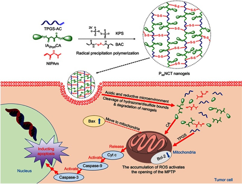
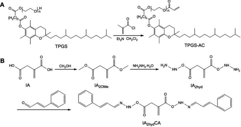
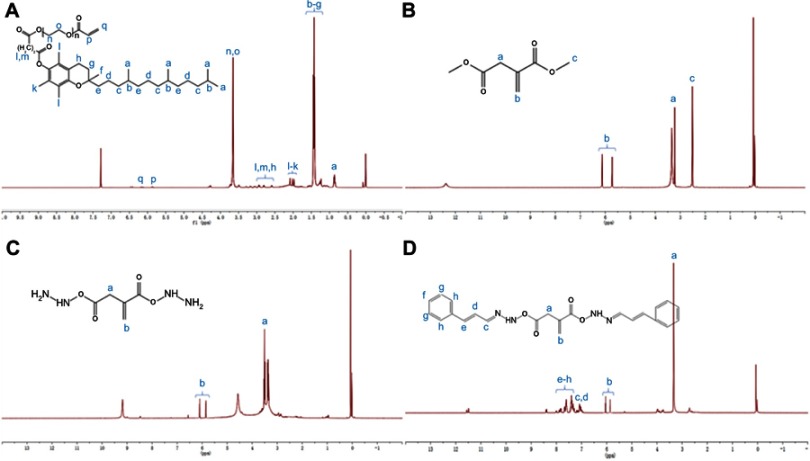
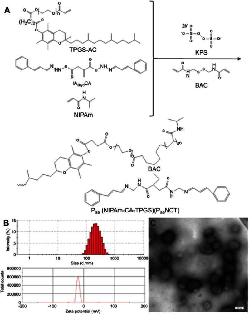
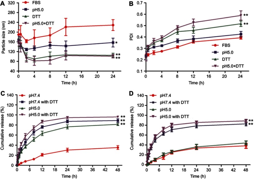
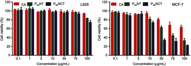
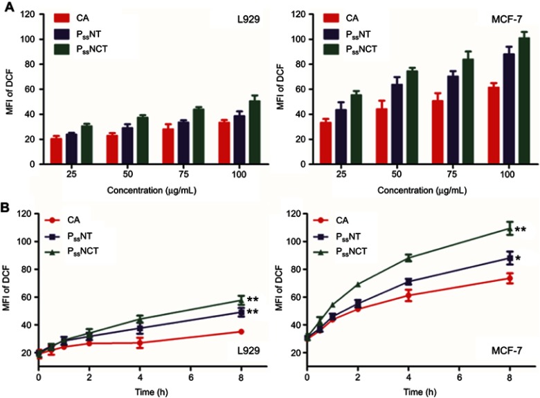
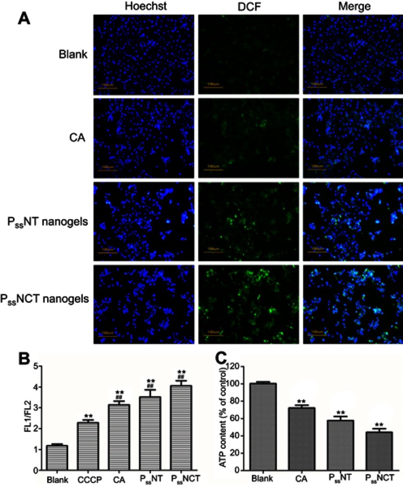
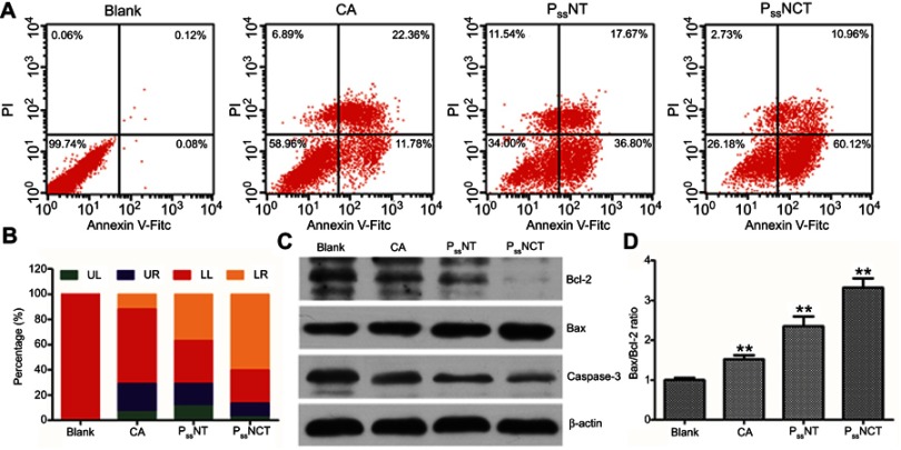
Purpose: The levels of reactive oxygen species (ROS) in tumor cells are much higher than that in normal cells, and rise rapidly under the influence of exogenous or endogenous inducing factors, eventually leading to the apoptosis of tumor cells. Therefore, this study prepared a dual pH/reducing-responsive poly (N-isopropylacrylamide-co-Cinnamaldehyde-co-D-α-tocopheryl polyethylene glycol 1000 succinate, PssNCT) nanogels, which employed two exogenous ROS inducers, cinnamaldehyde (CA) and D-α-tocopheryl polyethylene glycol 1000 succinate (TPGS), to selectively induce apoptosis by regulating ROS levels in tumor cells. Methods: The PssNCT nanogels were prepared by the free radical precipitation polymerization under the crosslink between pH-sensitive hydrazone and reducing-sensitive disulfide bonds, followed by the physicochemical and morphological characteristics investigations. Plasma stability, dual pH/reducing responsive degradation and in vitro release were also assessed. In cell experiments, cytotoxicity in different cells were first detected. The intracellular ROS levels and mitochondrial functions of tumor cells were then evaluated. Moreover, the apoptosis and western-blot assays were employed to verify the association between ROS levels elevation and apoptosis in tumor cells. Results: The nanogels exhibited a round-like hollow structure with the diameter smaller than 200nm. The nanogels were stable in plasma, while showed rapid degradation in acidic and reducing environments, thus achieving significant release of CA and TPGS in these media. Furthermore, the sufficient amplification of ROS signals was induced by the synergistically function of CA and TPGS on mitochondria, which resulted in the opening of the mitochondrial apoptotic pathway and enhanced cytotoxicity on MCF-7 cells. However, nanogels barely affected L929 cells owing to their lower intracellular ROS basal levels. Conclusion: The specific ROS regulation method achieved by these nanogels could be explored to selectively kill tumor cells according to the difference of ROS signals in different kinds of cells.

Keywords: TPGS; cinnamaldehyde; nanogels; oxidative stress; reactive oxygen species.


Conflict of interest statement

The authors report no conflicts of interest in this work.

Figures

Scheme 1
Scheme 1
Illustration of the preparation, dual pH/reducing-responsive intracellular degradation, ROS signal amplification, and induced apoptosis of PssNCT nanogels.
Figure 1
Figure 1
Synthesis of TPGS-AC (A) and IA2hydCA (B).
Figure 2
Figure 2
Characterization of polymers. (A) 1H-NMR spectrum of TPGS-CA recorded in DMSO-d6. (B) 1H-NMR spectrum of IA2OMe recorded in DMSO-d6. (C) 1H-NMR spectrum of IA2hyd recorded in DMSO-d6. (D) 1H-NMR spectrum of IA2hydCA recorded in DMSO-d6.
Figure 3
Figure 3
Synthesis and characterization of PssNCT nanogels. (A) Schematic synthesis of PssNCT nanogels. (B) Size distribution and Zeta potential of PssNCT nanogels determined by DLS. (C) TEM micrograph of PssNCT nanogels.
Figure 4
Figure 4
Plasma stability, dual pH/reducing-responsive degradation and in vitro release of PssNCT nanogels. (A) The particle size changes of PssNCT nanogels incubated with different media (mean±SD, n=3). *P<0.05, **P<0.01: significantly different from FBS. (B) The polydispersity index (PDI) changes of PssNCT nanogels incubated with different media (mean±SD, n=3). **P<0.01: significantly different from FBS. (C) In vitro release of CA from PssNCT nanogels in different media (mean±SD, n=3). **P<0.01: significantly different from pH 7.4 release media. (D) In vitro release of TPGS from PssNCT nanogels in different media (mean±SD, n=3). **P<0.01: significantly different from pH 7.4 release media.
Figure 5
Figure 5
In vitro cytotoxicity of free CA, PssNT, or PssNCT nanogels on L929 and MCF-7 cells in 24 hrs (mean±SD, n=3).
Figure 6
Figure 6
Changes of ROS levels in L929 and MCF-7 cells after incubating with free CA, PssNT, or PssNCT nanogels in different concentrations (A) or for different periods of time (B) (mean±SD, n=3). The concentrations of PssNT and PssNCT were fixed according to the concentration of CA. *P<0.05, **P<0.01: significantly different from the CA group.
Figure 7
Figure 7
Effect of intracellular ROS signal changes on mitochondrial function. (A) Observation of the intracellular ROS production in MCF-7 cells incubated with free CA, PssNT, or PssNCT nanogels for 8 hrs, using DCFH-DA as the detection probe. (blue for Hoechst, Green for DCF and scale bar=100 μm). (B) Changes in mitochondrial membrane potential after 8 hrs of incubation with free CA, PssNT, or PssNCT nanogels (mean±SD, n=3). The mitochondrial electron transport chain inhibitor carbonyl cyanide 3-chlorophenylhydrazone (CCCP) was set as the positive control. **P<0.01: significantly different from the blank group, ##P<0.01:significantly different from the CCCP group. (C) Changes in the intracellular ATP level of MCF-7 cells after incubated with free CA, PssNT, or PssNCT nanogels (mean ± SD, n=3). **P<0.01: significantly different from the blank group. In these experiments, the concentrations of PssNT and PssNCT were fixed at 100 μg/mL calculated by CA.
Figure 8
Figure 8
Effects of free CA, PssNT, or PssNCT nanogels on the apoptosis. (A) The apoptosis of MCF-7 cells induced by free CA, PssNT, or PssNCT nanogels for 24 hrs using the Annexin V-FITC/PI staining and analyzed by a flow cytometry. (B) The histogram of apoptosis of MCF-7 cells induced by free CA, PssNT, or PssNCT nanogels. (C) Changes in the expression of apoptosis-related proteins after incubated with free CA, PssNT, or PssNCT nanogels. (D) Changes in the Bax/Bcl-2 ratio after incubated with free CA, PssNT, or PssNCT nanogels (mean±SD, n=3). The concentrations of PssNT and PssNCT were fixed at 100 μg/mL calculated by CA. **P<0.01: significantly different from the blank group.

References

    1. Zhang Y, Shi ZL, Yang X, Yin ZF. Targeting of circulating hepatocellular carcinoma cells to prevent postoperative recurrence and metastasis. World J Gastroenterol. 2014;20(1):142–147. doi:10.3748/wjg.v20.i1.142 - DOI - PMC - PubMed
    1. Shrieve DC, Klish M, Wendland MM, Watson GA. Basic principles of radiobiology, radiotherapy, and radiosurgery. Neurosurg Clin N Am. 2004;15(4):467. doi:10.1016/S1042-3680(03)00074-3 - DOI - PubMed
    1. Wu CP, Hsieh CH, Wu YS. The emergence of drug transporter-mediated multidrug resistance to cancer chemotherapy. Mol Pharm. 2011;8(6):1996–2011. doi:10.1021/mp200261n - DOI - PubMed
    1. Malhotra V, Perry MC. Classical chemotherapy – mechanisms, toxicities and the therapeutic window. Cancer Biol Ther. 2003;2(4):S2–S4. - PubMed
    1. Infanger DW, Lynch ME, Fischbach C. Engineered culture models for studies of tumor-microenvironment interactions In: Yarmush ML, editor. Annual Review of Biomedical Engineering. Vol. 15 Palo Alto: Annual Reviews; 2013:29–53. - PMC - PubMed