. 2019 May 1;8(8):1608106.
doi: 10.1080/2162402X.2019.1608106. eCollection 2019.

Exposure to the antimicrobial peptide LL-37 produces dendritic cells optimized for immunotherapy

Affiliations

Exposure to the antimicrobial peptide LL-37 produces dendritic cells optimized for immunotherapy

Emily Gwyer Findlay et al. Oncoimmunology. .

Abstract

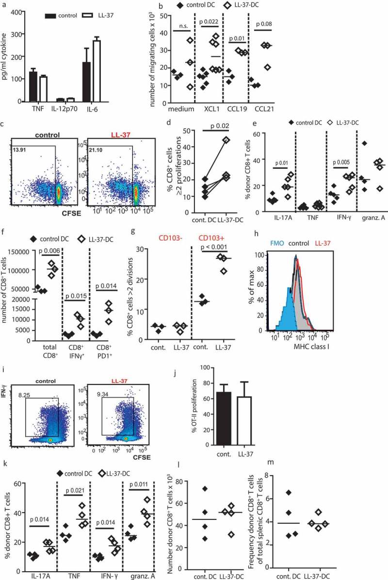
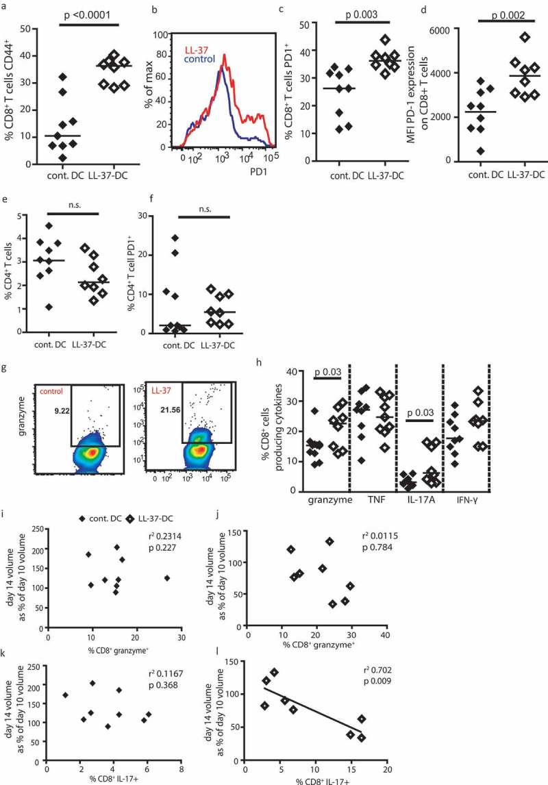
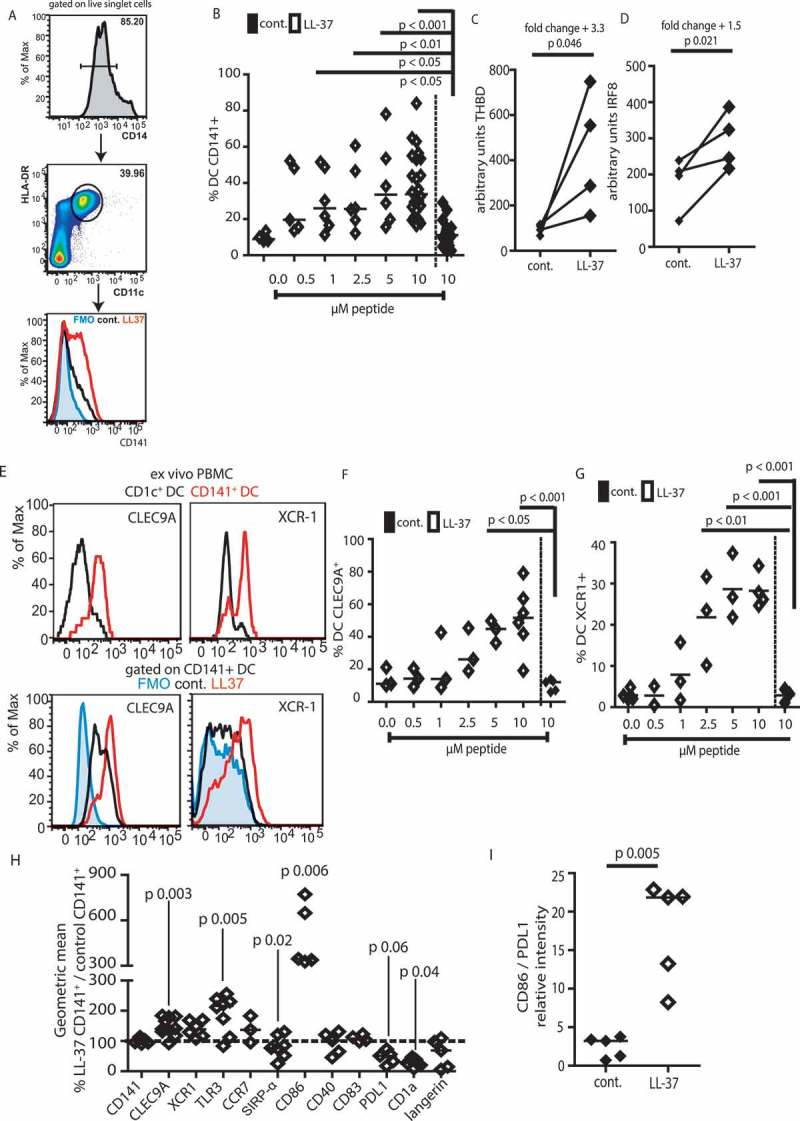
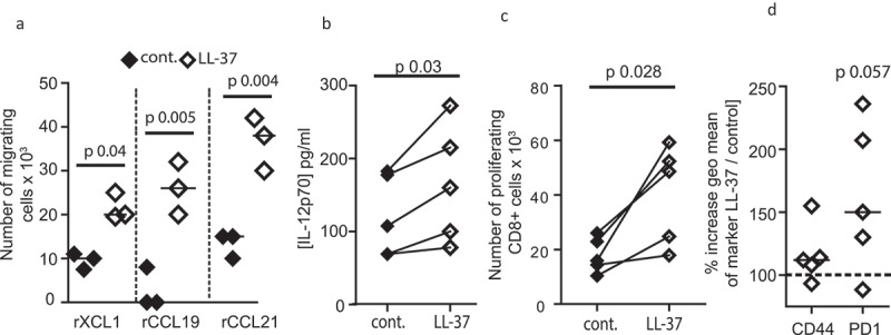
Immunization of patients with autologous, ex vivo matured dendritic cell (DC) preparations, in order to prime antitumor T-cell responses, is the focus of intense research. Despite progress and approval of clinical approaches, significant enhancement of these personalized immunotherapies is urgently needed to improve efficacy. We show that immunotherapeutic murine and human DC, generated in the presence of the antimicrobial host defense peptide LL-37, have dramatically enhanced expansion and differentiation of cells with key features of the critical CD103+/CD141+ DC subsets, including enhanced cross-presentation and co-stimulatory capacity, and upregulation of CCR7 with improved migratory capacity. These LL-37-DC enhanced proliferation, activation and cytokine production by CD8+ (but not CD4+) T cells in vitro and in vivo. Critically, tumor antigen-presenting LL-37-DC increased migration of primed, activated CD8+ T cells into established squamous cell carcinomas in mice, and resulted in tumor regression. This advance therefore has the potential to dramatically enhance DC immunotherapy protocols.

Keywords: CD103; CD141; CD86; CLEC9A; Immunotherapy; PD1; cancer; cathelicidin; cross-presentation; dendritic cells; host defense peptide.


Figures

Figure 1.
Figure 1.
LL-37 promotes generation of CD103+ DC. Bone marrow from adult C57Bl6/J mice was cultured for 7 days with 20 ng/mL GM-CSF (a–h) or 20 ng/mL Flt3-L (g–h) before CD103 expression on DC generated was assessed by flow cytometry. (a–c) DC were exposed to 10 µM LL-37 or scrambled control peptide; (d) LL-37 was added on days shown; (e, f) the proportion of DC and of CD103+ DC in cultures was assessed over time; (g–h) growth factors GM-CSF and Flt3-L were compared. Data shown are mean ± standard error or individual data points with line at median. Statistical tests used: (b) one-way ANOVA with Dunnett’s post-test comparing all to control, n = 4 −16 mice; (c) two-tailed t-test, n = 9; (d) one-way ANOVA with Dunnett’s post-test comparing all to control, n = 3; (e, f) paired two-tailed t-test, n = 3–5; (g, h) two-tailed t-test, n = 3–6.
Figure 2.
Figure 2.
LL-37-DC upregulates markers of cDC1 cells. (a, b) C57Bl6/J (WT) and Batf3−/- (KO) bone marrow was cultured for 7 days with 20 ng/mL GM-CSF and 10 µM LL-37 or control scrambled peptide. On day 7, CD103 expression was examined by flow cytometry. (c–f) Representative staining showing cDC1 markers on GM-CSF-derived WT LL-37-DC versus Flt3-L-generated DC. (g–k) other markers were expressed on the DC by flow cytometry. Data shown are mean ± standard error or individual data points with line at median. Statistical tests used: (h, i, j, k) two-tailed t-test (if necessary on raw data before conversion), n = 5–9; (f, g) two-way ANOVA with Bonferroni’s post-test comparing all groups to each other, n = 3–5.
Figure 3.
Figure 3.
LL-37 DC can cross-present antigen to CD8+ T cells. Bone marrow from adult C57Bl6/J mice was cultured for 7 days with 20 ng/mL GM-CSF. (a) production of inflammatory cytokines was assessed by ELISA. (b) Migration of the DC through 5-µM transwells was assessed. (c–e) DC were stimulated with 100 ng/mL Pam3Csk4, poly I:C and ovalbumin peptide for 3 h and their ability to stimulate OT-1 CD8+ cell proliferation (c, d) and cytokine production (e) was assessed before the experiment was repeated using whole ovalbumin protein in place of peptide (f). (g) On day 7 of culture DC were sorted by CD103 expression, exposed to whole ovalbumin protein and each population assessed for its ability to induce CD8+ T-cell proliferation. (h) On day 7 of DC culture MHC class I expression was assessed by flow cytometry. (i, j) antigen presentation using whole ovalbumin protein was examined with CD4+ OT-II cells. (k-m) OT-1 CD8+ T cells and stimulated DC were injected into WT mice and subsequently cytokine production (k), number (l) and frequency (m) of donor T cells in the spleen assessed. Data shown are mean ± standard error or individual data points with line at median. Statistical tests used: (b) two-tailed t-tests, n = 3–5 mice; (d, e, f, k) two-tailed t-tests, n = 3–6 mice over two experiments; (g) two-way ANOVA with Bonferroni post-test, n = 3 mice over two separate experiments.
Figure 4.
Figure 4.
LL-37-DC inducing regression of established tumors. Cells from the squamous cell carcinoma 7.1 cell line were cultured and 0.5 × 106 injected into each flank of FVB mice. On day 4, when tumors were palpable, 0.75 × 106 control-DC or LL-37-DC were injected subcutaneously, centrally in between the tumors. Tumor size was monitored twice weekly (a) until day 14 when mice were culled and examined (b, c, d). Tumors were disaggregated and total viable cell counts performed (e). CD8+ T-cell populations were identified by flow cytometry (f). (g) Cells from the SCC 6.2 line were used and tumor growth monitored for 14 days. Data shown are mean ± standard error or individual data points with line at median. Statistical tests used: (a) two-way repeated-measures ANOVA with Bonferroni’s post-test, n = 9 tumors treated with control DC and n = 10 with LL-37-DC; (e,f) two-tailed t-test, n = 9 tumors treated with control DC and n = 8 with LL-37-DC; (g) two-way repeated -measures ANOVA with Bonferroni's post-test, n=10.
Figure 5.
Figure 5.
Tumors treated with LL-37-DC contain granzyme+ PD-1+ CD8+ T cells. Cells from the squamous cell carcinoma 7.1 cell line were cultured and 0.5 × 106 injected into each flank of FVB mice. On day 4, when tumors were palpable, 0.75 × 106 control-DC or LL-37-DC were injected subcutaneously. (a-h) Tumors were disaggregated and T-cell populations assessed by flow cytometry. (i - l) correlation analyses were performed comparing cytokine production to volume alterations in tumors. Data are shown as individual data points with line at median. Statistical tests used: a, c, d, e, f, h) two-tailed t-tests, n = 9 tumors treated with control DC and n = 8 with LL-37-DC; i, j, k, l) Pearson correlation analysis, n = 9 tumors treated with control DC and n = 8 with LL-37-DC.
Figure 6.
Figure 6.
Human DC exposed to LL-37 upregulate CD141, CLEC9A, and XCR1. Monocytes were isolated from blood of healthy volunteers and incubated for 7 days with GM-CSF, IL-4 and 10 µM LL-37 or control peptide. On day 7, DC were identified as in (a) and flow cytometry (b) used to assess expression of CD141. (c–d) 24 h after incubation with LL-37 RNA was isolated from CD11c+ HLA-DR+ cells and expression of THBD and IRF8 examined. (e) Staining of cDC1 markers on LL-37-DC was compared to CD141+ DC isolated from blood. (f–i) other markers were examined on day 7 of culture. Data are shown as individual data points with line at median. Statistical tests used: (b, f, g) one-way ANOVA with Dunnett’s post-test comparing all to control, n = 3–24 donors; (c, d, h, i) two-tailed t-test, if necessary on raw data before conversion, n = 4–8 donors.
Figure 7.
Figure 7.
Human LL-37-DC induce CD8+ T-cell proliferation. Monocytes were isolated from blood of healthy volunteers and incubated for 7 days with GM-CSF, IL-4 and 10 µM LL-37 or control peptide. (a) On day 7, CD11c+ HLA-DR+ cells were sorted and migration through 5 µM transwells toward recombinant chemokines assessed; (b) sorted DC were stimulated with polyI:C and IL-12p70 production assessed by ELISA. (c, d) sorted DC were incubated with Epstein Barr Virus peptides and poly I:C before incubation with CFSE-labeled autologous CD8+ T cells. 60 h later T-cell proliferation (c) and activation (d) were assessed. Data are shown as individual data points with line at median. Statistical tests used: (a–d) paired two-tailed t-tests, if necessary on raw data before conversion, n = 3–5 donors.

References

    1. Saxena M, Balan S, Roudko V, Bhardwaj N.. Towards superior dendritic-cell vaccines for cancer therapy. Nat Biomed Eng. 2018;2(6):341–346. doi:10.1038/s41551-018-0250-x. - DOI - PMC - PubMed
    1. Sabado RL, Bhardwaj N. Cancer immunotherapy: dendritic-cell vaccines on the move. Nature. 2015;519(7543):300–301. doi:10.1038/nature14211. - DOI - PubMed
    1. Bol KF, Tel J, de Vries IJ, Figdor CG. Naturally circulating dendritic cells to vaccinate cancer patients. Oncoimmunology. 2013;2(3):e23431. doi:10.4161/onci.23431. - DOI - PMC - PubMed
    1. Kantoff PW, Higano CS, Shore ND, Berger ER, Small EJ, Penson DF, Redfern CH, Ferrari AC, Dreicer R, Sims RB, et al. Sipuleucel-T immunotherapy for castration-resistant prostate cancer. N Engl J Med. 2010;363(5):411–422. doi:10.1056/NEJMoa1001294. - DOI - PubMed
    1. Schreibelt G, Bol KF, Westdorp H, Wimmers F, Aarntzen EH, Duiveman-de Boer T, van de Rakt MW, Scharenborg NM, de Boer AJ, Pots JM, et al. Effective clinical responses in metastatic melanoma patients after vaccination with primary myeloid dendritic cells. Clin Cancer Res. 2016;22(9):2155–2166. doi:10.1158/1078-0432.CCR-15-2205. - DOI - PubMed

Publication types