. 2019 Aug 13:7:19.
doi: 10.1186/s41038-019-0157-0. eCollection 2019.

One-step approach for full-thickness skin defect reconstruction in rats using minced split-thickness skin grafts with Pelnac overlay

Affiliations

One-step approach for full-thickness skin defect reconstruction in rats using minced split-thickness skin grafts with Pelnac overlay

Tong Liu et al. Burns Trauma. .

Abstract

Background: Split-thickness skin grafting is the current gold standard for the treatment of traumatic skin loss. However, for patients with extensive burns, split-thickness skin grafting is limited by donor skin availability. Grafting split-thickness skin minced into micrografts increases the expansion ratio but may reduce wound repair quality. Dermal substitutes such as Pelnac can enhance the healing of full-thickness skin wounds, but their application currently requires two surgeries. The present study investigated whether it is possible to repair full-thickness skin defects and improve wound healing quality in a single surgery using Pelnac as an overlay of minced split-thickness skin grafts in a rat model.

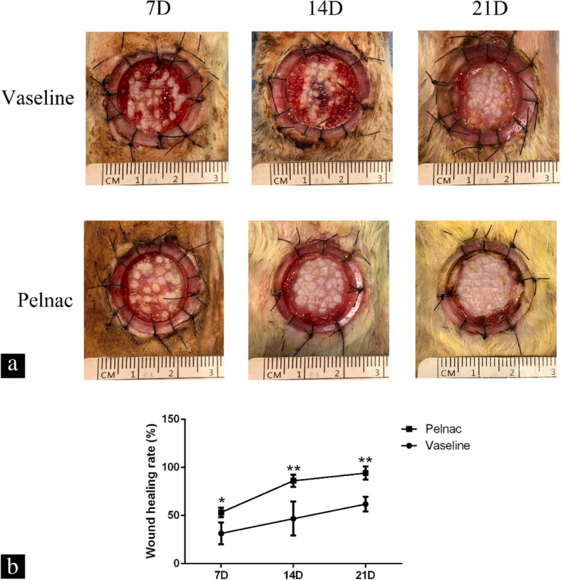
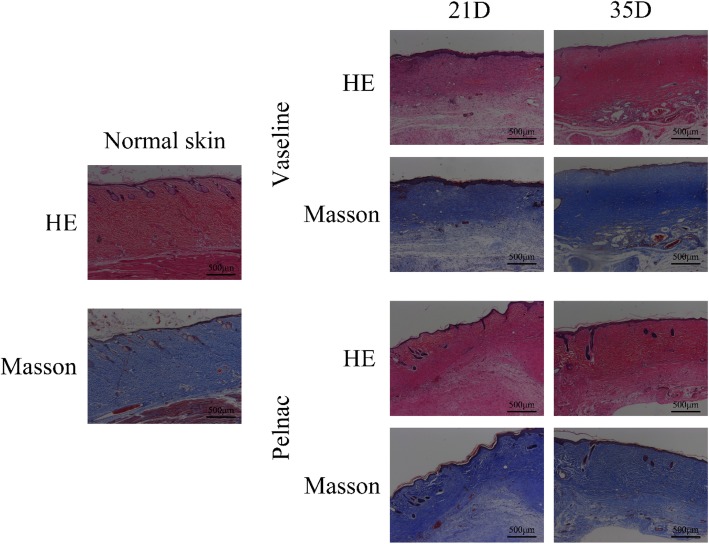
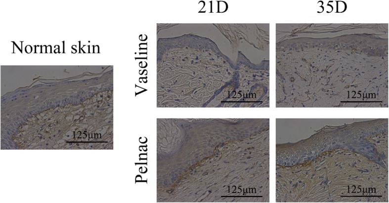
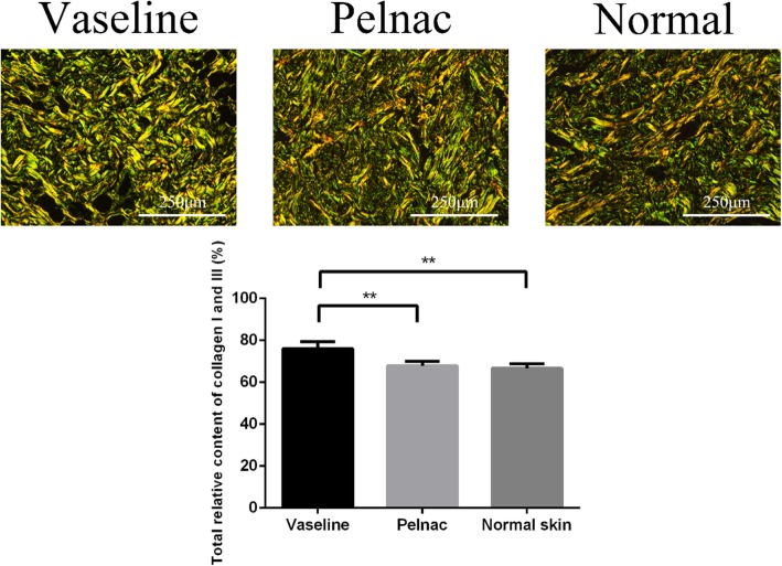
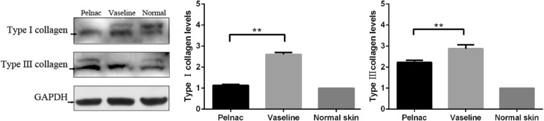
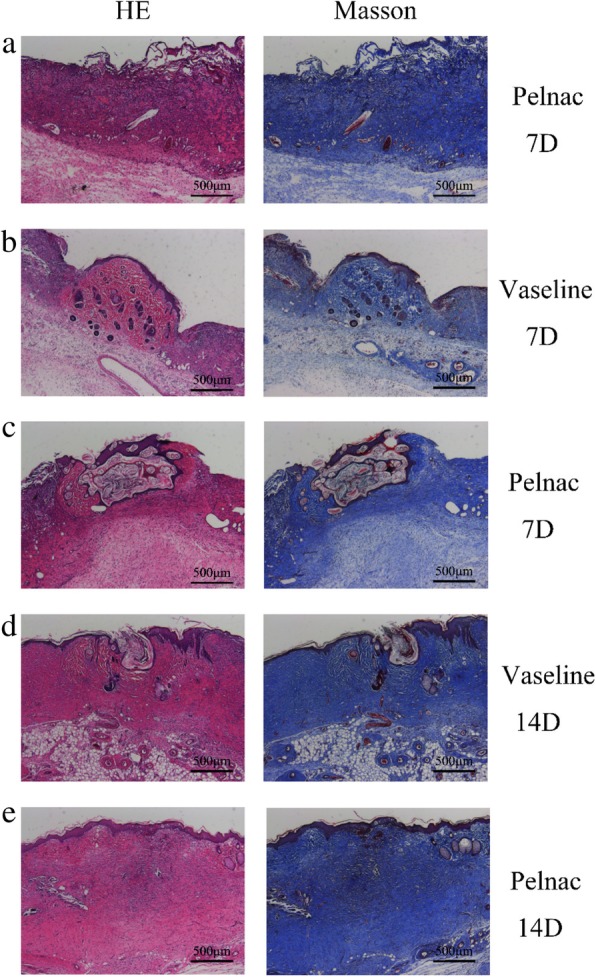
Methods: A full-thickness skin defect model was established using male Sprague-Dawley rats of 10 weeks old. The animals were randomly divided into control and experimental groups in which Vaseline gauze and Pelnac, respectively, were overlaid on minced split-thickness skin grafts to repair the defects. Wound healing rate and quality were compared between the two groups. For better illustration of the quality of wound healing, some results were compared with those obtained for normal skin of rats.

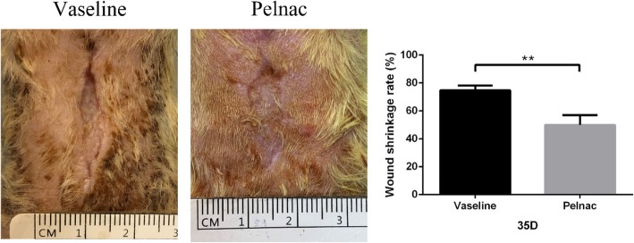
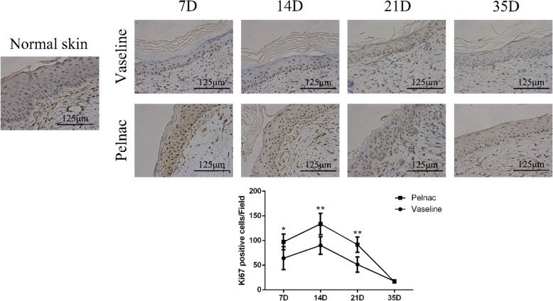
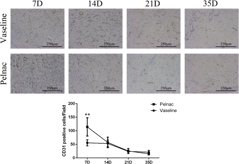
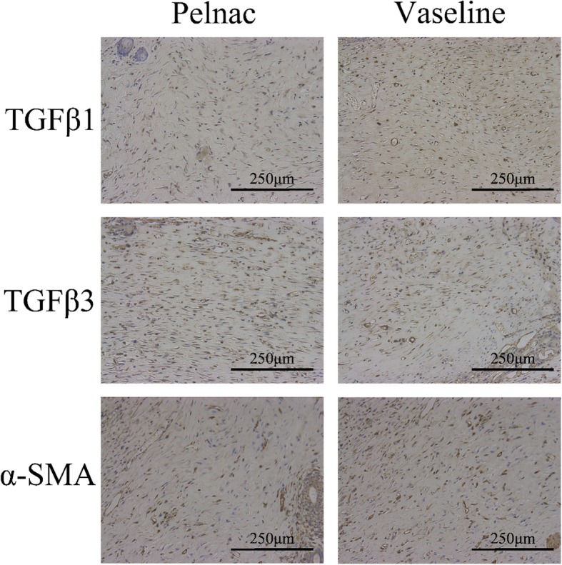
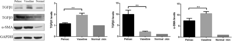
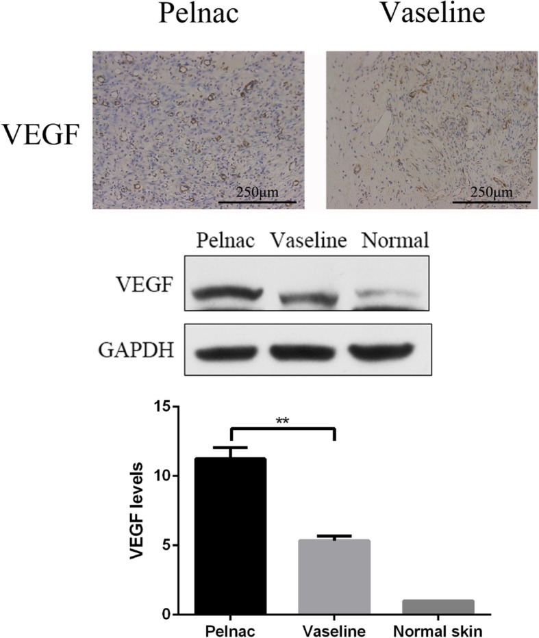
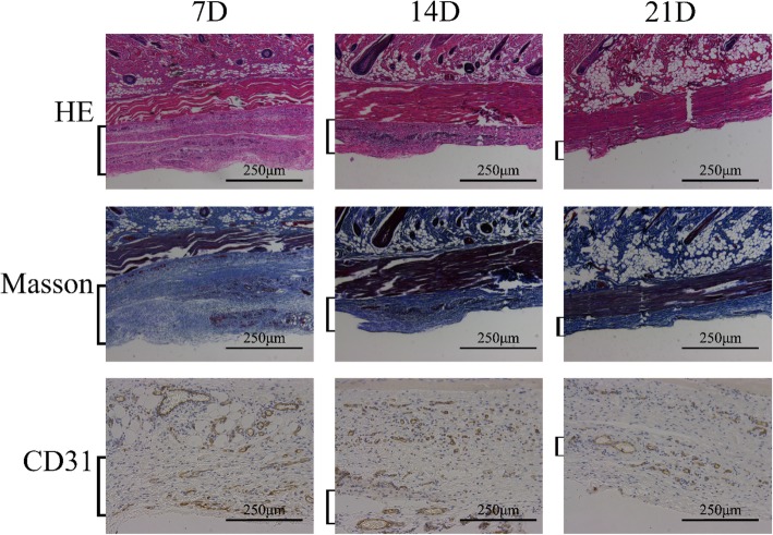
Results: We found that using Pelnac as an overlay for minced split-thickness skin grafts accelerated wound closure and stimulated cell proliferation and tissue angiogenesis. In addition, this approach enhanced collagen synthesis and increased the formation of basement membrane and dermis as well as the expression of growth factors related to wound healing while reducing scar formation.

Conclusions: Using minced split-thickness skin grafts overlaid with Pelnac enables the reconstruction of full-thickness skin defects in a single step and can increase the healing rate while improving the quality of wound healing.

Keywords: Full-thickness skin defect; Minced skin graft; Pelnac; Reconstruction; Skin wound healing; Split-thickness skin grafts.


Conflict of interest statement

Competing interestsThe authors declare that they have no competing interests.

Figures

Fig. 1
Fig. 1
Biopsy of split-thickness skin graft of rats showing the thickness and structure (× 50). Scale bars, 500 μm
Fig. 2
Fig. 2
Micrografts transplanted into full-thickness skin defects of rats and covered with Pelnac or Vaseline gauze. a Prepared micrografts were transplanted into the full-thickness wound in the back of a rat at a 1:5 expansion ratio. b In the experimental group, the micrograft was covered with Pelnac. c Local packing and compression were applied to prevent graft displacement
Fig. 3
Fig. 3
Wound size of rats at different time points post-operation. a Representative photographs of full-thickness skin wounds on days 7, 14, and 21 (7D, 14D, and 21D, respectively) after application of minced split-thickness skin grafts with Pelnac or Vaseline gauze as an overlay. b Statistical analysis of the wound healing rate in the two groups. 7D: Vaseline gauze group 31.51 ± 9.8%, Pelnac group 53.32 ± 4.3%; 14D: Vaseline gauze group 46.85 ± 15.3%, Pelnac group 86.19 ± 5.6%; 21D: Vaseline gauze group 62.00 ± 6.6%, Pelnac group 94.21 ± 5.9%. Data are presented as mean ± standard deviation. Error bars indicate standard deviation. Statistical analysis was performed by repeated-measures ANOVA. *p < 0.05, **p < 0.01
Fig. 4
Fig. 4
Wound shrinkage rate of rats on day 35 post-operation. Photographs of full-thickness skin wounds on day 35 after application of minced split-thickness skin grafts with Pelnac or Vaseline gauze as an overlay. Statistical analysis of the wound shrinkage rate in the two groups. Vaseline gauze group 74.78 ± 2.9%, Pelnac group 49.99 ± 6.1%. Data are presented as mean ± standard deviation. Error bars indicate standard deviation. Statistical analysis was performed by Student's t-test. **p < 0.01
Fig. 5
Fig. 5
Formation of the dermis in skin wounds of rats at different time points post-operation. Staining performed by hematoxylin-eosin (HE) and Masson’s trichrome staining 21 days (21D) and 35 days (35D) after surgery in Vaseline gauze and Pelnac groups (× 50). Scale bars, 500 μm
Fig. 6
Fig. 6
Type IV collagen deposition in the basement membrane of rats on days 21 and 35 post-operation. The deposition was monitored on day 21 (21D) and day 35 (35D), as determined by immunohistochemistry (× 400). Scale bars, 125 μm
Fig. 7
Fig. 7
Total relative collagen Ι and III content in Vaseline gauze and Pelnac groups on day 35 post-operation. Data are representative of day 35 and are compared to normal skin. (Top) collagen I and III was detected by Picrosirius red staining (× 200). Scale bars, 250 μm. (Bottom) statistical analysis of total relative collagen Ι and III content. Vaseline gauze group 76.06 ± 3.0%, Pelnac group 67.78 ± 2.0%, normal skin 66.65 ± 2.1%. Data are presented as mean ± standard deviation. Error bars indicate standard deviation. Statistical analysis was performed by one-way ANOVA. *p < 0.05, **p < 0.01
Fig. 8
Fig. 8
Pelnac as the overlay decreased collagen I and III expression in scars on day 35. (Left) collagen I and III levels in Pelnac or Vaseline gauze groups were detected by western blotting. (Middle and right) quantitative analysis of relative collagen I and III protein levels; the level in normal skin was set to 1. Data are presented as mean ± standard deviation. Error bars indicate standard deviation. Statistical analysis was performed by Student's t-test.*p < 0.05, **p < 0.01. GAPDH glyceraldehyde-3-phosphate dehydrogenase
Fig. 9
Fig. 9
Ki-67 expression in wound tissue of rats at different time points post-operation after treatment with Vaseline gauze or Pelnac as overlay. (Top) Ki-67 immunohistochemistry was performed on days 7, 14, 21, and 35 (7D, 14D, 21D, and 35D, respectively) (× 400). Scale bars, 125 μm. (Bottom) quantitative analysis of Ki67-positive cells. Data are presented as mean ± standard devistion. Error bars indicate standard deviation. Statistical analysis was performed by repeated-measures ANOVA. *p < 0.05, **p < 0.01
Fig. 10
Fig. 10
Cluster of differentiation 31 (CD31) expression in wound tissue of rats at different time points post-operation after treatment with Vaseline gauze or Pelnac as overlay. (Top) CD31 immunohistochemistry was performed on days 7, 14, 21, and 35 (7D, 14D, 21D, and 35D, respectively) (× 200). Scale bars, 250 μm. (Bottom) quantitative analysis of CD31-positive cells. Data are presented as mean ± standard devistion. Error bars indicate standard deviation. Statistical analysis was performed by repeated-measures ANOVA. *p < 0.05, **p < 0.01
Fig. 11
Fig. 11
Transforming growth factor (TGF)-β1, TGF-β3, and α-smooth muscle actin (α-SMA) expression in wound tissue after treatment with Vaseline gauze or Pelnac as an overlay was detected by immunohistochemistry on day 35 post-operation (× 200). Scale bars, 250 μm
Fig. 12
Fig. 12
Pelnac decreases transforming growth factor (TGF)-β1 and α-smooth muscle actin (α-SMA) and increases TGF-β3 expression in regenerated dermal tissue on day 35 post-operation. (Left) TGF-β1, TGF-β3, and α-SMA levels following treatment with Pelnac or Vaseline gauze as overlay were detected by western blotting. (Middle and right) quantitative analysis of relative protein levels; the level in normal skin was set to 1. Data are presented as mean ± standard devistion. Error bars indicate standard deviation. Statistical analysis was performed by Student's t-test. *p < 0.05, **p < 0.01. GAPDH glyceraldehyde-3-phosphate dehydrogenase
Fig. 13
Fig. 13
Pelnac increases vascular endothelial growth factor (VEGF) expression in rat wound tissue on day 7 post-operation. (Top) VEGF expression following treatment with Pelnac or Vaseline gauze as an overlay on day 7, as determined by immunohistochemistry (× 200). Scale bars, 250 μm. (Middle and bottom) VEGF expression following treatment with Pelnac or Vaseline gauze as determined by western blotting and quantitative analysis of expression levels. The level in normal skin was set to 1. Data are presented as mean ± standard devistion. Error bars indicate standard deviation. Statistical analysis was performed by Student's t-test.*p < 0.05, **p < 0.01
Fig. 14
Fig. 14
Effect of transplanted micrografts in full thickness skin defects with Pelnac or Vaseline gauze overlay on days 7 and 14. The figure shows Pelnac or Vaseline gauze overlay on collagen production in full-thickness skin defects on postoperative day 7 (7D) and day 14 (14D), as determined by hematoxylin-eosin (HE) and Masson’s trichrome staining (× 50). Scale bars, 500 μm. a Vascularization can be seen in the Pelnac group on day 7. b The minced split-thickness skin graft was attached to the wound base on day 7 in the Vaseline gauze group. c The minced split-thickness skin graft had reached the wound surface through Pelnac on day 7 in the Pelnac group. d The minced split-thickness skin graft had reached the wound surface on day 14 in the Vaseline gauze group. e The structure of the micrograft could not be seen in the Pelnac group on day 14
Fig. 15
Fig. 15
Vascularization of Pelnac after implantation under the dorsal skin of rats at different time points (square brackets). Vascularization after implantation under the dorsal skin of rats on postoperative days 7, 14, and 21 (7D, 14D, and 21D, respectively) visualized by hematoxylin-eosin (HE) and Masson’s trichrome staining and cluster of differentiation 31 (CD31) immunohistochemistry (× 200). Scale bars, 250 μm

References

    1. Huang YY. Topical antimicrobials for burn wound infections. Recent Pat Antiinfect Drug Discov. 2010;5:124–151. doi: 10.2174/157489110791233522. - DOI - PMC - PubMed
    1. Greenhalgh DG. Sepsis in the burn patient: a different problem than sepsis in the general population. Burns Trauma. 2017;5(1):23. doi: 10.1186/s41038-017-0089-5. - DOI - PMC - PubMed
    1. Czaika V, Alborova A, Richter H, Sterry W, Vergou T, Antoniou C, et al. Comparison of transepidermal water loss and laser scanning microscopy measurements to assess their value in the characterization of cutaneous barrier defects. Skin Pharmacol Physiol. 2012;25:39–46. doi: 10.1159/000330486. - DOI - PubMed
    1. Brusselaers N, Pirayesh A, Hoeksema H, Richters CD, Verbelen J, Beele H, et al. Skin replacement in burn wounds. J Trauma. 2010;68:490–501. doi: 10.1097/TA.0b013e3181c9c074. - DOI - PubMed
    1. Biswas A, Bharara M, Hurst C, Armstrong DG, Rilo H. The micrograft concept for wound healing: strategies and applications. J Diabetes Sci Technol. 2010;4:808–819. doi: 10.1177/193229681000400407. - DOI - PMC - PubMed