Randomized Controlled Trial
. 2020 May;104(5):1058-1069.
doi: 10.1097/TP.0000000000002917.

mTOR Inhibitor Therapy Diminishes Circulating CD8+ CD28- Effector Memory T Cells and Improves Allograft Inflammation in Belatacept-refractory Renal Allograft Rejection

Affiliations
Randomized Controlled Trial

mTOR Inhibitor Therapy Diminishes Circulating CD8+ CD28- Effector Memory T Cells and Improves Allograft Inflammation in Belatacept-refractory Renal Allograft Rejection

Cyd M Castro-Rojas et al. Transplantation. 2020 May.

Abstract

Background: Renal allograft rejection is more frequent under belatacept-based, compared with tacrolimus-based, immunosuppression. We studied kidney transplant recipients experiencing rejection under belatacept-based early corticosteroid withdrawal following T-cell-depleting induction in a recent randomized trial (Belatacept-based Early Steroid Withdrawal Trial, clinicaltrials.gov NCT01729494) to determine mechanisms of rejection and treatment.

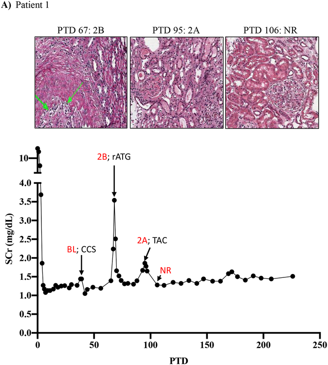
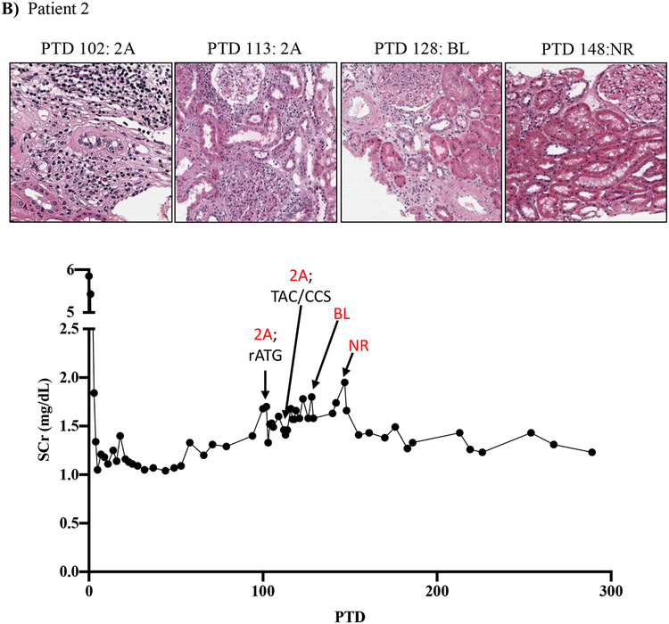
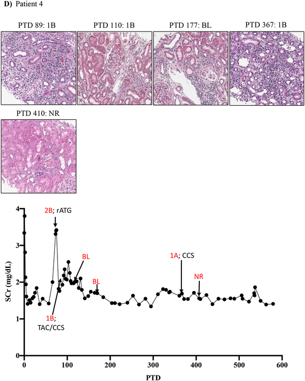
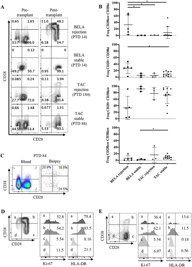
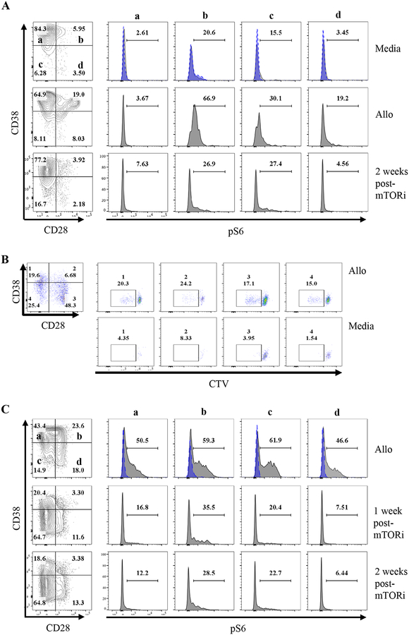
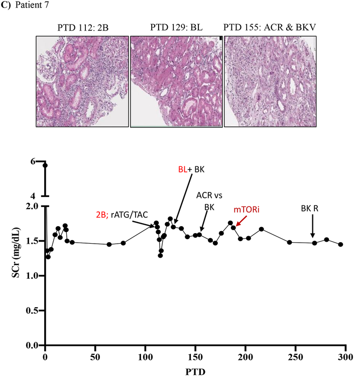
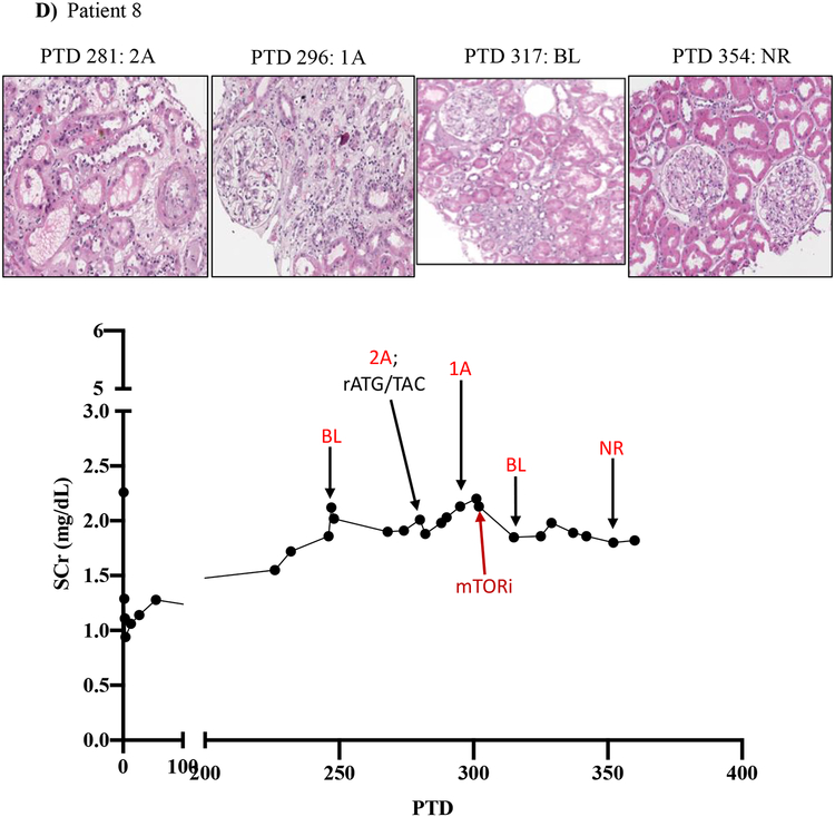
Methods: Peripheral mononuclear cells, serum creatinine levels, and renal biopsies were collected from 8 patients undergoing belatacept-refractory rejection (BRR). We used flow cytometry, histology, and immunofluorescence to characterize CD8 effector memory T cell (TEM) populations in the periphery and graft before and after mammalian target of rapamycin (mTOR) inhibition.

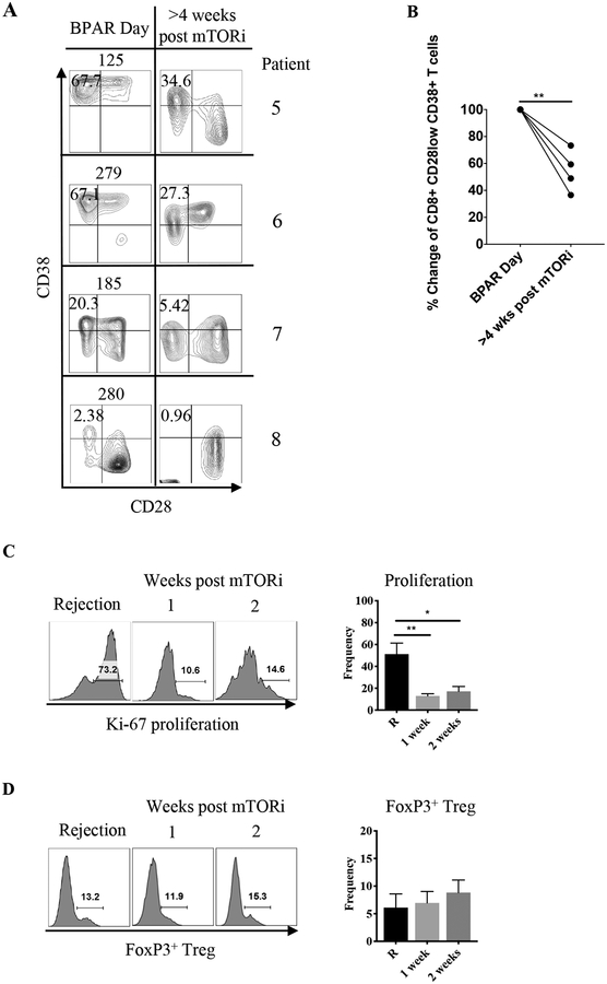
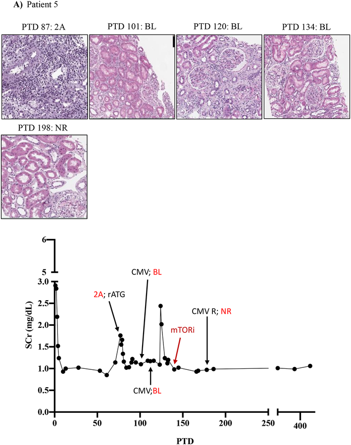
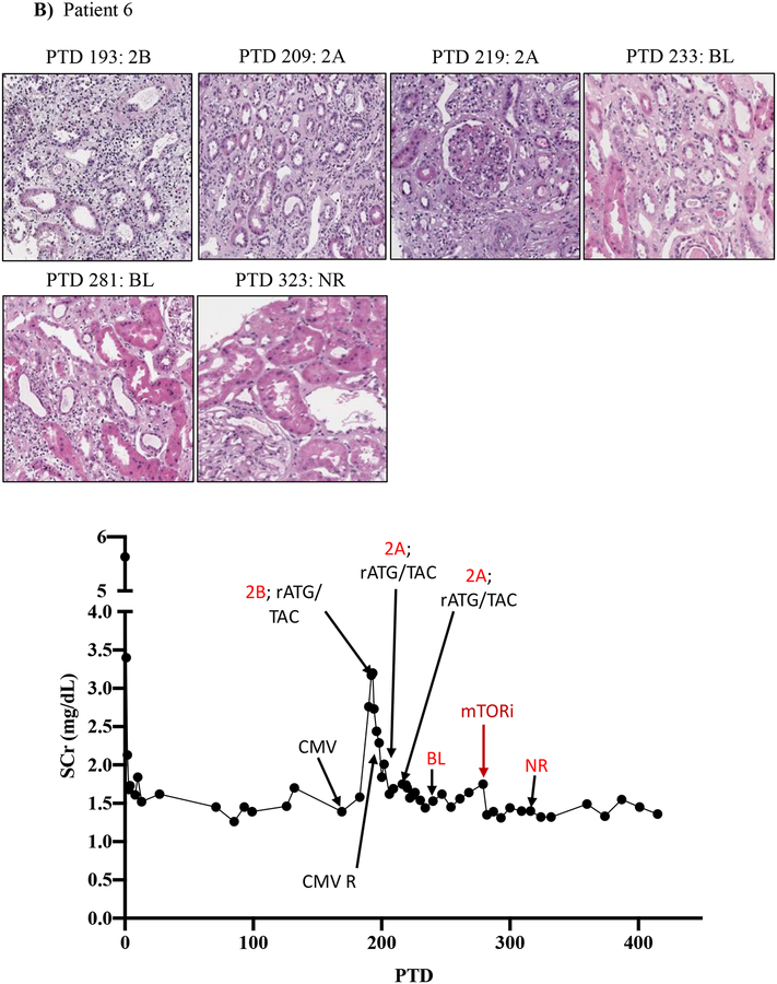
Results: Here, we found that patients with BRR did not respond to standard antirejection therapy and had a substantial increase in alloreactive CD8 T cells with a CD28/DR/CD38/CD45RO TEM. These cells had increased activation of the mTOR pathway, as assessed by phosphorylated ribosomal protein S6 expression. Notably, everolimus (an mTOR inhibitor) treatment of patients with BRR halted the in vivo proliferation of TEM cells and their ex vivo alloreactivity and resulted in their significant reduction in the peripheral blood. The frequency of circulating FoxP3 regulatory T cells was not altered. Importantly, everolimus led to rapid resolution of rejection as confirmed by histology.

Conclusions: Thus, while prior work has shown that concomitant belatacept + mTOR inhibitor therapy is effective for maintenance immunosuppression, our preliminary data suggest that everolimus may provide an available means for effecting "rescue" therapy for rejections occurring under belatacept that are refractory to traditional antirejection therapy with corticosteroids and polyclonal antilymphocyte globulin.


Conflict of interest statement

DISCLOSURE

The authors of this manuscript have no conflict of interest to disclose.

Figures

Figure 1:
Figure 1:. Resistance of belatacept-refractory rejection to standard of care immunosuppression.
A-D) Plots showing post-transplant serum creatinine, histology, Banff lesion score (red) and treatment course of four patients that underwent rejection under belatacept. Renal allograft pathology was determined by H&E staining (micrographs) at specified timepoints. A) Shows a 46-year-old male that received his first transplant from living donor after ESRD due to PCKD. Prior to transplant he had a cPRA of 0%. Initial immunosuppression included rATG induction, belatacept, MMF, and early corticosteroid withdrawal at 5 days post-transplant. B) Shows the history of a 56-year-old Caucasian male that received first kidney transplant after ESRD due to DM and had a 0% cPRA and non-prior HLA exposure history. Postoperatively, he received alemtuzumab induction with belatacept, MMF and early corticosteroid cessation at 5 days post-transplant. C) Shows a 53-year-old AA woman with ESRD due to necrotizing GN and the recipient of a first kidney transplant. She had a 0% cPRA with no prior history of HLA exposure. Immunosuppressive therapy included rATG induction, belatacept, MMF, and CCS cessation on PTD 5. D) Displays a 60-year-old male with ESRD due to DM that underwent kidney transplantation from a deceased donor and received alemtuzumab induction, belatacept, MMF, and CCS cessation at 5 days post-transplant. BL, borderline; CCS, corticosteroids; DM, diabetes mellitus; ESRD, end stage renal disease; GN, glomerulonephritis; MMF, mycophenolate mofetil; NR, no rejection; PCKD, polycystic kidney disease; rATG, rabbit anti-thymocyte globulin; TAC, tacrolimus.
Figure 2:
Figure 2:. Emergence of CD8+CD28CD38hi and CD8+CD28+CD38hi effector memory T cells in patients with belatacept-resistant allo-rejection.
A) CD8+ T cells gated from CD3+CD45RO+ were analyzed for the expression of CD28 and CD38 in stable and rejection patients under belatacept or tacrolimus. BELA rejection on patient 7 is PTD 14, which is the time of diagnosed rejection and prior to any SOC rejection therapy. TAC rejection depicts the timepoint of rejection diagnosis at PTD 184. Stable patients’ samples were chosen to match rejectors PTD. B) Frequencies of CD8+ CD45RO+ CD28+/− CD38+/− TEM populations in the rejector and stable patients. Open circle marks the CD8+ profile for patient 7 at PTD 185, after SOC rejection therapy. Results are presented as means ± standard deviation. C) At moment of confirmed rejection PTD 84 for patient 7, CD8+ TEM cells are present in the graft as shown by flow cytometric analysis of cells extracted from a previously frozen biopsy. D) Belatacept rejection CD8+ effector memory T cells and E) tacrolimus rejection CD8+ effector memory T cells were analyzed with Ki-67 for proliferation and HLA-DR for activation. BELA rejection n=4, BELA stable n=3, TAC rejection n=4, TAC stable n=10. *p<0.05.
Figure 3:
Figure 3:. Effector memory CD8+ T cells emerging under belatacept have increased p-RPS6 expression.
PBMCs isolated from patients 6 (A) and 7 (B, C) (n=2) undergoing rejection were analyzed for p-RPS6 before and after (1 and 2 weeks) mTORi. PBMCs were cultured alone, or with their respective donor APCs (allo) for 2 hours. Cells were then fixed and stained for CD45, CD3, CD8, CD38, CD28 and p-RPS6 phospho-flow analysis. Blue dashed-line histograms show isotype control. B) Allo reactive cells were detected by cell trace violet dilution in a mixed lymphocyte reaction assay in PBMCs of patient 7 at PTD 56 following rejection standard of care treatment were mixed with donor cells for 5 days and analyzed by flow cytometry.
Figure 4:
Figure 4:. Everolimus diminishes activated CD8+CD28CD38hi effector memory T cells.
A) Four patients (patients 5, 6, 7 and 8) presenting with belatacept–refractory rejection were treated with everolimus (n=4). PBMCs were analyzed for presence of CD38hi TEM cells pre- and post-everolimus therapy. Quadrants were drawn based on internal positive controls. B) Percent change at 4 weeks or more after everolimus treatment. Percent change was determined by calculating the decrease difference between pre and post, dividing the decrease by the original number and multiplying it by 100. C) Proliferation assessment by Ki-67 at rejection and 1 and 2 weeks post-everolimus. D) Effect of everolimus on FoxP3+ Tregs 1 and 2 weeks after treatment.
Figure 5.
Figure 5.. mTOR inhibition can rescue belatacept resistant rejections.
A-D) Diagrams show serum creatinine, histology, Banff lesion score (red), and treatment course of four patients that underwent rejection under belatacept and were treated with mTORi. Renal allograft pathology was determined by H&E stain analysis (micrographs) at specified timepoints. A) Shows a 50-year-old Caucasian female with ESRD secondary to hypertension that received a first kidney transplant from a deceased donor. She had a low cPRA (13%) and no evidence of pre-transplant DSA. The patient received rATG induction, belatacept, MMF, and early steroid withdrawal (5 days). B) Shows a 53-year-old unsensitized (cPRA 0%) Caucasian male with ESRD due to DM that received a first renal transplant from a living donor. Immunosuppression included alemtuzumab, belatacept, MMF, and steroid withdrawal at 5 days. CMV infection occurred on PTD 170 and required cessation of MMF therapy for resolution. C) Shows a 46-year-old unsensitized Caucasian male with focal segmental glomerulosclerosis that underwent a first kidney transplant from a living related one haplotype mismatched donor. Immunosuppression included rATG induction, belatacept, MMF, and early steroid cessation at 5 days. D) Shows a 44-year-old unsensitized Caucasian male with ESRD due to obstructive uropathy that underwent living related donor kidney transplant. Immunosuppression consistent of rATG induction, belatacept and MMF maintenance therapy with early steroid cessation on PTD 5. ACR, acute cellular rejection; BL, borderline; CCS, corticosteroids; DM, diabetes mellitus; ESRD, end stage renal disease; GN, glomerulonephritis; MMF, mycophenolate mofetil; NR, no rejection; rATG, rabbit Anti-thymocyte globulin; TAC, tacrolimus.

References

    1. Larsen CP, Pearson TC, Adams AB, et al. Rational development of LEA29Y (belatacept), a high-affinity variant of CTLA4-Ig with potent immunosuppressive properties. American journal of transplantation : official journal of the American Society of Transplantation and the American Society of Transplant Surgeons. 2005;5(3):443–453. - PubMed
    1. Talawila N, Pengel LH. Does belatacept improve outcomes for kidney transplant recipients? A systematic review. Transplant international : official journal of the European Society for Organ Transplantation. 2015;28(11):1251–1264. - PubMed
    1. Heher E, Markmann JF. The Clearer BENEFITS of Belatacept. The New England journal of medicine. 2016;374(4):388–389. - PubMed
    1. Vincenti F, Charpentier B, Vanrenterghem Y, et al. A phase III study of belatacept-based immunosuppression regimens versus cyclosporine in renal transplant recipients (BENEFIT study). American journal of transplantation : official journal of the American Society of Transplantation and the American Society of Transplant Surgeons. 2010;10(3):535–546. - PubMed
    1. Vincenti F, Rostaing L, Grinyo J, et al. Belatacept and Long-Term Outcomes in Kidney Transplantation. N Engl J Med. 2016;374(4):333–343. - PubMed

Publication types

MeSH terms

Associated data