

Textural features on 18F-FDG PET/CT and dynamic contrast-enhanced MR imaging for predicting treatment response and survival of patients with hypopharyngeal carcinoma
- PMID: 31415354
- PMCID: PMC6831375
- DOI: 10.1097/MD.0000000000016608
Textural features on 18F-FDG PET/CT and dynamic contrast-enhanced MR imaging for predicting treatment response and survival of patients with hypopharyngeal carcinoma
Abstract
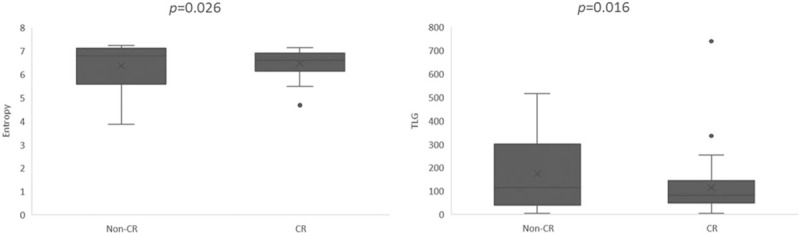
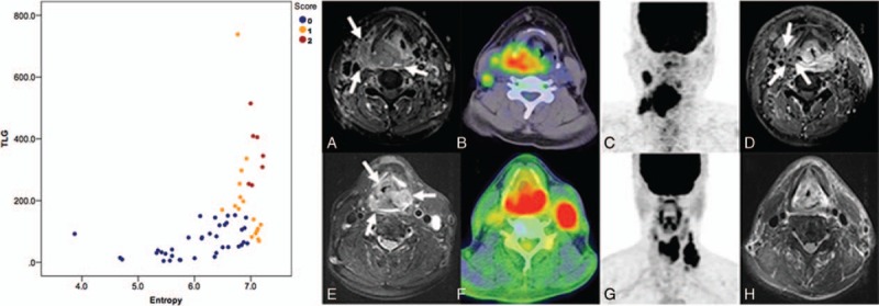
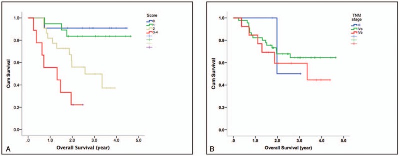
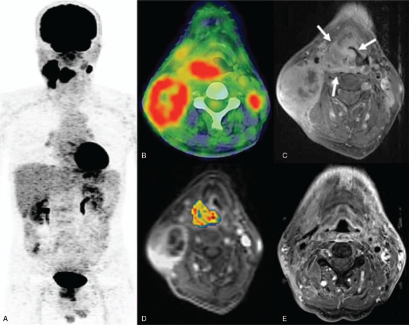
The utility of multimodality molecular imaging for predicting treatment response and survival of patients with hypopharyngeal carcinoma remains unclear. Here, we sought to investigate whether the combination of different molecular imaging parameters may improve outcome prediction in this patient group.Patients with pathologically proven hypopharyngeal carcinoma scheduled to undergo chemoradiotherapy (CRT) were deemed eligible. Besides clinical data, parameters obtained from pretreatment 2-deoxy-2-[fluorine-18]fluoro-D-glucose positron emission tomography/computed tomography (F-FDG PET/CT), dynamic contrast-enhanced (DCE) magnetic resonance imaging (MRI), and diffusion-weighted MRI were analyzed in relation to treatment response, recurrence-free survival (RFS), and overall survival (OS).A total of 61 patients with advanced-stage disease were examined. After CRT, 36% of the patients did not achieve a complete response. Total lesion glycolysis (TLG) and texture feature entropy were found to predict treatment response. The transfer constant (K), TLG, and entropy were associated with RFS, whereas K, blood plasma volume (Vp), standardized uptake value (SUV), and entropy were predictors of OS. Different scoring systems based on the sum of PET- or MRI-derived prognosticators enabled patient stratification into distinct prognostic groups (P <.0001). The complete response rate of patients with a score of 2 was significantly lower than those of patients with a score 1 or 0 (14.7% vs 58.9% vs 75.7%, respectively, P = .007, respectively). The combination of PET- and DCE-MRI-derived independent risk factors allowed a better survival stratification than the TNM staging system (P <.0001 vs .691, respectively).Texture features on F-FDG PET/CT and DCE-MRI are clinically useful to predict treatment response and survival in patients with hypopharyngeal carcinoma. Their combined use in prognostic scoring systems may help these patients benefit from tailored treatment and obtain better oncological results.
Conflict of interest statement
The authors report no conflicts of interest.
Figures
Similar articles
-
Dynamic contrast-enhanced MRI, diffusion-weighted MRI and 18F-FDG PET/CT for the prediction of survival in oropharyngeal or hypopharyngeal squamous cell carcinoma treated with chemoradiation.Eur Radiol. 2016 Nov;26(11):4162-4172. doi: 10.1007/s00330-016-4276-8. Epub 2016 Feb 24. Eur Radiol. 2016. PMID: 26911889
-
Clinical utility of multimodality imaging with dynamic contrast-enhanced MRI, diffusion-weighted MRI, and 18F-FDG PET/CT for the prediction of neck control in oropharyngeal or hypopharyngeal squamous cell carcinoma treated with chemoradiation.PLoS One. 2014 Dec 22;9(12):e115933. doi: 10.1371/journal.pone.0115933. eCollection 2014. PLoS One. 2014. PMID: 25531391 Free PMC article.
-
Prognostic value of 18 F-FDG PET/CT parameters in patients who undergo salvage treatments for recurrent squamous cell carcinoma of the larynx and hypopharynx.J Surg Oncol. 2018 Sep;118(4):644-650. doi: 10.1002/jso.25185. Epub 2018 Aug 21. J Surg Oncol. 2018. PMID: 30132891
-
Gastric cancer and image-derived quantitative parameters: Part 2-a critical review of DCE-MRI and 18F-FDG PET/CT findings.Eur Radiol. 2020 Jan;30(1):247-260. doi: 10.1007/s00330-019-06370-x. Epub 2019 Aug 7. Eur Radiol. 2020. PMID: 31392480 Free PMC article. Review.
-
Imaging Features of Anal Carcinoma after Chemoradiation.Radiographics. 2025 Apr;45(4):e240119. doi: 10.1148/rg.240119. Radiographics. 2025. PMID: 40080437 Review.
Cited by
-
A Systematic Review of PET Textural Analysis and Radiomics in Cancer.Diagnostics (Basel). 2021 Feb 23;11(2):380. doi: 10.3390/diagnostics11020380. Diagnostics (Basel). 2021. PMID: 33672285 Free PMC article. Review.
-
Diagnostic Accuracy of Combined PET/CT with MRI, 18F-FDG PET/MRI, and 18F-FDG PET/CT in Patients with Oropharyngeal and Hypopharyngeal Squamous Cell Carcinoma: A Systematic Review and Meta-Analysis.Contrast Media Mol Imaging. 2021 Apr 28;2021:6653117. doi: 10.1155/2021/6653117. eCollection 2021. Contrast Media Mol Imaging. 2021. PMID: 34007251 Free PMC article.
-
A systematic review and meta-analysis of predictive and prognostic models for outcome prediction using positron emission tomography radiomics in head and neck squamous cell carcinoma patients.Cancer Med. 2023 Aug;12(15):16181-16194. doi: 10.1002/cam4.6278. Epub 2023 Jun 24. Cancer Med. 2023. PMID: 37353996 Free PMC article.
-
Dynamic contrast-enhanced magnetic resonance imaging (DCE-MRI) for pretreatment prediction of neoadjuvant chemotherapy response in locally advanced hypopharyngeal cancer.Br J Radiol. 2020 Nov 1;93(1115):20200751. doi: 10.1259/bjr.20200751. Epub 2020 Sep 11. Br J Radiol. 2020. PMID: 32915647 Free PMC article.
-
Fully automated segmentation and radiomics feature extraction of hypopharyngeal cancer on MRI using deep learning.Eur Radiol. 2023 Sep;33(9):6548-6556. doi: 10.1007/s00330-023-09827-2. Epub 2023 Jun 20. Eur Radiol. 2023. PMID: 37338554 Free PMC article.
References
-
- Spector JG, Sessions DG, Haughey BH, et al. Delayed regional metastases, distant metastases, and second primary malignancies in squamous cell carcinomas of the larynx and hypopharynx. Laryngoscope 2001;111:1079–87. - PubMed
-
- Kuo P, Sosa JA, Burtness BA, et al. Treatment trends and survival effects of chemotherapy for hypopharyngeal cancer: analysis of the national cancer data base. Cancer 2016;122:1853–60. - PubMed
-
- Allal AS. Cancer of the pyriform sinus: trends towards conservative treatment. Bull Cancer 1997;84:757–62. - PubMed
-
- Hall SF, Groome PA, Irish J, et al. The natural history of patients with squamous cell carcinoma of the hypopharynx. Laryngoscope 2008;118:1362–71. - PubMed
Publication types
MeSH terms
Substances
LinkOut - more resources
Full Text Sources
Medical
Research Materials
