

Closed rupture of extensor tendon resulting from untreated Kienböck disease: A case report and a review of the literature
- PMID: 31415435
- PMCID: PMC6831435
- DOI: 10.1097/MD.0000000000016900
Closed rupture of extensor tendon resulting from untreated Kienböck disease: A case report and a review of the literature
Abstract
Rationale: Spontaneous closed extensor tendon rupture is a rare complication of Kienböck disease with only 23 cases reported in the English literature.
Patient concerns: We present a case of painless attritional rupture of the extensor tendons of the right fourth finger in a 69-year-old woman with Kienböck disease and review reported cases of Kienböck disease with subcutaneous closed tendon rupture.
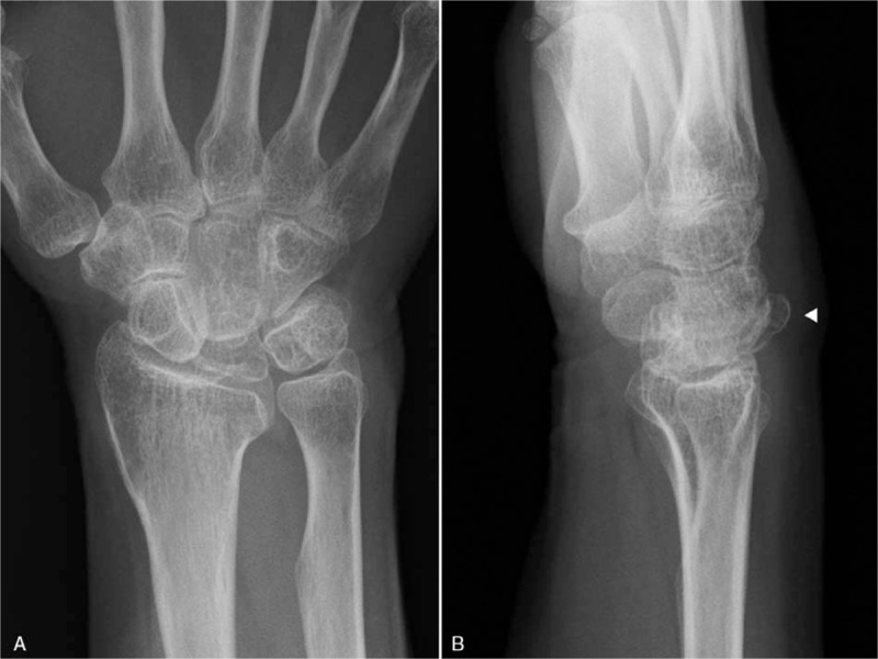
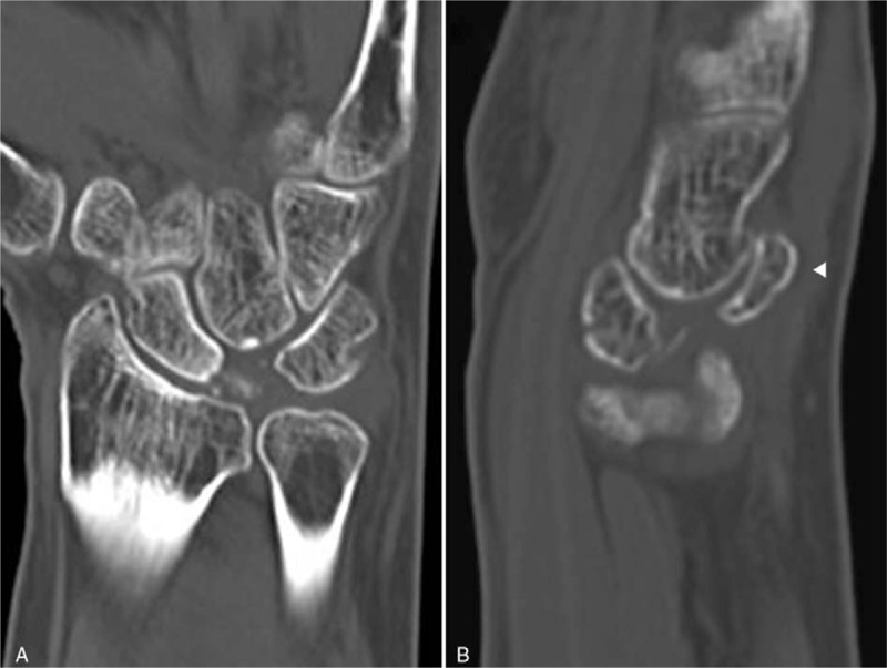
Diagnoses: Physical examination had shown mild painless swelling of the dorsum of the right hand. Plain radiographs showed a dorsally displaced fragment of collapsed lunate bone fracture (Lichtman grade IIIb). Although surgery was recommended, the patient did not desire surgery because she had no pain and no interference with the activities of daily living. Six months later, however, the patient returned to our hospital with complaints of loss of spontaneous extension of the fourth finger. CT and MRI showed aseptic necrosis and large dorsally displaced fragments of the lunate under the extensor tendons of the fingers, suggesting a subcutaneous fourth extensor tendon rupture.
Interventions: Surgery was performed to achieve functional recovery of the ring extensor and to prevent further subcutaneous tendon rupture. The extensor digitorum communis (EDC) of the ring finger was found to be ruptured and the EDCs to the third and fifth fingers were frayed due to attrition from the protrusion of the dorsal fragmented lunate bone. Inspection of the floor of the compartment revealed that the dorsally displaced fragment of the lunate bone had perforated the wrist capsule and protruded into the fourth compartment. The dorsal and volar fragments of the lunate bone were excised completely and scaphocapitate arthrodesis followed by the reconstruction of the fourth extensor tendon was performed.
Outcomes: A year after the surgery, radiography showed complete union of the scaphocapitate arthrodesis. The joint motion reached 45% of normal without any pain and there was full active extension of the fourth finger.
Lessons: Because dorsally displacement of collapsed lunate bone fragments is a risk factor for attritional closed rupture of tendons, radiography, and MRI are essential to diagnose and to treat any closed tendon rupture.
Conflict of interest statement
The authors declare that they have no conflict of interest. The authors alone are responsible for the content and writing of the paper.
Figures


References
-
- Papp SR, Athwal GS, Pichora DR. The rheumatoid wrist. J Am Acad Orthop Surg 2006;14:65–77. - PubMed
-
- Orljanski W, Gaterrer R, Schurz M, et al. Rupture of the extensor pollicis longus tendon after wrist trauma. Acta Chir Plast 2002;44:129–31. - PubMed
-
- Yamazaki H, Uchiyama S, Hata Y, et al. Extensor tendon rupture associated with osteoarthritis of the distal radioulnar joint. J Hand Surg Eur Vol 2008;33:469–74. - PubMed
-
- Valeri G, Ferrara C, Ercolani P, et al. Tendon involvement in rheumatoid arthritis of the wrist: MRI findings. Skeletal Radiol 2001;30:138–43. - PubMed
-
- Ariyoshi D, Imai K, Yamamoto S, et al. Subcutaneous tendon rupture of extensor tendons on bilateral wrists associated with calcium pyrophosphate dihydrate crystal deposition disease. Mod Rheumatol 2007;17:348–51. - PubMed
Publication types
MeSH terms
LinkOut - more resources
Full Text Sources
Medical
