. 2019 Aug 15:25:6110-6119.
doi: 10.12659/MSM.915782.

Plumbagin Restrains Hepatocellular Carcinoma Angiogenesis by Stromal Cell-Derived Factor (SDF-1)/CXCR4-CXCR7 Axis

Affiliations

Plumbagin Restrains Hepatocellular Carcinoma Angiogenesis by Stromal Cell-Derived Factor (SDF-1)/CXCR4-CXCR7 Axis

Jing Zhong et al. Med Sci Monit. .

Abstract

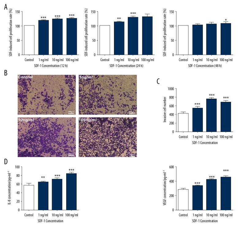
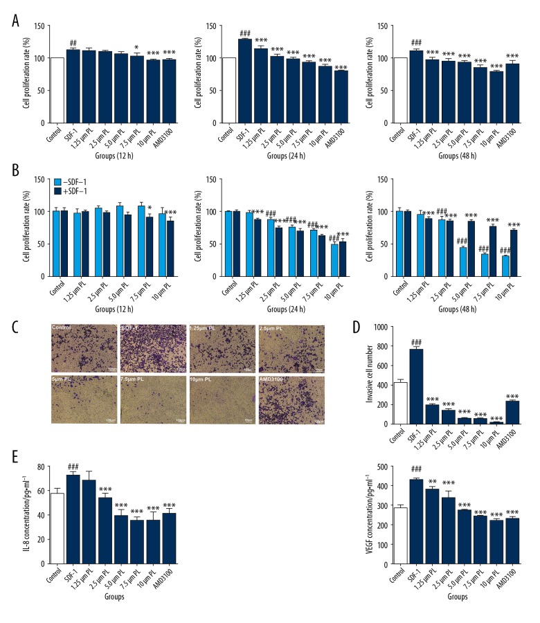
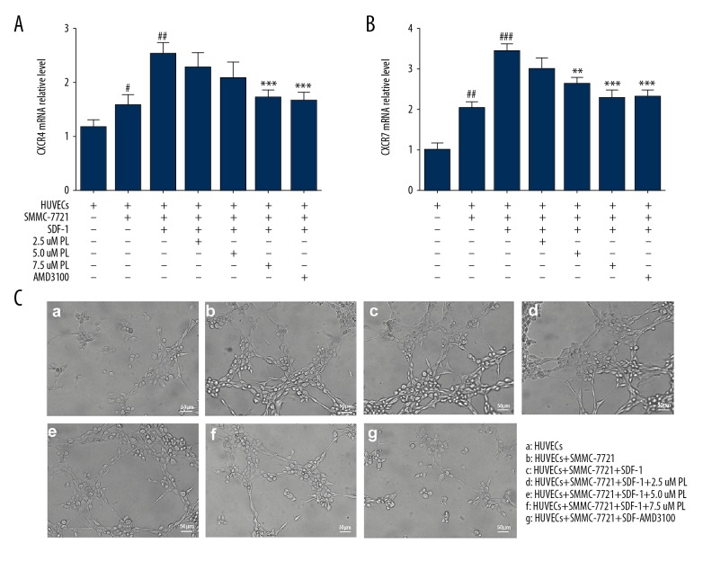
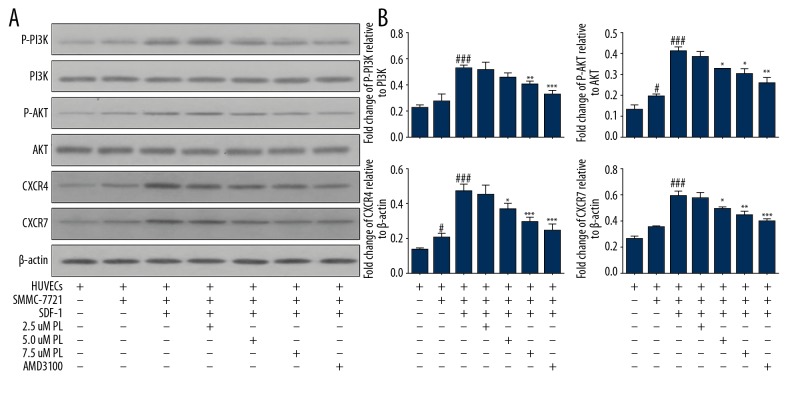
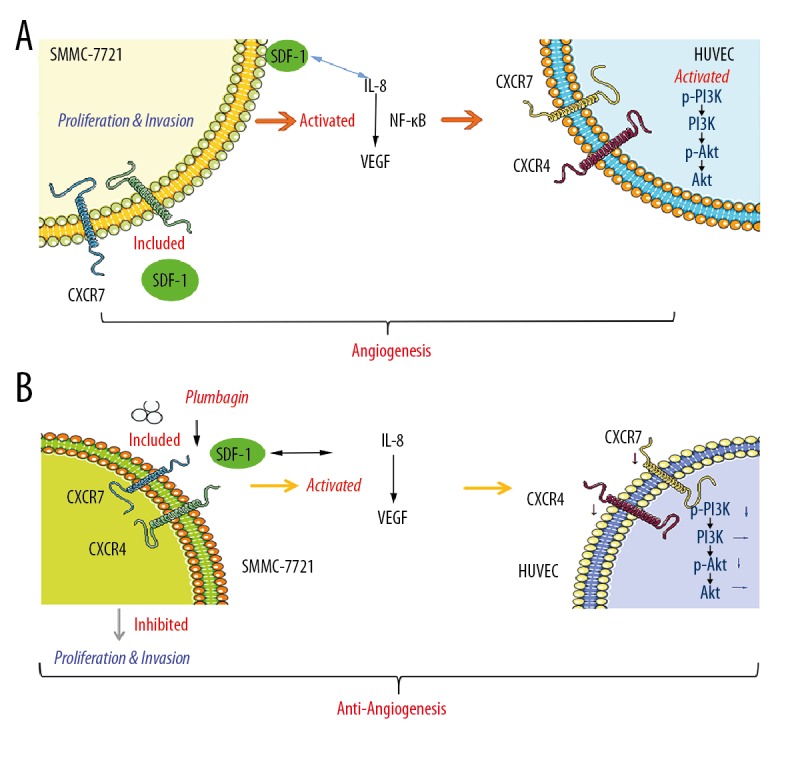
BACKGROUND Anti-angiogenic therapy has recently emerged as a highly promising therapeutic strategy for treating hepatocellular carcinoma (HCC). MATERIAL AND METHODS We assessed cellular proliferation, invasion, and activation of growth factors (VEGF and IL-8) with SDF-1 induced in the hepatocellular carcinoma cell line SMMC-7721, and this progression was limited by plumbagin (PL). The human umbilical vein endothelial cell line HUVEC was co-cultured with SDF-1-induced SMMC-7721, and the expressions of CXCR7, CXCR4, and PI3K/Akt pathways after PL treatment were detected by RT-PCR and Western blot analysis. RESULTS The treatment of the hepatoma cell line SMMC-7721 with SDF-1 resulted in enhanced secretion of the angiogenic factors, IL-8 and VEGF, and shows that these stimulatory effects are abolished by PL. The study further demonstrated that PL not only abolishes SDF-1-induced formation of endothelial tubes, but also inhibits expression of CXCR4 and CXCR7, and partially prevents activation of angiogenic signaling pathways. CONCLUSIONS The effect of PL on the SDF-1-CXCR4/CXCR7 axis has become an attractive target for inhibiting angiogenesis in hepatoma cells. Our results provide more evidence for the clinical application of PL as part of traditional Chinese medicine in modern cancer treatment.


Figures

Figure 1
Figure 1
Proliferation of SMMC-7721 cells treated with various concentrations of SDF-1 (1 ng/ml, 10 ng/ml, and 100 ng/ml) for 12 h, 24 h, or 48 h in a 5% CO2 incubator at 37°C. Each bar represents the mean value ± standard deviation (SD), * P<0.05; ** P<0.01; *** P<0.001, compared to the Control group (A). Microscopy of invasion of SMMC-7721 cells by different concentrations of SDF-1 (1 ng/ml, 10 ng/ml, and 100 ng/ml) (scale bar, 100 μm) (B). Numbers of invasive SMMC-7721 cells induced by different concentrations of SDF-1 (C). Effects of different concentrations of SDF-1 (1 ng/ml, 10 ng/ml, and 100 ng/ml) on secretion of IL-8 and VEGF in SMMC-7721 cells. Each bar represents the mean value ± standard deviation (SD), * P<0.05; ** P<0.01; *** P<0.001, compared to the Control group (D).
Figure 2
Figure 2
Cell proliferation of SDF-1 (10 ng/ml)-induced SMMC-7721 cells treated with various concentrations of PL (1.25 μm, 2.5 μm, 5 μm, 7.5 μm, and 10 μm) for 12 h, 24 h, or 48 h in a 5% CO2 incubator at 37°C. AMD3100 (100 ng/mL) was used as the positive control. Each bar represents the mean value ± standard deviation (SD), # P<0.05; ## P<0.01; ### P<0.001, compared to the Control group. * P<0.05; ** P<0.01; *** P<0.001, compared to the SDF-1 group. (A) In the absence of SDF-1, SMMC-7721 cells were treated with different concentrations of PL (1.25 μm, 2.5 μm, 5 μm, 7.5 μm, and 10 μm) for 12 h, 24 h, or 48 h, and their cell proliferation rate was measured. Cells were incubated in a 5% CO2 incubator at 37°C. P<0.05, ## P<0.01; ### P<0.001; Compared with the control group without SDF-1. * P<0.05; ** P<0.01; *** P<0.001, Compared with the control group with SDF-1 (B). Microscopy of invasion of SDF-1-induced SMMC-7721 cells by different concentrations of PL (1.25 μm, 2.5 μm, 5 μm, 7.5 μm, and 10 μm). AMD3100 (100 ng/mL) was used as the positive control (Scale bar, 100 μm) (C). Numbers of invasive cell SDF-1 induced SMMC-7721 cells by different concentrations of PL (D). Effects of different concentrations of PL (1.25 μm, 2.5 μm, 5 μm, 7.5 μm, and 10 μm) on secretion of IL-8 and VEGF in SMMC-7721 cells. AMD3100 (100 ng/mL) was used as the positive control. Each bar represents the mean value ± standard deviation (SD), # P<0.05; ## P<0.01; ### P<0.001, compared to the Control group. * P<0.05; ** P<0.01; *** P<0.001, compared to the SDF-1 group (E).
Figure 3
Figure 3
Effect of PL on the mRNA expression of CXCR4 and CXCR7 genes in SMMC-7721 cells co-cultured with HUVECs, grouped as follows: HUVECs only; HUVECs were co-cultured with SMMC-7721 cells; HUVECs were co-cultured with SMMC-7721 induced by SDF-1; the SMMC-7721 cells were treated with 2.5–7.5 μM PL as indicated; AMD3100 (100 ng/ml) was used as the positive control. After being exposed to the indicated concentrations of PL and AMD3100 for 24 h, the mRNA expression of CXCR4 and CXCR7 were analyzed using quantitative real-time PCR. Each bar represents the mean value ± standard deviation (SD). * P<0.05; ** P<0.01; *** P<0.001, compared to the HUVEC group. # P<0.05; ## P<0.01; ### P<0.001, compared to the SDF-1 group (A, B). Microscopically co-cultured HUVECs spontaneously form capillary-like structures on Matrigel, particularly SDF-1 induced SMMC-7721 cells. The number and continuity of capillary-like structures in HUVECs were significantly inhibited by 2.5–7.5 μM PL in a dose-dependent manner (scale bar, 50 μm) (C).
Figure 4
Figure 4
Expression of angiogenesis-related proteins detected by Western blotting after PL treatment, grouped as follows: HUVECs only; HUVECs were co-cultured with SMMC-7721 cells; HUVECs were co-cultured with SMMC-7721 induced by SDF-1; the SMMC-7721 cells were treated with 2.5–7.5 μM PL as indicated; AMD3100 (100 ng/ml) was used as the positive control. The expression of p-Akt, Akt, p-PI3K, PI3K, CXCR4, and CXCR7 proteins in the cells was analyzed by Western blotting using specific antibodies. Akt was used as an internal control for p-Akt, PI3K was used as an internal control for p-PI3K, and β-actin was used as an internal control for CXCR4 and CXCR7. Expression levels of each protein are indicated (A). PL inhibits protein expression. Each bar represents the mean value ± standard deviation (SD). * P<0.05; ** P<0.01; *** P<0.001, compared to the HUVEC group. # P<0.05; ## P<0.01; ### P<0.001, compared to the SDF-1 group (B).
Figure 5
Figure 5
SDF-1 induces angiogenesis-related signaling pathways in HUVECs co-cultured with SMMC-7721 hepatocellular carcinoma cells (A). PL inhibits the angiogenic SDF-1/CXCR4-CXCR7 axis activity of HUVECs co-cultured with SMMC-7721 hepatoma cells, downregulates the mechanism of PI3K/Akt pathway, and improves the microenvironment of hepatocellular carcinoma (B).

References

    1. Chen W, Zheng R, Zhang S, et al. Report of cancer incidence and mortality in china, 2010. China Cancer. 2013;40:5. - PubMed
    1. Mercedes F, David S, Jordi B, et al. Angiogenesis in liver disease. J Hepatol. 2009;50:604–20. - PubMed
    1. Liang CM, Chen L, Hu H, et al. Chemokines and their receptors play important roles in the development of hepatocellular carcinoma. World J Hepatol. 2015;7:1390–402. - PMC - PubMed
    1. Sumsakul W, Plengsuriyakarn T, Na-Bangchang K. Pharmacokinetics, toxicity, and cytochrome p450 modulatory activity of plumbagin. BMC Pharmacol Toxicol. 2016;17:50. - PMC - PubMed
    1. Wei YF, Jing-Qiang LI, Zhang ZW, et al. [Effects of plumbagin on cell cycle of human hepatic stellate cells stimulated by leptin and its related protein expression]. Chinese Traditional & Herbal Drugs. 2012;43:1776–780. [in Chinese]

MeSH terms