

Decellularized human periodontal ligament for periodontium regeneration
- PMID: 31415652
- PMCID: PMC6695143
- DOI: 10.1371/journal.pone.0221236
Decellularized human periodontal ligament for periodontium regeneration
Abstract
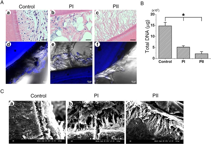
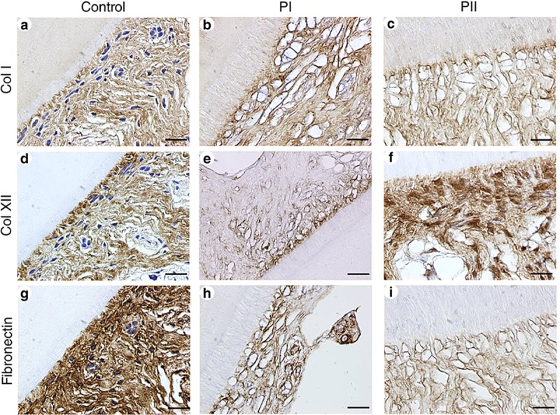
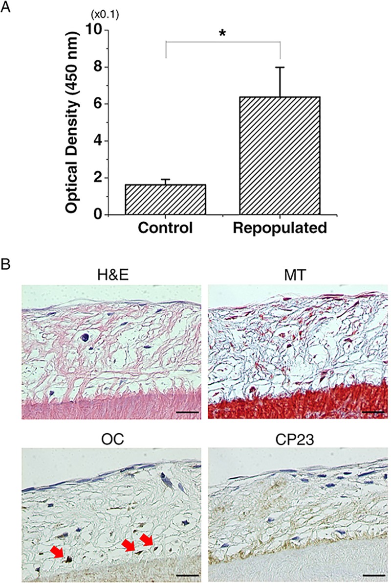
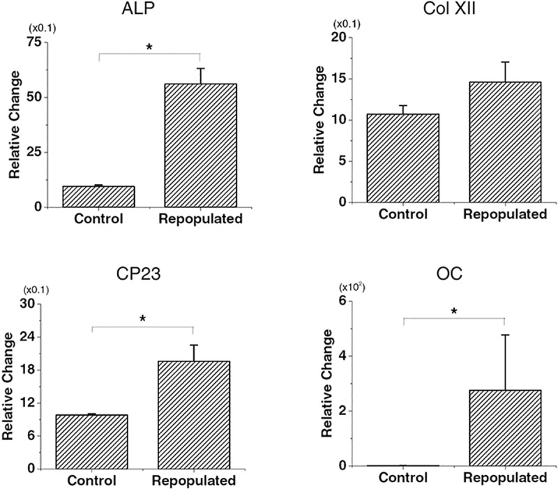
Regenerating the periodontal ligament (PDL) is a crucial factor for periodontal tissue regeneration in the presence of traumatized and periodontally damaged teeth. Various methods have been applied for periodontal regeneration, including tissue substitutes, bioactive materials, and synthetic scaffolds. However, all of these treatments have had limited success in structural and functional periodontal tissue regeneration. To achieve the goal of complete periodontal regeneration, many studies have evaluated the effectiveness of decellularized scaffolds fabricated via tissue engineering. The aim of this study was to fabricate a decellularized periodontal scaffold of human tooth slices and determine its regeneration potential. We evaluated two different protocols applied to tooth slices obtained from human healthy third molars. The extracellular matrix scaffold decellularized using sodium dodecyl sulfate and Triton X-100, which are effective in removing nuclear components, was demonstrated to preserve an intact structure and composition. Furthermore, the decellularized scaffold could support repopulation of PDL stem cells near the cementum and expressed cementum and periodontal-ligament-related genes. These results show that decellularized PDL scaffolds of human teeth are capable of inducing the proliferation and differentiation of mesenchymal stem cells, thus having regeneration potential for use in future periodontal regenerative tissue engineering.
Conflict of interest statement
The authors have declared that no competing interests exist.
Figures
Similar articles
-
Hierarchically patterned triple-layered gelatin-based electrospun membrane functionalized by cell-specific extracellular matrix for periodontal regeneration.Dent Mater. 2024 Jan;40(1):90-101. doi: 10.1016/j.dental.2023.10.022. Epub 2023 Nov 2. Dent Mater. 2024. PMID: 37923673
-
Periodontal tissue engineering with stem cells from the periodontal ligament of human retained deciduous teeth.J Periodontal Res. 2013 Feb;48(1):105-16. doi: 10.1111/j.1600-0765.2012.01509.x. Epub 2012 Aug 8. J Periodontal Res. 2013. PMID: 22881344
-
The effect of decellularized tissue engineered constructs on periodontal regeneration.J Clin Periodontol. 2018 May;45(5):586-596. doi: 10.1111/jcpe.12886. Epub 2018 Apr 17. J Clin Periodontol. 2018. PMID: 29500836
-
Periodontal Ligament Stem Cells: Regenerative Potency in Periodontium.Stem Cells Dev. 2019 Aug 1;28(15):974-985. doi: 10.1089/scd.2019.0031. Stem Cells Dev. 2019. PMID: 31215350 Review.
-
Developmental pathways of periodontal tissue regeneration: Developmental diversities of tooth morphogenesis do also map capacity of periodontal tissue regeneration?J Periodontal Res. 2019 Feb;54(1):10-26. doi: 10.1111/jre.12596. Epub 2018 Sep 12. J Periodontal Res. 2019. PMID: 30207395 Review.
Cited by
-
Weighted Gene Co-expression Network Analysis (WGCNA) of Wnt Signaling Related to Periodontal Ligament Formation: A Bioinformatics-Based Analysis.Cureus. 2024 Jul 2;16(7):e63639. doi: 10.7759/cureus.63639. eCollection 2024 Jul. Cureus. 2024. PMID: 39092323 Free PMC article.
-
Wnt/β-catenin Promotes Cementum Apposition in Periodontal Regeneration.J Dent Res. 2025 Feb;104(2):183-192. doi: 10.1177/00220345241286490. Epub 2024 Nov 25. J Dent Res. 2025. PMID: 39586793 Free PMC article.
-
3D-Printed Collagen-Based Waveform Microfibrous Scaffold for Periodontal Ligament Reconstruction.Int J Mol Sci. 2021 Jul 20;22(14):7725. doi: 10.3390/ijms22147725. Int J Mol Sci. 2021. PMID: 34299345 Free PMC article.
-
Tissue-specific biomarkers in gingival crevicular fluid are correlated with external root resorption caused by constant mechanical load: an in vivo study.Clin Oral Investig. 2021 Nov;25(11):6321-6333. doi: 10.1007/s00784-021-03932-6. Epub 2021 Apr 6. Clin Oral Investig. 2021. PMID: 33822289
-
MSCs-Derived Decellularised Matrix: Cellular Responses and Regenerative Dentistry.Int Dent J. 2024 Jun;74(3):403-417. doi: 10.1016/j.identj.2024.02.011. Epub 2024 Mar 16. Int Dent J. 2024. PMID: 38494389 Free PMC article. Review.
References
-
- McCulloch CA, Bordin S. Role of fibroblast subpopulations in periodontal physiology and pathology. J Periodontal Res. 1991;26(3 Pt 1):144–54. Epub 1991/05/01. . - PubMed
Publication types
MeSH terms
LinkOut - more resources
Full Text Sources
