Review
. 2019 Aug 14;20(16):3962.
doi: 10.3390/ijms20163962.

Endothelial Ca2+ Signaling, Angiogenesis and Vasculogenesis: just What It Takes to Make a Blood Vessel

Affiliations
Review

Endothelial Ca2+ Signaling, Angiogenesis and Vasculogenesis: just What It Takes to Make a Blood Vessel

Francesco Moccia et al. Int J Mol Sci. .

Abstract

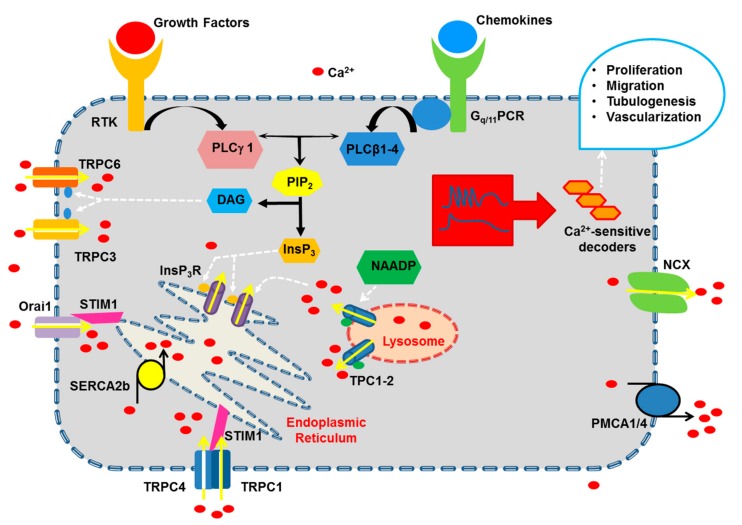
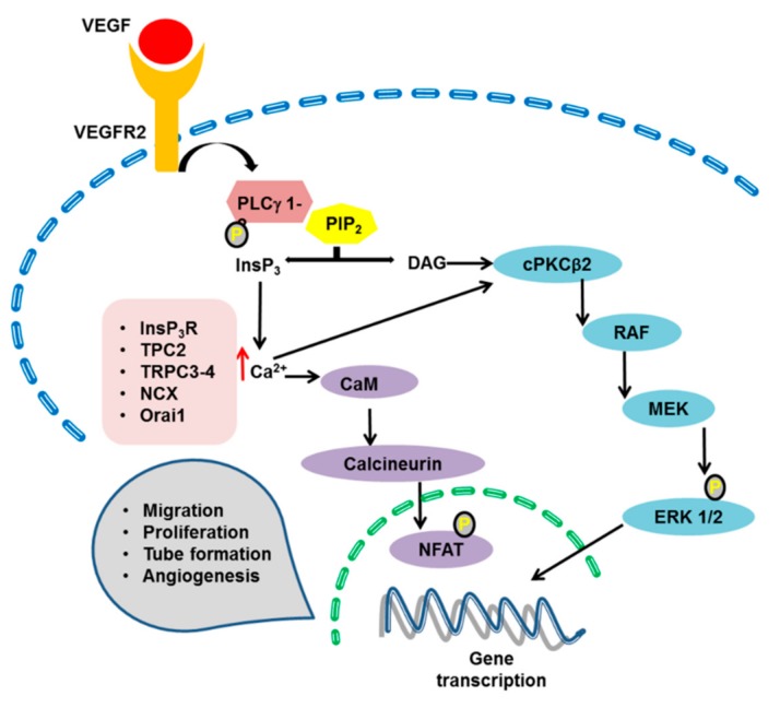
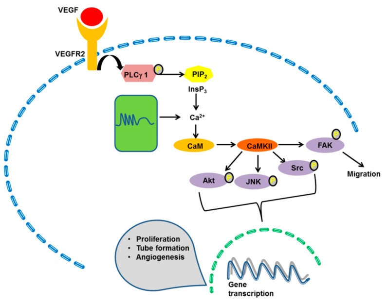
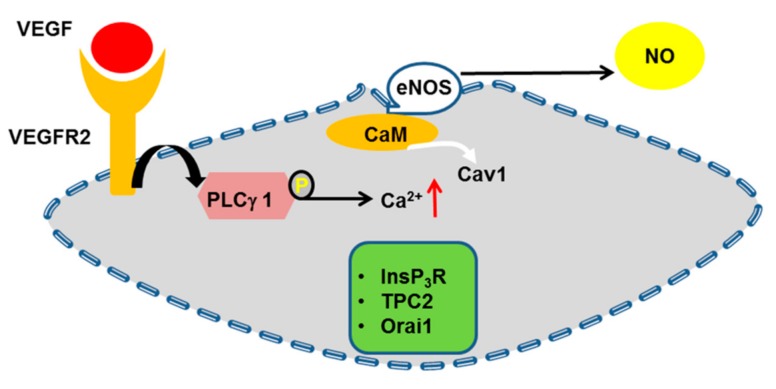
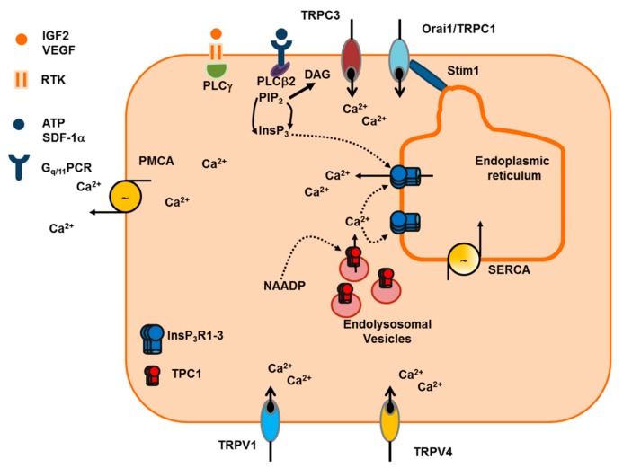
It has long been known that endothelial Ca2+ signals drive angiogenesis by recruiting multiple Ca2+-sensitive decoders in response to pro-angiogenic cues, such as vascular endothelial growth factor, basic fibroblast growth factor, stromal derived factor-1α and angiopoietins. Recently, it was shown that intracellular Ca2+ signaling also drives vasculogenesis by stimulation proliferation, tube formation and neovessel formation in endothelial progenitor cells. Herein, we survey how growth factors, chemokines and angiogenic modulators use endothelial Ca2+ signaling to regulate angiogenesis and vasculogenesis. The endothelial Ca2+ response to pro-angiogenic cues may adopt different waveforms, ranging from Ca2+ transients or biphasic Ca2+ signals to repetitive Ca2+ oscillations, and is mainly driven by endogenous Ca2+ release through inositol-1,4,5-trisphosphate receptors and by store-operated Ca2+ entry through Orai1 channels. Lysosomal Ca2+ release through nicotinic acid adenine dinucleotide phosphate-gated two-pore channels is, however, emerging as a crucial pro-angiogenic pathway, which sustains intracellular Ca2+ mobilization. Understanding how endothelial Ca2+ signaling regulates angiogenesis and vasculogenesis could shed light on alternative strategies to induce therapeutic angiogenesis or interfere with the aberrant vascularization featuring cancer and intraocular disorders.

Keywords: TRPC channels.; basic fibroblast growth factor; endothelial cells; endothelial colony forming cells; inositol-1,4,5-trisphosphate; nicotinic acid adenine dinucleotide phosphate; store-operated Ca2+ entry; stromal derived factor-1α; vascular endothelial growth factor.


Conflict of interest statement

The authors declare no conflict of interest.

Figures

Figure 1
Figure 1
The pro-angiogenic Ca2+ toolkit in vascular endothelial cells. Pro-angiogenic cues, such as growth factors and chemokines, bind to specific receptor tyrosine kinases (RTK) and Gq/11-protein Coupled Receptors (Gq/11PCR) thereby activating multiple phospholipase C (PLC) isoforms, which in turn cleave phosphatidylinositol 4,5-bisphosphate (PIP2) into inositol-1,4,5-trisphosphate (InsP3) and diacylglycerol (DAG). InsP3 triggers Ca2+ release from the endoplasmic reticulum (ER) through InsP3 receptors (InsP3R), while DAG stimulates extracellular Ca2+ entry through TRPC3 and TRPC6. However, the major Ca2+-entry pathway in vascular endothelial cells is provided by store-operated Ca2+ entry (SOCE), which is mainly mediated by the physical interaction between STIM1 and Orai1. In addition, SOCE may be sustained by the interplay among STIM1, Transient Receptor Potential (TRP) Canonical 1 (TRPC1) and TRPC4, with [35] or without the involvement of Orai1. Endogenous Ca2+ release may also be sustained by ryanodine receptors (RyR, not shown) and by endolysosomal two-pore channel 1-2 (TPC1-2), which are gated by nicotinic acid adenine dinucleotide phosphate (NAADP). Multiple Ca2+-transporting systems maintain resting Ca2+ concentration and clear cytosolic Ca2+ after the pro-angiogenic signal. These include Sarco-Endoplasmic Reticulum Ca2+-ATPase 2a (SERCA2a), Plasma Membrane Ca2+-ATPase 1 (PMCA1) and PMCA4, and Na+/Ca2+ exchanger (NCX). Please, see the text for a more detailed description of how the endothelial Ca2+ toolkit is recruited by pro-angiogenic cues.
Figure 2
Figure 2
Ca2+-dependent activation of the extracellular signal-regulated kinase (ERK) pathway and of the Nuclear Factor of Activated T-cells (NFAT). VEGF binding to VEGFR2 triggers an increase in intracellular Ca2+ concentration that may stimulate the ERK1/2 phosphorylation cascade or NFAT nuclear translocation. VEGF-induced endothelial Ca2+ signals may be mediated by multiple Ca2+-entry/release pathways, depending on species and vascular bed. The Ca2+ response to VEGF may recruit the Ca2+-dependent PKCβ2 (cPKCβ2), which engages the downstream RAF1–MEK–ERK1/2 cascade to induce gene expression. An increase in [Ca2+]i is required to promote cPKCβ2 translocation to the plasma membrane, where it is activated by DAG. Moreover, VEGF-induced endothelial Ca2+ signals may be sensed by calmodulin (CaM), which in turn activates calcineurin to dephosphorylate NFAT, thereby inducing its nuclear translocation. See Figure 1, Section 4.1 (ERK) and Section 4.3 (calcineurin and NFAT) for further details.
Figure 3
Figure 3
The Ca2+-dependent activation of Ca2+/Calmodulin (CaM)-dependent protein kinase 2 (CaMKII). Endothelial Ca2+ oscillations recruit CaMKII, which, in turns, stimulate angiogenesis by phosphorylating multiple targets, as widely illustrated in Section 4.4. VEGF-induced endothelial Ca2+ oscillations are sensed by CaM, which in turn stimulates CaMKII to phosphorylate multiple targets, including FAK to promote endothelial cell migration and Akt, JNK and Src to induce gene expression. The Ca2+ entry/release pathways that are recruited by VEGF to engage endothelial CaMKII are yet to be fully elucidated.
Figure 4
Figure 4
The Ca2+-dependent activation of the endothelial nitric oxide (NO) synthase (eNOS). VEGF binding to VEGFR2 causes an increase in intracellular Ca2+ concentration that displaces caveolin 1 (CaV1) from eNOS, thereby removing the tonic inhibition and inducing NO release. VEGF may impinge on several Ca2+ entry/release pathways to engage eNOS, including InsP3R, TPC2 and Orai1. See Figure 1 and Section 4.6. for further details.
Figure 5
Figure 5
The Ca2+ toolkit in endothelial colony forming cells (ECFC). Growth factors, such as VEGF, and chemokines, such as stromal derived factor-1α (SDF-1α), bind to specific RTK and Gq/11PCR, thereby activating multiple PLC isoforms, which in turn cleave PIP2 into InsP3 and DAG. InsP3 triggers ER-dependent Ca2+ release through InsP3R, while DAG gates TRPC3 exclusively in umbilical cord blood-derived ECFCs. SOCE is the major Ca2+ entry pathway also in ECFC, in which it is mediated by the dynamic interplay among STIM1, Orai1 and TRPC1. Endogenous Ca2+ release is further supported by NAADP, which evokes EL Ca2+ release through TPC1. SERCA and PMCA contribute to maintain resting Ca2+ levels and clear cytosolic Ca2+ after a pro-angiogenic Ca2+ signal. Pro-angiogenic Ca2+ signals may also be delivered by TRP Vanilloid 1 (TRPV1) and TRPV4. See Section 5.2. (SDF-1α) and Section 5.3. (VEGF and NAADP) for further details.

References

    1. Herbert S.P., Stainier D.Y. Molecular control of endothelial cell behaviour during blood vessel morphogenesis. Nat. Rev. Mol. Cell Biol. 2011;12:551–564. doi: 10.1038/nrm3176. - DOI - PMC - PubMed
    1. Ramasamy S.K., Kusumbe A.P., Adams R.H. Regulation of tissue morphogenesis by endothelial cell-derived signals. Trends Cell Biol. 2015;25:148–157. doi: 10.1016/j.tcb.2014.11.007. - DOI - PMC - PubMed
    1. Aird W.C. Spatial and temporal dynamics of the endothelium. J. Thromb. Haemost. 2005;3:1392–1406. doi: 10.1111/j.1538-7836.2005.01328.x. - DOI - PubMed
    1. Galley H.F., Webster N.R. Physiology of the endothelium. Br. J. Anaesth. 2004;93:105–113. doi: 10.1093/bja/aeh163. - DOI - PubMed
    1. Cahill P.A., Redmond E.M. Vascular endothelium—Gatekeeper of vessel health. Atherosclerosis. 2016;248:97–109. doi: 10.1016/j.atherosclerosis.2016.03.007. - DOI - PMC - PubMed

MeSH terms

LinkOut - more resources