

Comparative analysis of mammalian sperm ultrastructure reveals relationships between sperm morphology, mitochondrial functions and motility
- PMID: 31416446
- PMCID: PMC6696699
- DOI: 10.1186/s12958-019-0510-y
Comparative analysis of mammalian sperm ultrastructure reveals relationships between sperm morphology, mitochondrial functions and motility
Abstract
Background: Sperm morphology mainly refers to the shape of the head, the length of the flagellar segments, including the midpiece, principal piece and end piece, and the size of the accessory structures, including axonemes, outer dense fibers (ODFs), mitochondrial sheath (MS) and fibrous sheath (FS). Across species, there is considerable diversity in morphology. An established theory posits that the length of the sperm flagellum, especially the length of the midpiece, is a critical factor influencing sperm metabolism and velocity. However, our understanding of the relationships between sperm ultrastructures and the sperm flagellar length is incomplete.
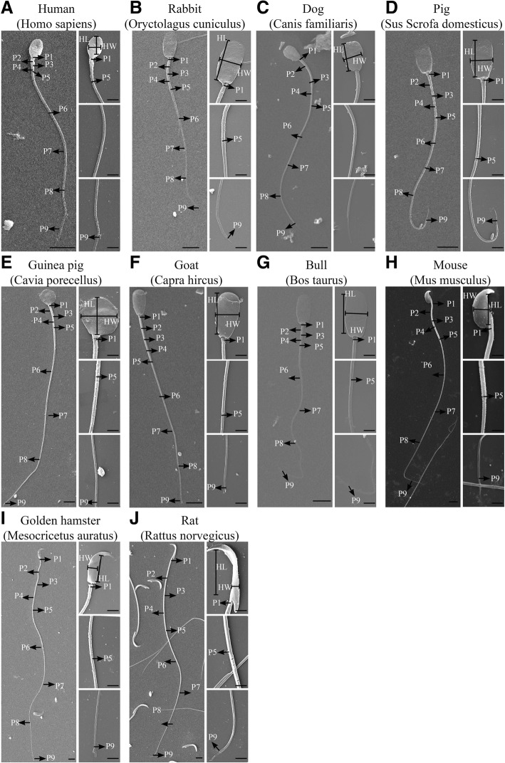
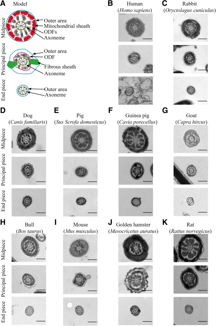
Methods: The morphologies of sperm from 10 mammalian species, human, mouse, rat, dog, rabbit, goat, pig, bull, guinea pig and golden hamster, were examined by scanning electron microscopy (SEM) and transmission electron microscopy (TEM). According to the SEM and TME images, the length of sperm heads and flagellar segments, the cross-sectional areas of the accessory structures and flagella and the width of sperm heads were measured using Image J software. The variation tendencies (referred to as slope) of the accessory structures along flagella were calculated by the linear regression method. Mitochondrial functions were measured using commercial kits. The velocities of sperm were measured using CASA software.
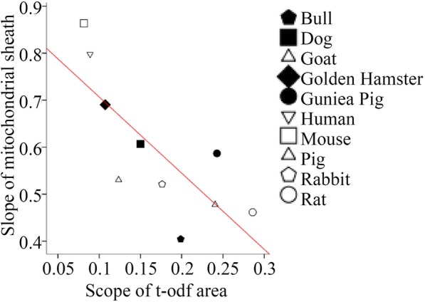
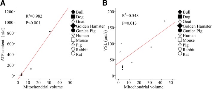
Results: The three-dimensional morphologies of sperm from 10 species and the slopes of internal accessory structures along flagella were obtained. The width of the axoneme tapered slightly from the base to the tip of the sperm flagellum, and slopes of the axonemes correlated negatively with the variability in flagellar length across species. Additionally, the cross-sectional areas of the ODFs and/or the MS were positively correlated with the lengths of the midpiece, principal piece, and total flagellum, as well as with sperm velocities. Mitochondrial volumes were positively correlated with ATP content and sperm swimming velocities.
Conclusions: Our results not only show the relationship between sperm internal structures, flagellar length and sperm physiology but also provide sizes of mitochondria and ODFs as new targets with which to study the regulation of sperm length and velocity.
Keywords: Axoneme; Fibrous sheath; Flagellar lengths; Mitochondrial function; Mitochondrial sheath; Outer dense fibers; Sperm motility.
Conflict of interest statement
The authors declare that they have no competing interests.
Figures
References
-
- Mortimer D. The functional anatomy of the human spermatozoon: relating ultrastructure and function. Mol Hum Reprod. 2018;24(12):567–592. - PubMed
Publication types
MeSH terms
Grants and funding
LinkOut - more resources
Full Text Sources
