. 2019 Sep 13;294(37):13833-13849.
doi: 10.1074/jbc.RA119.009910. Epub 2019 Aug 15.

Ega3 from the fungal pathogen Aspergillus fumigatus is an endo-α-1,4-galactosaminidase that disrupts microbial biofilms

Affiliations

Ega3 from the fungal pathogen Aspergillus fumigatus is an endo-α-1,4-galactosaminidase that disrupts microbial biofilms

Natalie C Bamford et al. J Biol Chem. .

Abstract

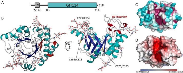
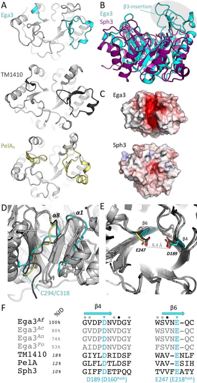
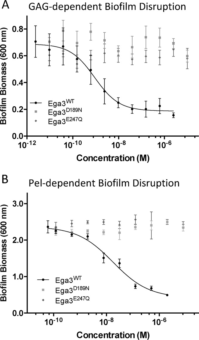
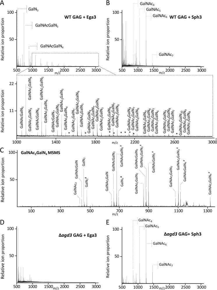
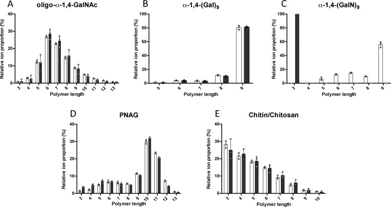
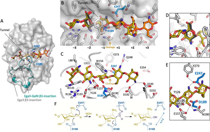
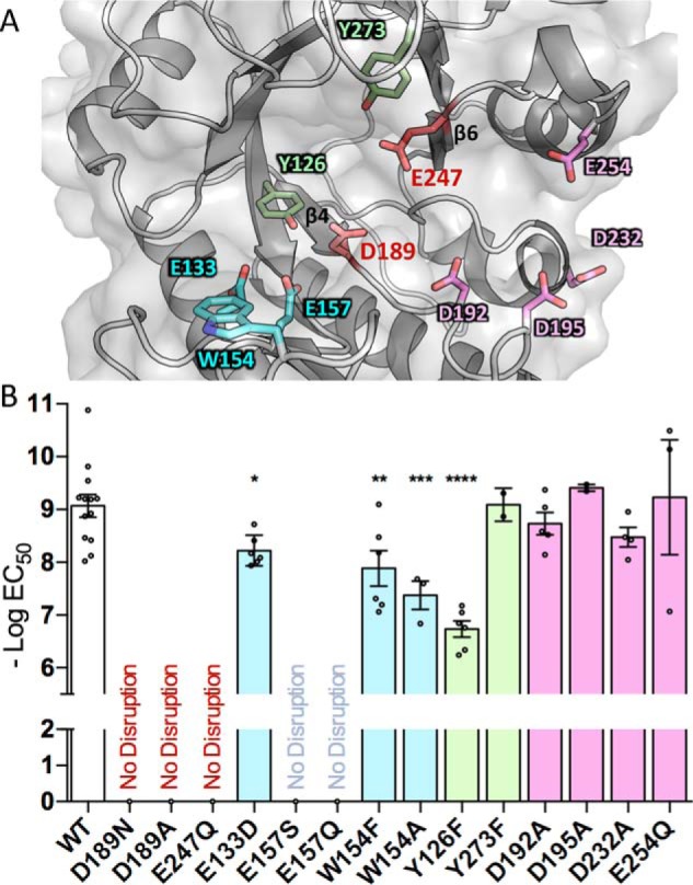
Aspergillus fumigatus is an opportunistic fungal pathogen that causes both chronic and acute invasive infections. Galactosaminogalactan (GAG) is an integral component of the A. fumigatus biofilm matrix and a key virulence factor. GAG is a heterogeneous linear α-1,4-linked exopolysaccharide of galactose and GalNAc that is partially deacetylated after secretion. A cluster of five co-expressed genes has been linked to GAG biosynthesis and modification. One gene in this cluster, ega3, is annotated as encoding a putative α-1,4-galactosaminidase belonging to glycoside hydrolase family 114 (GH114). Herein, we show that recombinant Ega3 is an active glycoside hydrolase that disrupts GAG-dependent A. fumigatus and Pel polysaccharide-dependent Pseudomonas aeruginosa biofilms at nanomolar concentrations. Using MS and functional assays, we demonstrate that Ega3 is an endo-acting α-1,4-galactosaminidase whose activity depends on the conserved acidic residues, Asp-189 and Glu-247. X-ray crystallographic structural analysis of the apo Ega3 and an Ega3-galactosamine complex, at 1.76 and 2.09 Å resolutions, revealed a modified (β/α)8-fold with a deep electronegative cleft, which upon ligand binding is capped to form a tunnel. Our structural analysis coupled with in silico docking studies also uncovered the molecular determinants for galactosamine specificity and substrate binding at the -2 to +1 binding subsites. The findings in this study increase the structural and mechanistic understanding of the GH114 family, which has >600 members encoded by plant and opportunistic human pathogens, as well as in industrially used bacteria and fungi.

Keywords: Aspergillus; biofilm; carbohydrate biosynthesis; carbohydrate processing; enzyme mechanism; exopolysaccharide matrix; galactosaminogalactan (GAG); glycoside hydrolase; glycoside hydrolase 114 (GH114); protein structure; virulence factor.


Conflict of interest statement

The authors declare that they have no conflicts of interest with the contents of this article

Figures

Figure 1.
Figure 1.
Structure of Ega3 reveals a modified (β/α)8-barrel fold with a deep highly-conserved cleft. A, predicted domain arrangement of Ega3. B, crystal structure of (β/α)8-barrel fold of Ega3 shown in cartoon representation. The (β/α)-barrel is colored in blue (β-strands) and teal (α-helices) with the five N-glycans that could be built into the electron density displayed as gray sticks. The secondary structure elements of the β3-insertion are shown in dark red, and the three disulfide bonds are shown in yellow. The missing elements typically found in a (β/α)8-barrel, β5, α1, and α8, are labeled in bold and italic. C, surface representation colored from variable in teal to conserved in fuchsia, as calculated by Consurf (91). D, electrostatic surface, calculated using APBS in PyMOL, shows a highly negatively charged cleft (+10 kT to −10 kT) (60).
Figure 2.
Figure 2.
Ega3 is structurally similar to PelAh and TM1410. A, tertiary structure alignment of Ega3 (cyan), PelAh (yellow, PDB 5TCB (18)), and the hypothetical protein TM1410 from T. maritima (gray, PDB 2AAM). β3-insertion and structural elements following β6 are found in each structure and are colored. B, tertiary structure alignment of Ega3 (teal) with Sph3 (purple, PDB 5C5G (16)). The β3-insertion is circled and is not present in Sph3. C, comparison of surface electrostatics between Ega3 and Sph3 as done in Fig. 1. D, aligned structure from A emphasizing the lack of helical structure after β8 of the barrel. E, highly-conserved aspartic and glutamic acid residues occur at the end of β4 and β6, respectively, and are 6.4 Å apart in Ega3. F, sequence alignment of A. fumigatus Ega3 (Ega3Af) and its orthologues from Aspergillus clavatus (Ega3Ac), Aspergillus niger (Ega3An), and Fusarium oxysporum (Ega3Fo) with TM1410, PelAh, and Sph3 showing the degree of amino acid conservation surrounding the putative catalytic residues (blue). Secondary structure is represented in blue for Ega3 above the corresponding residues. Sequence identity to Ega3 is listed for the entire sequence with sequence conservation represented by colored dots; Ega3 orthologues were calculated by ClustalOmega; and TM1410, PelAh, and Sph3 sequence identities were determined through the secondary structure alignments in Coot.
Figure 3.
Figure 3.
Ega3 treatment disrupts A. fumigatus and P. aeruginosa biofilm. Effects of the treatment with the indicated hydrolases on Af293 biofilms (A) and.Pel-dependent P. aeruginosa PA14 biofilms (B). Residual biofilm biomass was quantified by crystal violet staining.
Figure 4.
Figure 4.
Ega3 is specific for GalN-containing regions of GAG. MALDI-TOF MS spectra of oligosaccharide products released by treatment of secreted GAG from WT A. fumigatus after treatment with 1 μm Sph3 (A) or 1 μm Ega3 (B) are shown. C, MS-MS spectra of the reduced and propionylated tetra-deacetylated galactosamine hexasaccharide species produced from Ega3 treatment. Ions are labeled with their monosaccharide composition, with the numerical subscript indicating the number of sugar units present, and # indicates the reducing end of the oligosaccharide., MALDI-TOF MS spectra of secreted GAG purified from the A. fumigatus Δagd3 mutant after treatment with 1 μm Ega3 (D) or Sph3 (E). All ions corresponding to oligosaccharides are labeled with monosaccharide composition or with an asterisk for the low intensity ions.
Figure 5.
Figure 5.
Ega3 is an endo-α-1,4-galactosaminidase. MS analyses of oligosaccharide substrates before (white bars) and after treatment with 10 μm Ega3 for 24 h (black bars) are shown. Substrates include the following: A, purified oligo-α-1,4-GalNAc isolated from GAG biofilms; B, synthesized α-1,4-(Gal)9; C, synthesized α-1,4-(GalN)9; D, synthesized oligo-β-1,6-GlcNAc (PNAG); and E, chitin/chitosan. Statistically significant differences between pre- and post-treatment oligosaccharide profiles were observed only for α-1,4-(GalN)9 in C.
Figure 6.
Figure 6.
Ega3 binds galactosamine using a flexible loop to create a substrate-binding tunnel. A, cartoon representation of the Ega3–GalN complex with transparent surface representation. B, alignment of apo-Ega3 (cyan) and Ega3–GalN (dark teal) showing the movement of the β3-insertion containing Glu-133, Trp-154, and Glu-157. C, galactosamine-binding site with the |FoFc| omit map contoured at 3.0 σ. Residues that interact with galactosamine are in orange, and the putative catalytic acidic residues are in teal. D, comparison of the flexible loop contained in the β3-insertion of Ega3 (teal), Ega3–GalN (dark teal), PelAh (yellow, PDB 5TCB), and TM1410 (gray, PDB 2AAM). The unknown ligand in TM1410 is in red. E, sequence alignment of the β3-insertion of Ega3 orthologues, and TM1410 and PelAh based on the structural alignment. Residues in β-strands are in blue, and helices (310 and α) in yellow are shown in cartoon representation of the Ega3 2° structure above. Residues that bind galactosamine are shown in red. Sequence identity was calculated by ClustalOmega for Ega3 proteins, whereas TM1410, PelAh, and Sph3 sequence identity was determined through the structural alignments in Coot.
Figure 7.
Figure 7.
In silico docking of α-1,4-(GalN)5 reveals six substrate-binding subsites. A, transparent surface representation of Ega3 structure with the galactosamine (dark gray) found in the crystal structure and the top two scoring conformations (no. 1 is yellow and no. 2 is orange). The β3-insertion is shown as a cartoon labeled with an arrowhead indicating the change between the apo structure (gray) and Ega3–GalN (teal). Putative catalytic residues are in blue. B, cartoon representation of the Ega3–GalN structure (white) and putative catalytic residues (blue). The galactosamine (dark gray) found in the crystal structure aligns with the top two scoring conformations (no. 1 is yellow and no. 2 is orange). The subsites are numbered with the putative site of cleavage between −1 and +1. C, side view of the lowest energy conformer (yellow) with residues that participate in binding the oligosaccharide labeled. Dashed lines indicate H-bonds and salt bridges to ligand amines. All interaction distances are less than 3.1 Å. D, saccharide in subsite −2 overlaps with the galactosamine (dark gray) in the Ega3–GalN structure and has an identical hydrogen bond network. The hydrophobic pocket created by Leu-88 and Leu-311 is indicated by the dashed light orange lines. E, Ega3 active site with the hydrogen bond network is indicated by the dashed black lines. The catalytic nucleophile, Asp-189, is aligned to attack the anomeric carbon (red dashed line). F, proposed mechanism of Ega3 with D189 acting as the catalytic nucleophile.
Figure 8.
Figure 8.
Ega3 requires conserved acidic residues for GAG hydrolysis and anti-biofilm activity. A, cartoon representation of Ega3 showing the residues mutated in this study. B, point mutations of the conserved amino acids in the putative substrate-binding cleft were assayed for activity against A. fumigatus biofilms. No disruption indicates no activity against biofilms at the highest concentrations tested (100 μm). Text and bar colors correspond to the residue coloring as depicted in A. Bars indicate the mean of 2–8 assays performed in triplicate with the standard deviation of the logEC50. Statistical significance as compared with WT was determined using one-way analysis of variance with Dunn's multiple comparison. *, p < 0.05; **, p < 0.01; ***, p < 0.001, ****, p < 0.0001.

References

    1. Kaur S., and Singh S. (2014) Biofilm formation by Aspergillus fumigatus. Med. Mycol. 52, 2–9 10.3109/13693786.2013.819592 - DOI - PubMed
    1. Brown G. D., Denning D. W., Gow N. A., Levitz S. M., Netea M. G., and White T. C. (2012) Hidden killers: human fungal infections. Sci. Transl. Med. 4, 165rv13 10.1126/scitranslmed.3004404 - DOI - PubMed
    1. Stevens D. A., Moss R. B., Kurup V. P., Knutsen A. P., Greenberger P., Judson M. A., Denning D. W., Crameri R., Brody A. S., Light M., Skov M., Maish W., Mastella G., Participants in the Cystic Fibrosis Foundation Consensus Conference (2003) Allergic bronchopulmonary aspergillosis in cystic fibrosis–state of the art: Cystic Fibrosis Foundation Consensus Conference. Clin. Infect. Dis. 37, Suppl., 3, S225–S264 10.1086/376525 - DOI - PubMed
    1. Knutsen A. P., and Slavin R. G. (1990) Cystic Fibrosis (Richard B.S., ed) Vol 1, pp. 103–118, Humana Press, Totowa, NJ: 10.1007/978-1-4612-0475-6_7 - DOI
    1. Gresnigt M. S., Bozza S., Becker K. L., Joosten L. A., Abdollahi-Roodsaz S., van der Berg W. B., Dinarello C. A., Netea M. G., Fontaine T., De Luca A., Moretti S., Romani L., Latge J. P., and van de Veerdonk F. L. (2014) A polysaccharide virulence factor from Aspergillus fumigatus elicits anti-inflammatory effects through induction of interleukin-1 receptor antagonist. PLoS Pathog. 10, e1003936 10.1371/journal.ppat.1003936 - DOI - PMC - PubMed

Publication types

MeSH terms

LinkOut - more resources