. 2019 May;149(5):641-649.
doi: 10.4103/ijmr.IJMR_45_17.

Effect of cell density on formation of three-dimensional cartilaginous constructs using fibrin & human osteoarthritic chondrocytes

Affiliations

Effect of cell density on formation of three-dimensional cartilaginous constructs using fibrin & human osteoarthritic chondrocytes

B S Shamsul et al. Indian J Med Res. 2019 May.

Abstract

Background & objectives: Seeding density is one of the major parameters affecting the quality of tissue-engineered cartilage. The objective of this study was to evaluate different seeding densities of osteoarthritis chondrocytes (OACs) to obtain the highest quality cartilage.

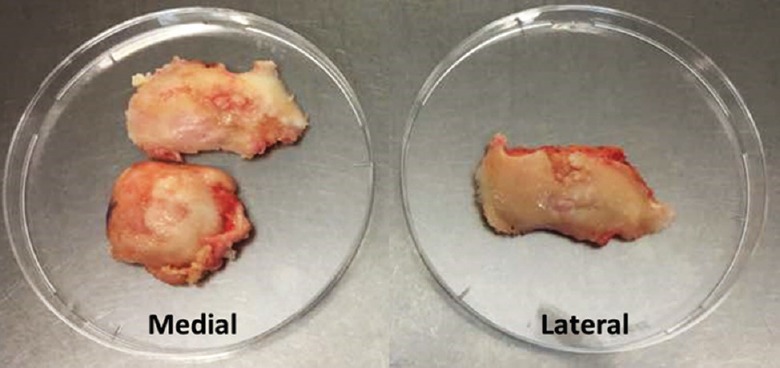
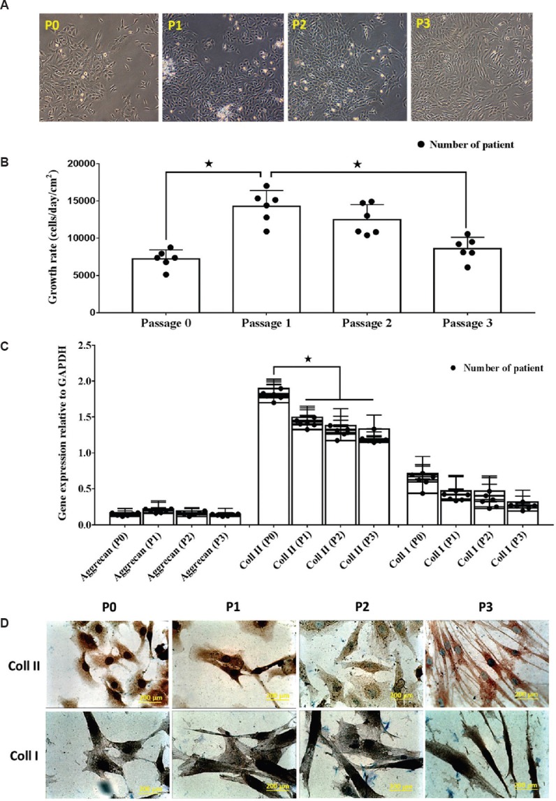
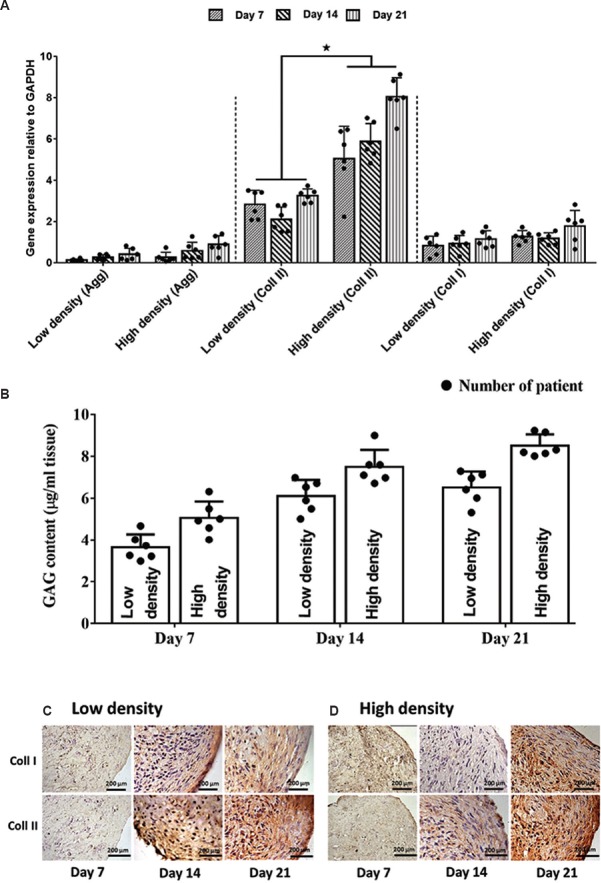
Methods: The OACs were expanded from passage 0 (P0) to P3, and cells in each passage were analyzed for gross morphology, growth rate, RNA expression and immunochemistry (IHC). The harvested OACs were assigned into two groups: low (1×10[7] cells/ml) and high (3×10[7] cells/ml) cell density. Three-dimensional (3D) constructs for each group were created using polymerised fibrin and cultured for 7, 14 and 21 days in vitro using chondrocyte growth medium. OAC constructs were analyzed with gross assessments and microscopic evaluation using standard histology, IHC and immunofluorescence staining, in addition to gene expression and biochemical analyses to evaluate tissue development.

Results: Constructs with a high seeding density of 3×10[7] cells/ml were associated with better quality cartilage-like tissue than those seeded with 1×10[7] cells/ml based on overall tissue formation, cell association and extracellular matrix distribution. The chondrogenic properties of the constructs were further confirmed by the expression of genes encoding aggrecan core protein and collagen type II.

Interpretation & conclusions: Our results confirmed that cell density was a significant factor affecting cell behaviour and aggregate production, and this was important for establishing good quality cartilage.

Keywords: Cartilage; chondrocytes; collagen-fibrin; osteoarthritis; seeding density.


Conflict of interest statement

None

Figures

Fig. 1
Fig. 1
Gross view of the collected samples.
Fig. 2
Fig. 2
(A). Photomicrographs (×40) of human osteoarthritis chondrocytes at day 7 from P0, P1, P2 and P3. After seven days of in vitro culture, all chondrocytes demonstrated polygonal morphology. (B) The growth rate of cultured chondrocytes decreased over successive passages *P<0.05. (C) Gene expression for collagen types I and II reduced after several passages, whereas aggrecan gene expression was consistently expressed. (D) Prominent staining of collagen type II in P0 detected by immunocytochemistry, which became weaker by P3. Collagen type I staining was observed for all passages.
Fig. 3
Fig. 3
(A) Cytofluorometry with propidium iodide staining showing diploid DNA content in cultured chondrocytes, suggesting that cultured chondrocytes maintained 100 per cent diploidy in the defined chondrocyte growth medium, with no evidence of aneuploidy, haploidy or tetraploidy. The X-axis represents the relative fluorescence intensity proportional to the DNA content. (B) Percentage of cells in the G1, G2 and S phases over successive passages. S phase index indicated that chondrocytes were actively proliferating during monolayer cultivation P<0.05 compared to P0.
Fig. 4
Fig. 4
(A&B) In vitro osteoarthritis chondrocyte-fibrin constructs derived from cells seeded at low and high densities at days 7, 14 and 21, showing cells of various shapes and sizes. Positive Safranin O (magnification: 200μm) staining confirmed the presence of cartilage-specific proteoglycan (C). The diameter of the high-density seeding construct was significantly bigger compared to that of the low-density construct at days 14 and 21 (*P<0.05), with a difference of 2.1 and 1.6 mm, respectively (D&E). Image showing the cell distribution and aggregates in the fibrin construct seeded with chondrocytes at low and high densities at different time points (day 7, 14 and 21).
Fig. 5
Fig. 5
(A) Significant (*P<0.05) expression of collagen type II (coll II), particularly in high-density seeded cells. All evaluated genes showed progressive expression throughout the culture period. Constructs showed strong expression of collagen type II in the pericellular matrix area and throughout the extracellular matrix, confirming an immature cartilage phenotype at days 14 and 21 for both groups glyceraldehyde phosphate dehydrogenase (GAPDH) and glycosaminoglycan (GAG) (B). Proteoglycan deposition showed no significant differences between constructs (C&D). Immunohistochemistry staining. Collagen type I and II (coll I & II) deposition was also detected in both constructs at day 7, 14 and 21 in low and high seeding chondrocytes fibrin construct.

References

    1. Goldring MB. Chondrogenesis, chondrocyte differentiation, and articular cartilage metabolism in health and osteoarthritis. Ther Adv Musculoskelet Dis. 2012;4:269–85. - PMC - PubMed
    1. Li H, Davison N, Moroni L, Feng F, Crist J, Salter E, et al. Evaluating osteoarthritic chondrocytes through a novel 3-dimensional in vitro system for cartilage tissue engineering and regeneration. Cartilage. 2012;3:128–40. - PMC - PubMed
    1. Brittberg M LA. Treatment of deep cartilage defects in the knee autologous chondrocytes transplantation. N Engl J Med. 1994;331:889–95. - PubMed
    1. Freed LE, Vunjak-Novakovic G. Culture of organized cell communities. Adv Drug Deliv Rev. 1998;33:15–30. - PubMed
    1. Phull AR, Eo SH, Abbas Q, Ahmed M, Kim SJ. Applications of chondrocyte-based cartilage engineering: An overview. Biomed Res Int. 2016;2016:1879837. - PMC - PubMed