. 2019 Aug 16;10(9):622.
doi: 10.1038/s41419-019-1847-z.

Inhibition of IRE1α RNase activity reduces NLRP3 inflammasome assembly and processing of pro-IL1β

Affiliations

Inhibition of IRE1α RNase activity reduces NLRP3 inflammasome assembly and processing of pro-IL1β

Aaron Talty et al. Cell Death Dis. .

Erratum in

Abstract

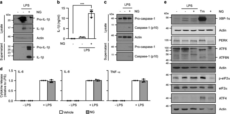
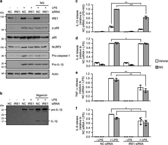
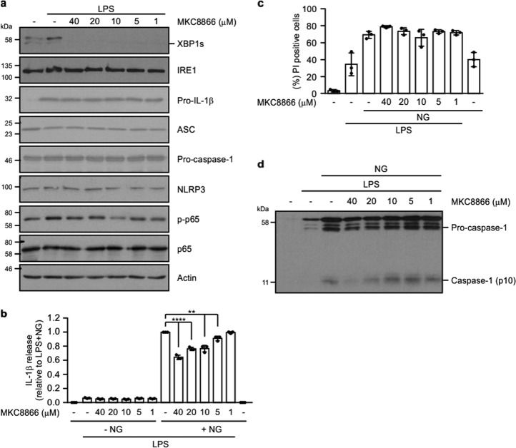
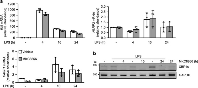
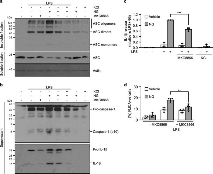
The inflammasome is a multiprotein complex assembled in response to Pathogen Associated Molecular Patterns (PAMPs) and Danger Associated Molecular Patterns (DAMPs). Inflammasome activation occurs through a two-step mechanism, with the first signal facilitating priming of inflammasome components while the second signal triggers complex assembly. Once assembled, the inflammasome recruits and activates pro-caspase-1, which in turn processes pro-interleukin (IL)-18 and pro-IL-1β into their bio-active forms. Owing to its key role in the regulation of innate immune responses, the inflammasome has emerged as a therapeutic target for the treatment of inflammatory conditions. In this study we demonstrate that IRE1α, a key component of the Unfolded Protein Response, contributes to assembly of the NLRP3 inflammasome. Blockade of IRE1α RNase signaling lowered NLRP3 inflammasome assembly, caspase-1 activation and pro-IL-1β processing. These results underscore both the importance and potential therapeutic relevance of targeting IRE1α signaling in conditions of excessive inflammasome formation.


Conflict of interest statement

A.S. and A.M.G are co-founders and shareholders of Cell Stress Discoveries Ltd. Q.Z. and J.B.P. are employees and shareholders of Fosun Orinove PharmaTech Inc. The remaining authors declare that they have no conflict of interest.

Figures

Fig. 1
Fig. 1. IRE1α-XBP1 axis of the UPR is selectively activated upon TLR4 stimulation.
THP-1 cells were primed with either 1 μg/ml LPS alone for 24 h or 1 μg/ml LPS for 24 h followed by addition of 10 μM nigericin (NG) for 45 min. a Processing of pro-IL-1β was analysed in cell lysates and conditioned medium by immunoblotting for full-length pro-IL-1β and processed p17 IL-1β. b Levels of IL-1β were quantified in conditioned medium from untreated, LPS, LPS and NG-treated THP-1 cells by ELISA (n = 3). c Processing of caspase-1 was analysed in cell lysates and conditioned medium by immunoblotting for pro-caspase-1 and processed p10 caspase-1. d IL-6, IL-8, and TNF-α levels were quantified in conditioned medium from untreated, LPS, LPS and NG, and NG-only treated THP-1 cells by ELISA (n = 3). e UPR markers XBP1s, PERK, ATF6, phospho-eIF2α, total eIF2α, and ATF4 were analysed by immunoblotting in THP-1 post treatment with LPS alone or LPS and NG. Tunicamycin (Tm)-treated THP-1 cells served as a positive control for the UPR activation. Actin was used as a loading control. ***P < 0.001 based on a Student’s t test. Error bars represent SD.
Fig. 2
Fig. 2. Knockdown of IRE1α reduces LPS-induced cytokine production.
THP-1 cells transfected with either noncoding (NC) or IRE1α targeting siRNA were treated with either 1 μg/ml LPS alone for 24 h or 1 μg/ml LPS for 24 h followed by addition of 10 μM nigericin (NG) for 45 min. a Cell lysates were analysed via immunoblotting for IRE1α, phospho-p65, total p65, NLRP3, pro-caspase-1 and pro-IL-1β. Actin was used as a loading control. b Cell conditioned medium was analysed via immunoblotting for pro-IL-1β processing. cf Levels of IL-1β, IL-8, TNF-α, and IL-6 were assayed in cell conditioned medium by ELISA (n = 3). *P < 0.05, **P < 0.01, and ***P < 0.001 based on a Student’s t test. Error bars represent SD.
Fig. 3
Fig. 3. MKC8866 reduces LPS-induced IRE1α signaling and suppresses NLRP3 inflammasome activation.
a THP-1 cells were treated with 1 μg/ml LPS alone or in combination with indicated concentrations of IRE1α inhibitor (MKC8866) for 24 h following which cell lysates were collected and analysed via immunoblotting for XBP1s, IRE1α, pro-IL-1β, ASC, pro-caspase-1, NLRP3, phospho-p65 and total p65. Actin was used as a loading control. b Conditioned medium from THP-1 cells treated with 1 μg/ml LPS alone or in combination with 10 μM nigericin (NG) in the presence or absence of indicated concentrations of MKC8866 were analysed by ELISA for IL-1β secretion (n = 3). c Propidium iodide (PI) uptake was assessed in THP-1 cells following treatment with 1 μg/ml LPS alone or in combination with 10 μM nigericin (NG) plus the indicated concentrations of MKC8866 (n = 3). d Conditioned medium from b was immunoblotted for pro- and cleaved caspase-1. **P < 0.01 and ****P < 0.0001 based on a Student’s t test. Error bars represent SD.
Fig. 4
Fig. 4. Addition of MKC8866 does not impact upon LPS-mediated increases in CASP1, IL1β, or NLRP3 transcript.
THP-1 cells were treated with 1 μg/ml LPS in the presence or absence of 20 μM MKC8866 for the indicated times after which RNA was extracted. a Q-PCR was carried out to assess relative expression of IL1β and NLRP3 and CASP1 transcripts (n = 3). Error bars represent SD. b RT-PCR was carried out to assess XBP1 splicing and GAPDH mRNA expression.
Fig. 5
Fig. 5. Addition of MKC8866 reduces NLRP3 inflammasome assembly.
THP-1 cells were treated with either 1 μg/ml LPS alone or in combination with 20 μM MKC8866 for 24 h followed by addition of 10 μM nigericin (NG) alone or in combination with 100 mM KCl for 45 min after which cells and conditioned medium were harvested. Immunoblotting was used to determine a ASC oligomerization status and b caspase-1 (pro- and cleaved p10) and IL-1β processing. c IL-1β levels were quantified by ELISA in THP-1 conditioned medium following indicated treatments (n = 3). d The percentage of THP-1 cells displaying caspase-1-like activity following the indicated treatments was assessed by the FLICA assay (n = 4). **P < 0.01 and ***P < 0.001 based on a Student’s t test. Error bars represent SD.
Fig. 6
Fig. 6. TXNIP expression decreases in THP-1 cells upon LPS treatment.
THP-1 cells were treated for the indicated times with 1 μg/ml LPS alone or in combination with 20 μM MKC8866 and after which a TXNIP expression was determined by Q-PCR (n = 3) and b by immunoblotting. Error bars represent SD.
Fig. 7
Fig. 7. IRE1α RNase inhibition reduces activation of the NLRP3 inflammasome in primary PBMCs.
PBMCs isolated from healthy individuals were treated for 2 h with either 0.5 ng/ml LPS alone or in combination with 20 μM MKC8866 followed by addition of 5 mM ATP for 45 min following which cell lysates and conditioned medium was collected. a Levels of IL-1β in cell conditioned medium were quantified by ELISA (n = 3). b IRE1α RNase activity was assessed by monitoring levels of XBP1 splicing via RT-PCR. Tunicamycin (Tm) treatment served as a positive control for XBP1 splicing. c Immunoblotting for caspase-1 (pro- and p10) and IL-1β (pro- and p17) was performed using PBMC conditioned medium collected following the indicated treatments. *P < 0.05 based on a Student’s t test. Error bars represent SD.

References

    1. Cox JS, Shamu CE, Walter P. Transcriptional induction of genes encoding endoplasmic reticulum resident proteins requires a transmembrane protein kinase. Cell. 1993;73:1197–1206. doi: 10.1016/0092-8674(93)90648-A. - DOI - PubMed
    1. Mori K, Ma W, Gething MJ, Sambrook J. A transmembrane protein with a cdc2+/CDC28-related kinase activity is required for signaling from the ER to the nucleus. Cell. 1993;74:743–756. doi: 10.1016/0092-8674(93)90521-Q. - DOI - PubMed
    1. Harding HP, Zhang Y, Ron D. Protein translation and folding are coupled by an endoplasmic-reticulum-resident kinase. Nature. 1999;397:271–274. doi: 10.1038/16729. - DOI - PubMed
    1. Haze K, et al. Identification of the G13 (cAMP-response-element-binding protein-related protein) gene product related to activating transcription factor 6 as a transcriptional activator of the mammalian unfolded protein response. Biochem. J. 2001;355:19–28. doi: 10.1042/bj3550019. - DOI - PMC - PubMed
    1. Ron D, Walter P. Signal integration in the endoplasmic reticulum unfolded protein response. Nat. Rev. Mol. Cell Biol. 2007;8:519–529. doi: 10.1038/nrm2199. - DOI - PubMed

Publication types

MeSH terms