. 2019 Aug 15;10(1):3684.
doi: 10.1038/s41467-019-11592-0.

Alternate subunit assembly diversifies the function of a bacterial toxin

Affiliations

Alternate subunit assembly diversifies the function of a bacterial toxin

Casey C Fowler et al. Nat Commun. .

Abstract

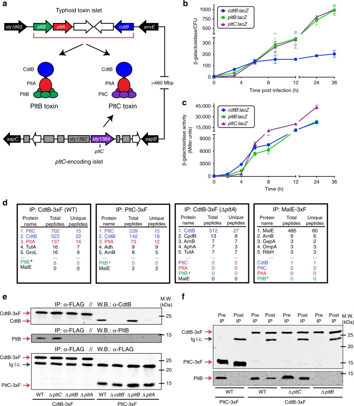
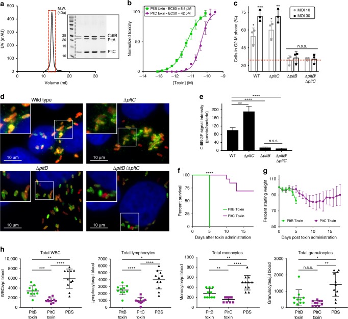
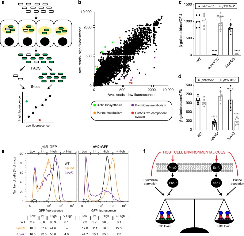
Bacterial toxins with an AB5 architecture consist of an active (A) subunit inserted into a ring-like platform comprised of five delivery (B) subunits. Salmonella Typhi, the cause of typhoid fever, produces an unusual A2B5 toxin known as typhoid toxin. Here, we report that upon infection of human cells, S. Typhi produces two forms of typhoid toxin that have distinct delivery components but share common active subunits. The two typhoid toxins exhibit different trafficking properties, elicit different effects when administered to laboratory animals, and are expressed using different regulatory mechanisms and in response to distinct metabolic cues. Collectively, these results indicate that the evolution of two typhoid toxin variants has conferred functional versatility to this virulence factor. More broadly, this study reveals a new paradigm in toxin biology and suggests that the evolutionary expansion of AB5 toxins was likely fueled by the plasticity inherent to their structural design coupled to the functional versatility afforded by the combination of homologous toxin components.


Conflict of interest statement

The authors declare no competing interests.

Figures

Fig. 1
Fig. 1
S. Typhi produces two distinct typhoid toxins with common active but different delivery subunits. a Illustration of the S. Typhi typhoid toxin genomic locus, as well as a distant locus that encodes pltC (sty1364), an orphan pertussis-like toxin delivery subunit that exhibits homology to pltB. b, and c Expression of pltC, pltB and cdtB over time under conditions that stimulate typhoid toxin gene expression. The β-galactosidase activity in the pltC:lacZ, pltB:lacZ and cdtB:lacZ S. Typhi reporter strains was measured at the indicated time points following infection of Henle-407 cells b or growth in TTIM medium c. Values indicate the mean ±S.D. for three independent samples. d Interaction of PltC with PltA/CdtB in S. Typhi grown in TTIM. Cell lysates from S. Typhi strains encoding cdtB-3xFLAG, pltC-3xFLAG, cdtB-3xFLAG (in ΔpltA background), or malE-3xFLAG were immunoprecipitated with an anti-FLAG antibody and interacting proteins were identified using LC/MS/MS. For each sample the number of peptides for the five most abundant proteins recovered and for all typhoid toxin subunits (color-coded according to panel a) are shown. *Detection of PltB with the LC–MS/MS protocol even for purified typhoid toxin preparations is inefficient. e S. Typhi produces both PltB- and PltC-typhoid toxins within infected human cells. Henle-407 cells were infected with S. Typhi wild type or the indicated mutant strains encoding 3xFLAG epitope-tagged CdtB or PltC (as indicated) and 24 hs post-infection the interaction of the indicated toxin components were probed by co-immunoprecipitation and western blot analysis. f PltB forms a complex with CdtB, but not with PltC. The interaction of the indicated toxin components in cell lysates of the indicated strains encoding 3xFLAG epitope tagged CdtB or PltC were probed by anti-FLAG co-immunoprecipitation and western blot analysis. Whole cell lysates (Pre IP) and immunoprecipitated samples (post IP) were probed using an anti-FLAG antibody as a control (top blot) and an anti-PltB antibody (bottom blot) to identify PltB interactions with CdtB or PltC in the indicated strains. Ig. l. c.: Immunoglobulin light chain detected by the secondary antibody. Source data are provided as a Source Data file
Fig. 2
Fig. 2
The PltB- and PltC-typhoid toxins exhibit different biological properties. a Gel filtration chromatography and SDS-PAGE/Coomassie blue (inset) analyses of purified PltC-typhoid toxin. Lanes on gel represent individual chromatographic fractions (red box) containing purified toxin. b PltC-typhoid toxin elicits G2/M cell cycle arrest in human epithelial cells. Purified PltB- or PltC-typhoid toxins were added to the culture medium of Henle-407 cells at the indicated concentrations and 48h after, cells were fixed and analyzed by flow cytometry to evaluate toxicity as indicated in Materials and Methods. The data shown are the mean normalized toxicity ± S.D. for three independent experiments. c PltC-typhoid toxin does not induce G2/M arrest in S. Typhi-infected cells. Henle-407 cells were infected with the indicated strains at a multiplicity of infection (MOI) of 10 or 30, as indicated, and 48h post-infection cells were collected and the percentage of cells in G2/M phase was determined as described for panel b. Mean values±S.D. are shown for three independent experiments assayed in duplicate (6 total samples). Asterisks denote statistically significant levels G2/M cell cycle arrest compared to mock infected cells (red dotted line) as determined by unpaired two-tailed t-tests. d, e PltC-typhoid toxin is not packaged into vesicle transport carriers. Henle-407 cells were infected with the indicated S. Typhi strains encoding 3x-FLAG epitope-tagged CdtB and 48 hs post-infection the cells were fixed and stained with DAPI (blue), α-FLAG (green), and α-S. Typhi LPS (red) antibodies. Typhoid toxin-containing export vesicles, which appear as green puncta d, were quantified by image analysis e as indicated in Materials and methods. Values are from >25 images (~100 infected cells) taken in two independent experiments and represent the mean relative ratios ± S.E.M. Asterisks denote the statistical significance of the indicated pairwise comparisons determined using unpaired two-tailed t-tests. fh The PltB- and PltC-typhoid toxins elicit different effects when administered to mice. Highly purified preparations of PltB- (2μg) or PltC-typhoid toxins (10μg) were administered to C57BL/6 mice. For one group of mice, their survival f and body weight (mean ± S.D.) g was recorded at the indicated times. The remaining mice were killed at four days post-toxin administration and a blood sample was collected and analyzed to quantify the indicated cell types (mean ± S.D.) h. WBCs, white blood cells. The Mantel-Cox test was used for statistical analysis of mouse survival and Brown-Forsythe and Welch ANOVA coupled with Dunnett’s T3 multiple comparisons tests were used to statistically compare the indicated samples for the blood analysis. For all panels, ****p < 0.0001, ***p < 0.001, **p < 0.01, *p < 0.05, n.s.s. not statistically significant. Source data are provided as a Source Data file
Fig. 3
Fig. 3
Distinct regulatory mechanisms and metabolic cues control the expression of PltB and PltC. a Schematic representation of the FAST-INseq genetic screen used to identify S. Typhi genes that influence pltC expression in infected host cells. A large library of random transposon mutants was generated in the S. Typhi pltC:gfp strain and used to infect Henle-407 cells. Sixteen hours post-infection the bacteria were isolated and sorted by FACS into pools exhibiting high and low GFP fluorescence. INseq was then used to identify mutants that stimulate (green dot in plot) or reduce (red dot) pltC expression during infection. b Overview of the results of the FAST-INseq screen. Plot shows the normalized numbers of sequencing reads of transposon insertions within each S. Typhi gene in the high fluorescence vs. low fluorescence pools. c, d Expression levels of pltB:lacZ and pltC:lacZ reporters in infected human cells for wild-type S. Typhi (WT) and the indicated deletion mutant strains. Henle-407 cells were infected with the indicated strains for 24 h, after which the β-galactosidase activity from bacterial lysates was measured and normalized to the numbers of CFU recovered. Values indicate mean values  ± S.D. for six independent determinations taken over two separate experiments. Asterisks denote statistically significant differences relative to the corresponding wild-type sample determined using unpaired two-tailed t-tests. ****p < 0.0001, *p < 0.05, n.s.s. not statistically significant. e Flow cytometry analysis of pltB:gfp and pltC:gfp expression of the indicated S. Typhi strains 24 h post-infection. Histograms show the GFP fluorescence intensities of individual bacteria for the indicated strains. Gates were established to show the percentage of bacteria exhibiting high, low and intermediate (int) fluorescence. The percentage of bacteria with fluorescence intensities within these gates is shown (bottom). Gating strategy provided in Supplementary Fig. 5b. f Overview of the identified factors that differentially affect the expression of pltB and pltC and thus are likely to be important for controlling relative abundance of the two typhoid toxins produced by S. Typhi upon encountering different environments during infection

References

    1. Beddoe T, Paton A, Le Nours J, Rossjohn J, Paton J. Structure, biological functions and applications of the AB5 toxins. Trends Biochem. Sci. 2010;35:411–418. doi: 10.1016/j.tibs.2010.02.003. - DOI - PMC - PubMed
    1. Merritt E, Hol W. AB5 toxins. Curr. Opin. Struct. Biol. 1995;5:165–171. doi: 10.1016/0959-440X(95)80071-9. - DOI - PubMed
    1. Dougan G, Baker S. Salmonella enterica serovar Typhi and the pathogenesis of typhoid fever. Annu Rev. Microbiol. 2014;68:317–336. doi: 10.1146/annurev-micro-091313-103739. - DOI - PubMed
    1. Parry CM, Hien TT, Dougan G, White NJ, Farrar JJ. Typhoid fever. N. Engl. J. Med. 2002;347:1770–1782. doi: 10.1056/NEJMra020201. - DOI - PubMed
    1. Spano S, Ugalde JE, Galan JE. Delivery of a Salmonella Typhi exotoxin from a host intracellular compartment. Cell Host Microbe. 2008;3:30–38. doi: 10.1016/j.chom.2007.11.001. - DOI - PubMed

Publication types