. 2019 Aug 15;9(1):11914.
doi: 10.1038/s41598-019-48316-9.

In vitro exercise model using contractile human and mouse hybrid myotubes

Affiliations

In vitro exercise model using contractile human and mouse hybrid myotubes

Weijian Chen et al. Sci Rep. .

Abstract

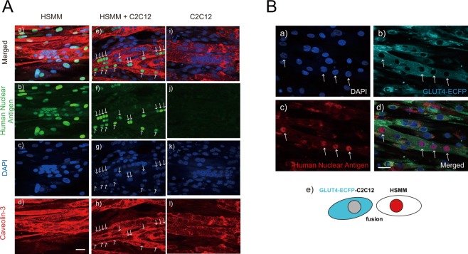
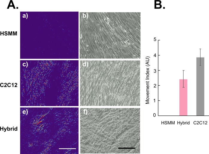
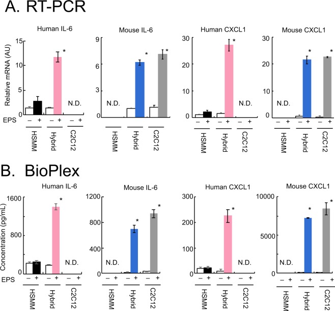
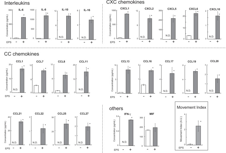
Contraction of cultured myotubes with application of electric pulse stimulation (EPS) has been utilized for investigating cellular responses associated with actual contractile activity. However, cultured myotubes derived from human subjects often exhibit relatively poor EPS-evoked contractile activity, resulting in minimal contraction-inducible responses (i.e. myokine secretion). We herein describe an "in vitro exercise model", using hybrid myotubes comprised of human myoblasts and murine C2C12 myoblasts, exhibiting vigorous contractile activity in response to EPS. Species-specific analyses including RT-PCR and the BioPlex assay allowed us to separately evaluate contraction-inducible gene expressions and myokine secretions from human and mouse constituents of hybrid myotubes. The hybrid myotubes, half of which had arisen from primary human satellite cells obtained from biopsy samples, exhibited remarkable increases in the secretions of human cytokines (myokines) including interleukins (IL-6, IL-8, IL-10, and IL16), CXC chemokines (CXCL1, CXCL2, CXCL5, CXCL6, CXCL10), CC chemokines (CCL1, CCL2, CCL7, CCL8, CCL11, CCL13, CCL16, CCL17, CCL19, CCL20, CCL21, CCL22, CCL25, CCL27), and IFN-γ in response to EPS-evoked contractile activity. Together, these results indicate that inadequacies arising from human muscle cells are effectively overcome by fusing them with murine C2C12 cells, thereby supporting the development of contractility and the resulting cellular responses of human-origin muscle cells. Our approach, using hybrid myotubes, further expands the usefulness of the "in vitro exercise model".


Conflict of interest statement

The authors declare no competing interests.

Figures

Figure 1
Figure 1
Status of fusion between human and mouse muscle cells. (A) After 7–8 days of differentiation, the differentiated myotubes derived from human alone (HSMM, panels a–d), human-origin and mouse-origin cells in a mixture (HSMM + C2C12, panels e–h), and murine C2C12 cells alone (panels i–l) were fixed and then analyzed for myotubular fusion status by using anti-human nuclear antigen (anti-HNA) and anti-Caveolin 3 antibodies, as described in the Materials and Methods. DAPI was used for nuclear staining. Scale bar = 25 μm. Three independent experiments were performed and representative images are presented. Note that both anti-HNA-positive and -negative nuclei are observed in a myotube from the mixed culture (HSMM + C2C12) (panels eg). (B) Differentiated myotubes that arose from HSMM and GLUT4-ECFP-expressing C2C12 cells (panel e) were fixed and then analyzed for their myotubular fusion status by using GLUT4-ECFP (panel b) and anti- HNA antibody (panel c). Three independent experiments were performed and representative images are presented. Note that the myotube expressing GLUT4-ECFP (panel b) contains anti-HNA-positive nuclei (panel d, allows). DAPI was used for nuclear staining (panel a). Scale bar = 25 μm.
Figure 2
Figure 2
Evaluation of contractile activity of cultured myotubes. After 7–8 days of differentiation, EPS (20 V/25 mm, 1-Hz frequency, 4-ms duration) was applied to differentiated myotubes originating from human samples alone (HSMM, panels a and b), murine C2C12 cells alone (panels c and d), and a mixture of the two (hybrid, panels e and f) for 16 h. Images of myotubes were then taken sequentially during EPS at the last 15 min. of the total EPS session (Supplementary Movies S1–S3). (A) The pseudo-colored differential images (panels a, c and e), reflecting contractile area and ability, were produced as described in the Materials and Methods. The bright-field images of the same area (panels b, d, and f) are also presented. Scale bar = 250 μm. Three independent experiments were performed and representative images are presented. (B) The Movement Index was determined as described in the Materials and Methods (n = 3; *P < 0.05).
Figure 3
Figure 3
Contraction-inducible upregulation of the expressions and secretions of myokines. After 7–8 days of differentiation, the differentiated myotubes originating from human samples alone (HSMM), human-origin and mouse-origin cells in a mixture (HSMM + C2C12), and murine C2C12 cells alone were treated with or without EPS at 20 V/25 mm, 1 Hz, 4-ms duration for 16 h. (A) Total RNA was extracted and the relative abundances of mRNAs for mouse IL-6 and CXCL1 as well as human IL-6 and CXCL1 were evaluated by real-time PCR analysis. Data normalized using mouse GAPDH or human RPLP0 transcripts were averaged over 3 independent experiments (*P < 0.05). (B) Conditioned media were collected and concentrations of human IL-6 and CXCL1 as well as mouse IL-6 and CXCL1 were evaluated employing the BioPlex assay (*P < 0.05, n = 3).
Figure 4
Figure 4
Secretion of contraction-inducible myokines from hybrid myotubes comprised of C2C12 cells and human muscle cells obtained from a biopsy sample. After 7–8 days of differentiation, the differentiated myotubes originating from murine C2C12 cells and human primary myoblasts obtained from a biopsy sample (seeded in the 4 individual wells of 2 distinct culture plates) were treated with (n = 4) or without (n = 4) EPS at 20 V/25 mm, 1 Hz, 4-ms duration for 16 h. Conditioned media were collected and subjected to the 40-Plex BioPlex assay (*P < 0.05, n = 4). Two independent experiments using the same cell stock sample were performed, and representative results are shown. The Movement Index is also shown in the BOX.

References

    1. Nedachi T, Fujita H, Kanzaki M. Contractile C2C12 myotube model for studying exercise-inducible responses in skeletal muscle. American journal of physiology. Endocrinology and metabolism. 2008;295:E1191–1204. doi: 10.1152/ajpendo.90280.2008. - DOI - PubMed
    1. Farmawati A, et al. Characterization of contraction-induced IL-6 up-regulation using contractile C2C12 myotubes. Endocr J. 2013;60:137–147. doi: 10.1507/endocrj.EJ12-0316. - DOI - PubMed
    1. Tarum J, Folkesson M, Atherton PJ, Kadi F. Electrical pulse stimulation: an in vitro exercise model for the induction of human skeletal muscle cell hypertrophy. A proof-of-concept study. Experimental physiology. 2017;102:1405–1413. doi: 10.1113/ep086581. - DOI - PubMed
    1. Orfanos Z, et al. Breaking sarcomeres by in vitro exercise. Sci Rep. 2016;6:19614. doi: 10.1038/srep19614. - DOI - PMC - PubMed
    1. Nieuwoudt S, et al. In vitro contraction protects against palmitate-induced insulin resistance in C2C12 myotubes. American journal of physiology. Cell physiology. 2017;313:C575–C583. doi: 10.1152/ajpcell.00123.2017. - DOI - PMC - PubMed

Publication types