. 2019 Aug 15;9(1):11875.
doi: 10.1038/s41598-019-48370-3.

Non-professional phagocytosis: a general feature of normal tissue cells

Affiliations

Non-professional phagocytosis: a general feature of normal tissue cells

Jacob C Seeberg et al. Sci Rep. .

Erratum in

Abstract

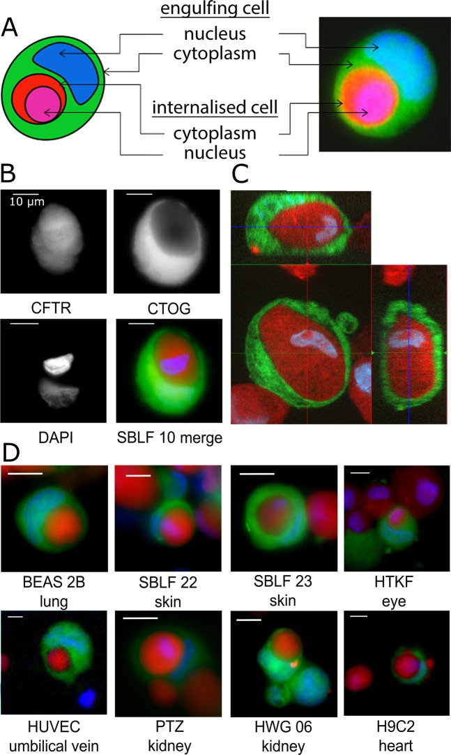
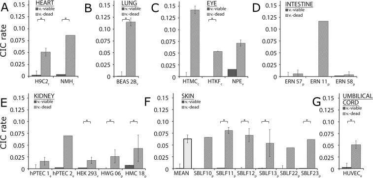
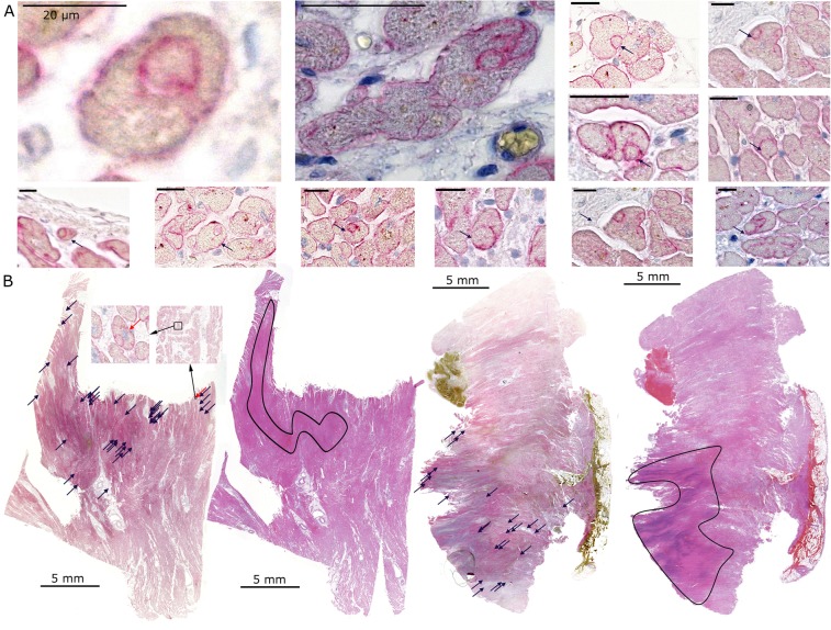
Non-professional phagocytosis by cancer cells has been described for decades. Recently, non-professional phagocytosis by normal tissue cells has been reported, which prompted us to take a closer look at this phenomenon. Non-professional phagocytosis was studied by staining cultured cells with live-cell staining dyes or by staining paraffin-embedded tissues by immunohistochemistry. Here, we report that each of 21 normal tissue cell lines from seven different organs was capable of phagocytosis, including ex vivo cell cultures examined before the 3rd passage as well as the primary and virus-transformed cell lines. We extended our analysis to an in vivo setting, and we found the occurrence of non-professional phagocytosis in healthy skin biopsies immediately after resection. Using dystrophin immunohistochemistry for membrane staining, human post-infarction myocardial tissue was assessed. We found prominent signs of non-professional phagocytosis at the transition zone of healthy and infarcted myocardia. Taken together, our findings suggest that non-professional phagocytosis is a general feature of normal tissue cells.


Conflict of interest statement

The authors declare no competing interests.

Figures

Figure 1
Figure 1
A schematic view and representative CIC images. The engulfing green cell contains an internalised, round, red cell and a crescent-shaped compressed nucleus (A). A CIC structure assembled by the internalised CFTR (red)-labelled cell and the engulfing CTOG (green)-labelled cell. The nucleus is labelled with DAPI and appears blue (B). A z-stack image of an SBLF 10 fibroblast cell (green) engulfing a dead cell (red). The nuclei are stained blue. The left panel is a xy optical slice taken at approximately the midpoint of the cell. The top left is an xz cross-section, and the right is a yz cross-section (C). Eight representative CIC images derived from different cell lines are displayed (D). Scale bars in are 10 µm.
Figure 2
Figure 2
The cell-in-cell rates in 21 different cell lines of different tissues, comparing the viable cells incubated with dead cells or with other viable cells. The CICs in cell lines obtained from the heart (A), lung (B), eye (C), intestine (D), kidney (E), skin (F) and umbilical cord (G). p indicates cell lines of primary origin that were cultivated for less than ten passages; e indicates ex vivo cell lines used prior to the fifth passage; v marks commercially available, virus-transformed cell lines; c indicates commercially available cell lines. *Indicates p < 0.05.
Figure 3
Figure 3
Non-professional phagocytes in the skin. Skin biopsies were immunohistologically stained for the cell membrane protein E-cadherin (brown) and for cell nuclei (blue) (A). A schematic view and examples of cell-in-cell structures in skin tissue are shown (B). Scale bars in (A) are 200 µm, 50 µm, and 20 µm; scale bars in (B) are 10 µm.
Figure 4
Figure 4
Non-professional phagocytes in heart tissue. Examples of CIC structures in the myocardium stained by dystrophin and haemalaun (A). A human post-infarction myocardium slide was stained for the cell membrane protein dystrophin and the neighbouring slide was stained with haematoxylin and eosin (B). The CIC-like structures were marked on the dystrophin slide (blue arrows). Afterwards, the infarction area was detected in the haematoxylin and eosin-stained slides (black outline). Inset images show two magnifications of the indicated area. The red arrows mark an exemplary CIC-like structure. The scale bars in (A) are 20 µm and in (B) are 5 mm.

References

    1. Fais S. Cannibalism: a way to feed on metastatic tumors. Cancer Lett. 2007;258:155–64. doi: 10.1016/j.canlet.2007.09.014. - DOI - PubMed
    1. Xia P, Wang S, Guo Z, Yao X. Emperipolesis, entosis and beyond: dance with fate. Cell Res. 2008;18:705–7. doi: 10.1038/cr.2008.64. - DOI - PubMed
    1. Overholtzer M, et al. A nonapoptotic cell death process, entosis, that occurs by cell-in-cell invasion. Cell. 2007;131:966–79. doi: 10.1016/j.cell.2007.10.040. - DOI - PubMed
    1. Schwegler M, et al. Prognostic value of homotypic cell internalization by nonprofessional phagocytic cancer cells. Biomed Res Int. 2015;2015:359392. doi: 10.1155/2015/359392. - DOI - PMC - PubMed
    1. Schwegler M, et al. Clearance of primary necrotic cells by non-professional phagocytes. Biol Cell. 2015;107:372–87. doi: 10.1111/boc.201400090. - DOI - PubMed