

Assessing the role of surface glycans of extracellular vesicles on cellular uptake
- PMID: 31417177
- PMCID: PMC6695415
- DOI: 10.1038/s41598-019-48499-1
Assessing the role of surface glycans of extracellular vesicles on cellular uptake
Abstract
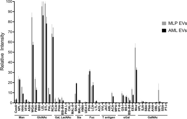
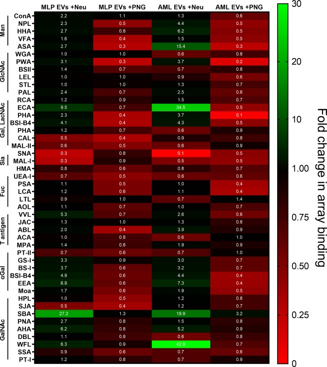
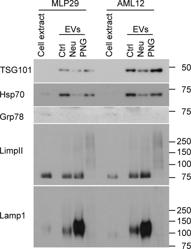
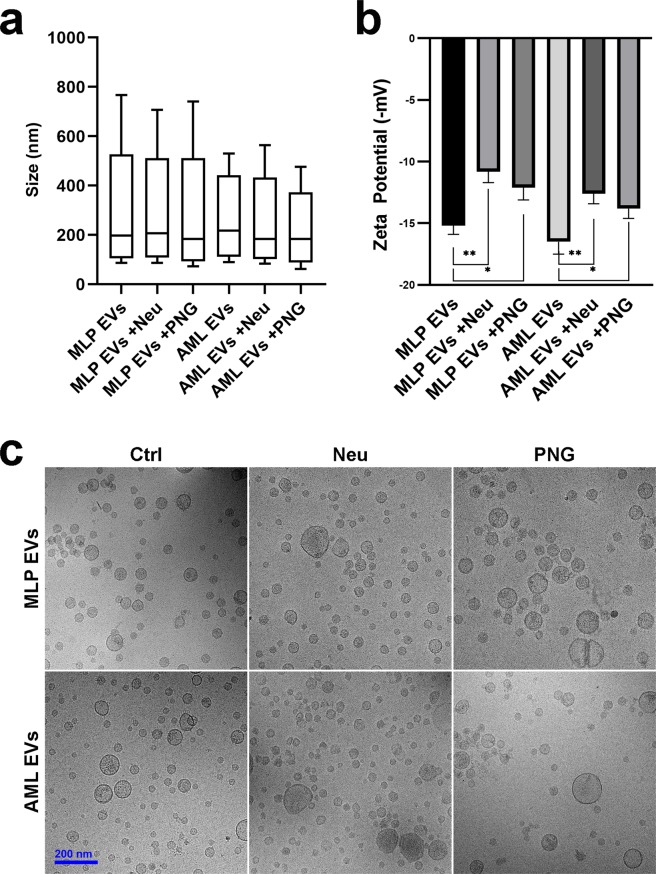
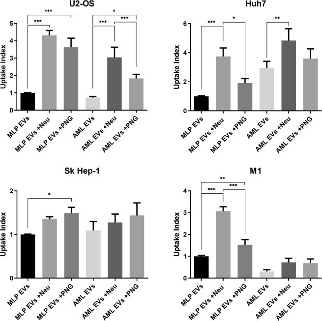
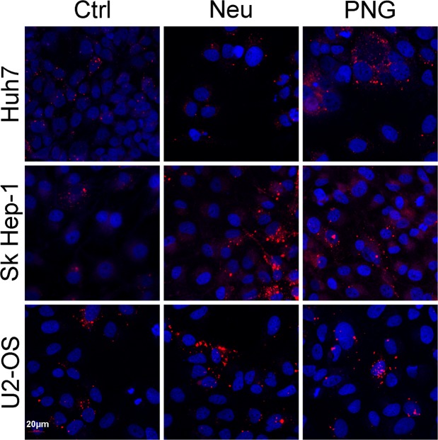
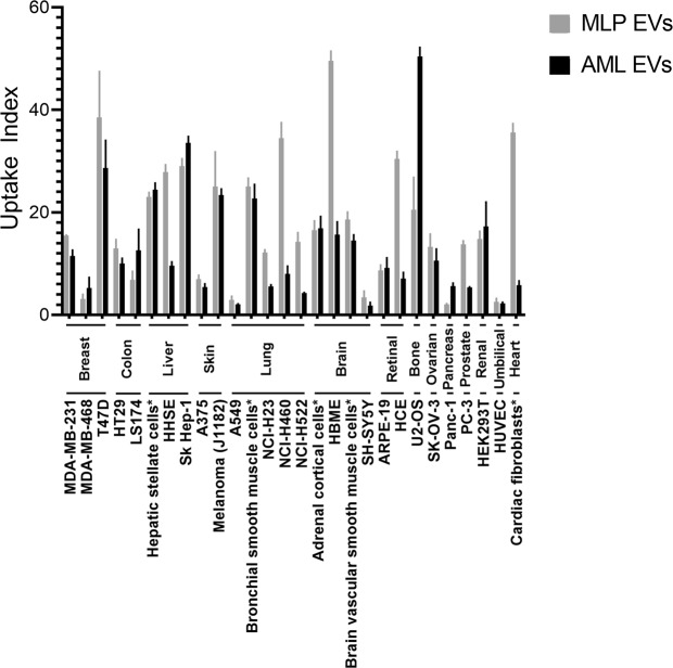
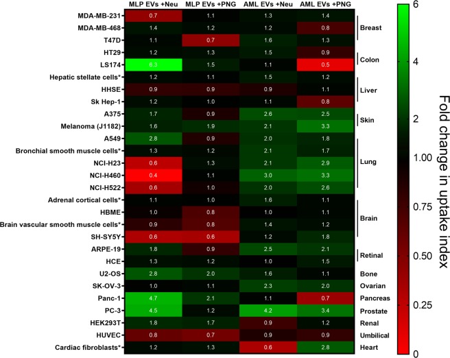
Extracellular vesicles (EVs) are important mediators of cell-cell communication in a broad variety of physiological contexts. However, there is ambiguity around the fundamental mechanisms by which these effects are transduced, particularly in relation to their uptake by recipient cells. Multiple modes of cellular entry have been suggested and we have further explored the role of glycans as potential determinants of uptake, using EVs from the murine hepatic cell lines AML12 and MLP29 as independent yet comparable models. Lectin microarray technology was employed to define the surface glycosylation patterns of EVs. Glycosidases PNGase F and neuraminidase which cleave N-glycans and terminal sialic acids, respectively, were used to analyze the relevance of these modifications to EV surface glycans on the uptake of fluorescently labelled EVs by a panel of cells representing a variety of tissues. Flow cytometry revealed an increase in affinity for EVs modified by both glycosidase treatments. High-content screening exhibited a broader range of responses with different cell types preferring different vesicle glycosylation states. We also found differences in vesicle charge after treatment with glycosidases. We conclude that glycans are key players in the tuning of EV uptake, through charge-based effects, direct glycan recognition or both, supporting glycoengineering as a toolkit for therapy development.
Conflict of interest statement
Drs. Roura-Ferrer, Martinez and Gamiz are employees of Innoprot, S.L. and performed the high-content screening experiments described within. All other authors declare no competing interests.
Figures
Similar articles
-
Assessment of Surface Glycan Diversity on Extracellular Vesicles by Lectin Microarray and Glycoengineering Strategies for Drug Delivery Applications.Small Methods. 2022 Feb;6(2):e2100785. doi: 10.1002/smtd.202100785. Epub 2021 Dec 1. Small Methods. 2022. PMID: 35174988
-
Role of Glycans in Equine Endometrial Cell Uptake of Extracellular Vesicles Derived from Amniotic Mesenchymal Stromal Cells.Int J Mol Sci. 2025 Feb 19;26(4):1784. doi: 10.3390/ijms26041784. Int J Mol Sci. 2025. PMID: 40004247 Free PMC article.
-
N-Glycans in Immortalized Mesenchymal Stromal Cell-Derived Extracellular Vesicles Are Critical for EV-Cell Interaction and Functional Activation of Endothelial Cells.Int J Mol Sci. 2022 Aug 23;23(17):9539. doi: 10.3390/ijms23179539. Int J Mol Sci. 2022. PMID: 36076936 Free PMC article.
-
Glycosylation on extracellular vesicles and its detection strategy: Paving the way for clinical use.Int J Biol Macromol. 2025 Mar;295:139714. doi: 10.1016/j.ijbiomac.2025.139714. Epub 2025 Jan 9. Int J Biol Macromol. 2025. PMID: 39798737 Review.
-
Glycosylation of extracellular vesicles: current knowledge, tools and clinical perspectives.J Extracell Vesicles. 2018 Mar 4;7(1):1442985. doi: 10.1080/20013078.2018.1442985. eCollection 2018. J Extracell Vesicles. 2018. PMID: 29535851 Free PMC article. Review.
Cited by
-
Cell-specific targeting of extracellular vesicles through engineering the glycocalyx.J Extracell Vesicles. 2022 Dec;11(12):e12290. doi: 10.1002/jev2.12290. J Extracell Vesicles. 2022. PMID: 36463392 Free PMC article.
-
Recommendations for reproducibility of cerebrospinal fluid extracellular vesicle studies.J Extracell Vesicles. 2024 Jan;13(1):e12397. doi: 10.1002/jev2.12397. J Extracell Vesicles. 2024. PMID: 38158550 Free PMC article. Review.
-
Exploiting Manipulated Small Extracellular Vesicles to Subvert Immunosuppression at the Tumor Microenvironment through Mannose Receptor/CD206 Targeting.Int J Mol Sci. 2020 Aug 31;21(17):6318. doi: 10.3390/ijms21176318. Int J Mol Sci. 2020. PMID: 32878276 Free PMC article. Review.
-
Exosomes in Head and Neck Squamous Cell Carcinoma.Front Oncol. 2019 Sep 18;9:894. doi: 10.3389/fonc.2019.00894. eCollection 2019. Front Oncol. 2019. PMID: 31620359 Free PMC article. Review.
-
Fostering "Education": Do Extracellular Vesicles Exploit Their Own Delivery Code?Cells. 2021 Jul 9;10(7):1741. doi: 10.3390/cells10071741. Cells. 2021. PMID: 34359911 Free PMC article. Review.
References
Publication types
MeSH terms
Substances
LinkOut - more resources
Full Text Sources
