Review
. 2019 Jul 30:7:535.
doi: 10.3389/fchem.2019.00535. eCollection 2019.

Cellulose Nano-Films as Bio-Interfaces

Affiliations
Review

Cellulose Nano-Films as Bio-Interfaces

Vikram Singh Raghuwanshi et al. Front Chem. .

Abstract

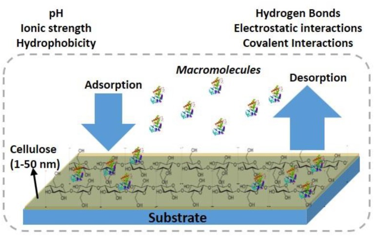
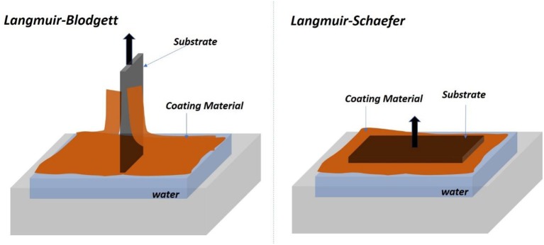
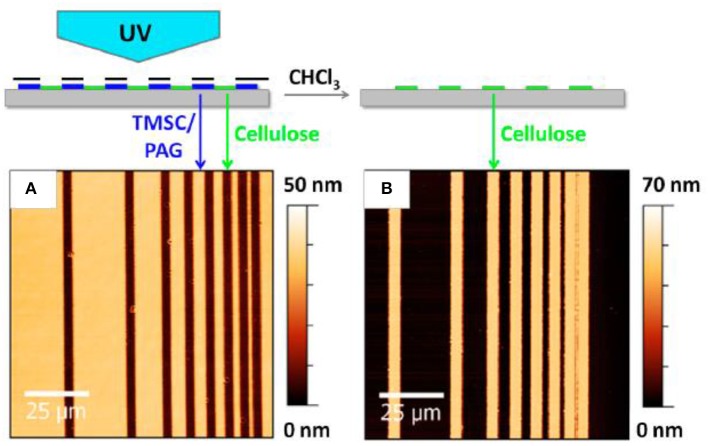
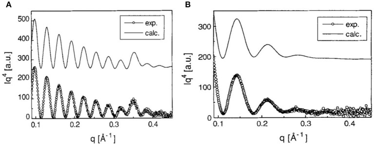
Cellulose, the most abundant polymer on earth, has enormous potential in developing bio-friendly, and sustainable technological products. In particular, cellulose films of nanoscale thickness (1-100 nm) are transparent, smooth (roughness <1 nm), and provide a large surface area interface for biomolecules immobilization and interactions. These attractive film properties create many possibilities for both fundamental studies and applications, especially in the biomedical field. The three liable-OH groups on the monomeric unit of the cellulose chain provide schemes to chemically modify the cellulose interface and engineer its properties. Here, the cellulose thin film serves as a substrate for biomolecules interactions and acts as a support for bio-diagnostics. This review focuses on the challenges and opportunities provided by engineering cellulose thin films for controlling biomolecules interactions. The first part reviews the methods for preparing cellulose thin films. These are by dispersing or dissolving pure cellulose or cellulose derivatives in a solvent to coat a substrate using the spin coating, Langmuir-Blodgett, or Langmuir-Schaefer method. It is shown how different cellulose sources, preparation, and coating methods and substrate surface pre-treatment affect the film thickness, roughness, morphology, crystallinity, swelling in water, and homogeneity. The second part analyses the bio-macromolecules interactions with the cellulose thin film interfaces. Biomolecules, such as antibodies and enzymes, are adsorbed at the cellulose-liquid interface, and analyzed dry and wet. This highlights the effect of film surface morphology, thickness, crystallinity, water intake capacity, and surface pre-treatment on biomolecule adsorption, conformation, coverage, longevity, and activity. Advance characterization of cellulose thin film interface morphology and adsorbed biomolecules interactions are next reviewed. X-ray and neutron scattering/reflectivity combined with atomic force microscopy (AFM), quartz crystal microbalance (QCM), microscopy, and ellipsometer allow visualizing, and quantifying the structural morphology of cellulose-biomolecule interphase and the respective biomolecules conformations, kinetics, and sorption mechanisms. This review provides a novel insight on the advantages and challenges of engineering cellulose thin films for biomedical applications. This is to foster the exploration at the molecular level of the interaction mechanisms between a cellulose interface and adsorbed biomolecules with respect to adsorbed molecules morphology, surface coverage, and quantity. This knowledge is to engineer a novel generation of efficient and functional biomedical devices.

Keywords: biomolecule; cellulose; characterization; diagnostics; interface; thin film.


Figures

Figure 1
Figure 1
Schematic of antibodies adsorption/desorption at the cellulosic thin film interface.
Figure 2
Figure 2
Schematic of the Langmuir-Blodgett (Left) and Langmuir-Schaefer (Right) set of forming film from the air/water interface.
Figure 3
Figure 3
Atomic force microscope image of the cellulose thin before (A) and after (B) regeneration. Reproduced with permission from Wolfberger et al. (2014).
Figure 4
Figure 4
(A) X-ray reflectivity curves of multilayers of TMSC films at the Si substrate. (B) X-ray reflectivity curves are from the cellulose thin film regenerated from TMSC by HCl acid hydrolysis. Dotted are the experimental curves and solid line are the modeling curves. Reproduced with permission from Schaub et al. (1993).
Figure 5
Figure 5
(A) AFM image of the deuterated bacterial cellulose (DBC) film dissolved in the ionic liquid. (B) AFM image of the cellulose film regenerated from the DBC in the HCl acid hydrolysis. Reproduced with permission from Su et al. (2016).
Figure 6
Figure 6
AFM height images of LS films of CNC transferred at different surface pressures, (A) 45 mN/m and (B) 60 mN/m. Reproduced with permission from Habibi et al. (2010a).
Figure 7
Figure 7
X-ray photoelectron carbon spectra of untreated (A) and washed ramie (B) cellulose nanocrystalline films. Reproduced with permission from Habibi et al. (2007).
Figure 8
Figure 8
AFM images and the corresponding surface profiles for (a) mostly crystalline cellulose film, (b) amorphous cellulose film on polystyrene coated gold substrates. The scan size is 1 mm2 and the z-range is 15 nm. Reproduced with permission from Tammelin et al. (2015).
Figure 9
Figure 9
ATR-FTIR spectra of cellulose and TMSC films showing different vibrational bonds. Reproduced with permission from Maver et al. (2015).
Figure 10
Figure 10
QCM-D measurement of the water absorption in amorphous and crystalline cellulose using thin films. Change in frequency (A) and change in dissipation (B) as a function of time upon exposure to water for more crystalline and highly amorphous cellulose films. (f0 = 5 MHz, n = 3, f3/n). Reproduced with permission from Tammelin et al. (2015).
Figure 11
Figure 11
(A) XPS wide scans from trimethylsilylcellulose (TMSC) and cellulose films hydrolyzed from TMSC with 2 M HCl for 1 min. (B) Hydrolysis of TMSC films with 0.5 M HCl followed with XPS. The carbon emission is resolved to contributions illustrating the decline of silicon bonded carbon from TMSC, as the hydrolysis proceeds. Reproduced with permission from Kontturi et al. (2003).
Figure 12
Figure 12
(A) NR curves of deuterated cellulose and IgG adsorbed deuterated cellulose. (B) SLD profile with thickness from the substrate interface obtained by fitting the NR curves. Reproduced with permission from Raghuwanshi et al. (2017a).
Figure 13
Figure 13
QCM-D data for the adsorption of human IgG on EDC/NHS activated NFC-film in the presence of conjugated antihuman IgG. Conjugation of IgG resulted in a small shift in frequency. Reproduced with permission from Orelma et al. (2012).
Figure 14
Figure 14
Optical images of human blood before (left) and after (middle) drying and the contact angle sessile drop image (right), respectively, on the (a–c) glass slide surface; (d–f) cellulose acetate surface; (g–i) regenerated cellulose surface. The before and after drying images were taken in transmittance mode. Reproduced with permission from Prathapan et al. (2018).
Figure 15
Figure 15
SPR sensogram on the adsorption of 0.1 mg/mL human IgG on cellulose. (a) CMC-modified cellulose (b), and chitosan modified cellulose (c), from aqueous solutions of pH 5.0, 6.2, 7.4, and 8.0 (the respective curves follow the same order from top down). Corresponding calculated adsorbed mass is indicated as a function of pH in the inserts (calculated from SPR data modeling). Reproduced with permission from Orelma et al. (2011).

References

    1. Ahola S., Myllytie P., Österberg M., Teerinen T., Laine J. (2008). Effect of polymer adsroption on cellulose nanofibril water binding capacity and aggregation. Bioresources 3, 1315–1328.
    1. Arcot L. R., Chen X., Wenchao X., Johansson L. S., Rojas O. J. (2015). Paper-based plasmon-enhanced protein sensing by controlled nucleation of silver nanoparticles on cellulose. Cellulose 22, 4027–4034. 10.1007/s10570-015-0783-z - DOI
    1. Ashkar R., Bilheux H. Z., Bordallo H., Briber R., Callaway D. J. E., Cheng X. L., et al. (2018). Neutron scattering in the biological sciences: progress and prospects. Acta Crystal. Sect. D Struct. Biol. 74, 1129–1168. 10.1107/S2059798318017503 - DOI - PubMed
    1. Atalla R. H., Vanderhart D. L. (1984). Native cellulose - a composite of 2 distinct crystalline forms. Science 223, 283–285. 10.1126/science.223.4633.283 - DOI - PubMed
    1. Aulin C., Ahola S., Josefsson P., Nishino T., Hirose Y., Osterberg M., et al. (2009). Nanoscale cellulose films with different crystallinities and mesostructures-their surface properties and interaction with water. Langmuir 25, 7675–7685. 10.1021/la900323n - DOI - PubMed