Review
. 2019 Jul 30:7:143.
doi: 10.3389/fcell.2019.00143. eCollection 2019.

Autophagic Control of Skin Aging

Affiliations
Review

Autophagic Control of Skin Aging

Leopold Eckhart et al. Front Cell Dev Biol. .

Abstract

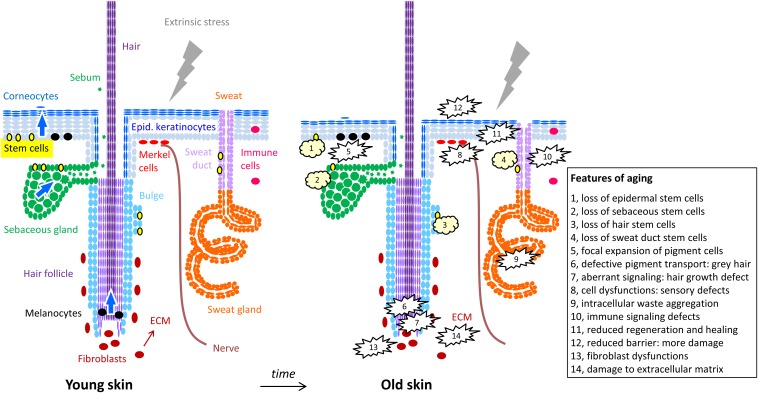
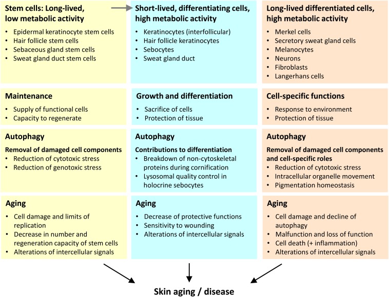
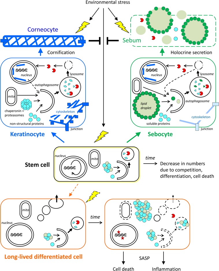
The skin forms the barrier to the environment. Maintenance of this barrier during aging requires orchestrated responses to variable types of stress, the continuous renewal of the epithelial compartment, and the homeostasis of long-lived cell types. Recent experimental evidence suggests that autophagy is critically involved in skin homeostasis and skin aging is associated with and partially caused by defects of autophagy. In the outer skin epithelium, autophagy is constitutively active during cornification of keratinocytes and increases the resistance to environmental stress. Experimental suppression of autophagy in the absence of stress is tolerated by the rapidly renewing epidermal epithelium, whereas long-lived skin cells such as melanocytes, Merkel cells and secretory cells of sweat glands depend on autophagy for cellular homeostasis and normal execution of their functions during aging. Yet other important roles of autophagy have been identified in the dermis where senescence of mesenchymal cells and alterations of the extracellular matrix (ECM) are hallmarks of aging. Here, we review the evidence for cell type-specific roles of autophagy in the skin and their differential contributions to aging.

Keywords: aging; autophagy; epidermis; hair; keratinocytes; melanocytes; skin; sweat gland.


Figures

FIGURE 1
FIGURE 1
Cells of the skin and their changes during aging. The structure of the skin is schematically depicted with oval symbols representing cells. Molecular and cellular features of aging are indicated in the right panel and in the box. For simplification, the diversity of immune cells and dermal cell populations are not depicted here.
FIGURE 2
FIGURE 2
Three categories of cells play different autophagy-dependent roles in skin aging. We propose that the aging of the skin is driven by changes in the distinct types of skin cells that we categorize according to lifetime (inversely correlated to turnover) and differentiation (execution of specific function within the tissue context). The main roles of autophagy and aging-associated changes are summarized for each cell category.
FIGURE 3
FIGURE 3
Roles of autophagy in skin cells. The contributions of autophagy to cornification, holocrine secretion, and homeostasis of stem cells and long-lived differentiated cells (exemplified by a sweat gland secretory cell) are schematically shown. Time-dependent changes, such as a decline of autophagy, affect mainly stem cells and long-lived differentiated cells. Short-lived keratinocytes and sebocytes are continuously generated from stem cells and then differentiate rapidly. Autophagy contributes to the formation of corneocytes and sebum, which protect the living cells of the skin against stress factors from the environment. The conversion of stem cells into (short-lived) differentiating keratinocytes and sebocytes occurs throughout life while the conversion of stem cells into long-lived differentiated secretory cells occurs rarely in adult life. Note that specialized stem cells exist in the interfollicular epidermis and in skin appendages whereas only one stem cell is shown in this simplified schematic drawing.

References

    1. Akinduro O., Sully K., Patel A., Robinson D. J., Chikh A., McPhail G., et al. (2016). Constitutive autophagy and nucleophagy during epidermal differentiation. J. Invest. Dermatol. 136 1460–1470. 10.1016/j.jid.2016.03.016 - DOI - PubMed
    1. Antunes F., Pereira G. J., Jasiulionis M. G., Bincoletto C., Smaili S. S. (2018). Nutritional shortage augments cisplatin-effects on murine melanoma cells. Chem. Biol. Interact. 281 89–97. 10.1016/j.cbi.2017.12.027 - DOI - PubMed
    1. Balaburski G. M., Hontz R. D., Murphy M. E. (2010). p53 and ARF: unexpected players in autophagy. Trends Cell Biol. 20 363–369. 10.1016/j.tcb.2010.02.007 - DOI - PMC - PubMed
    1. Bejarano E., Murray J. W., Wang X., Pampliega O., Yin D., Patel B., et al. (2018). Defective recruitment of motor proteins to autophagic compartments contributes to autophagic failure in aging. Aging Cell 17:e12777. 10.1111/acel.12777 - DOI - PMC - PubMed
    1. Bellei B., Pitisci A., Ottaviani M., Ludovici M., Cota C., Luzi F., et al. (2013). Vitiligo: a possible model of degenerative diseases. PLoS One 8:e59782. 10.1371/journal.pone.0059782 - DOI - PMC - PubMed

LinkOut - more resources