Review
. 2019 Aug 15;21(4):81.
doi: 10.1007/s10544-019-0412-y.

A review of microsampling techniques and their social impact

Affiliations
Review

A review of microsampling techniques and their social impact

Benson U W Lei et al. Biomed Microdevices. .

Abstract

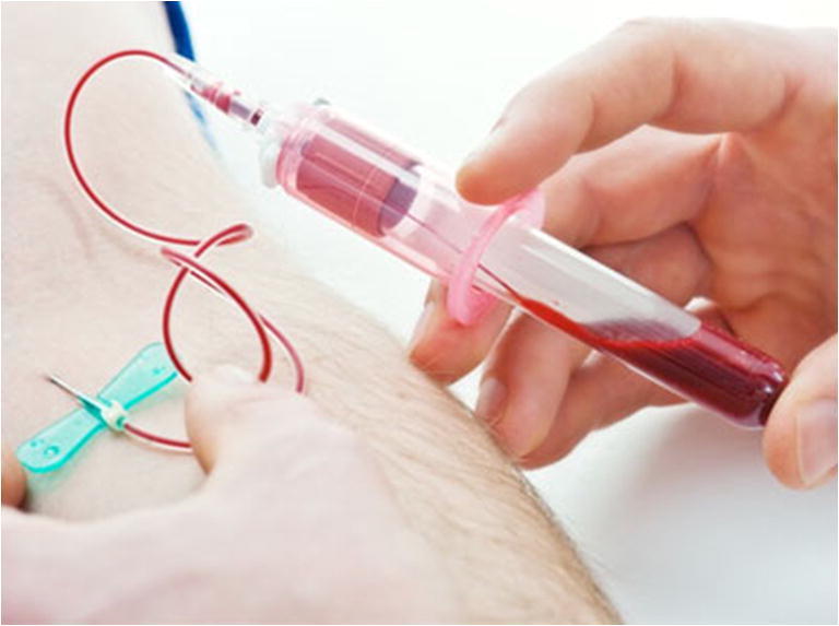
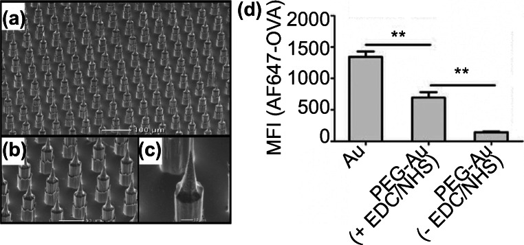
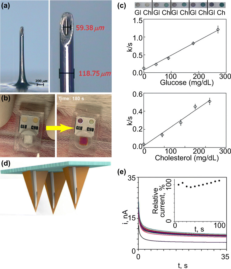
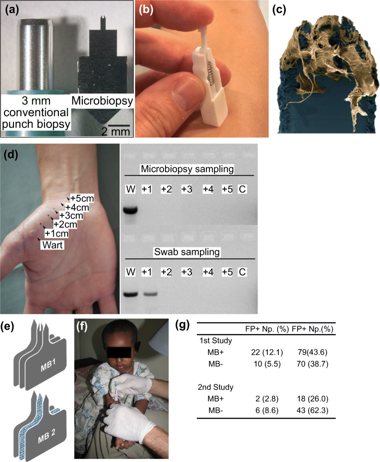
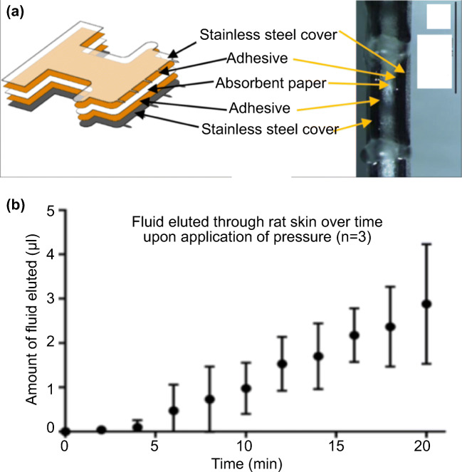
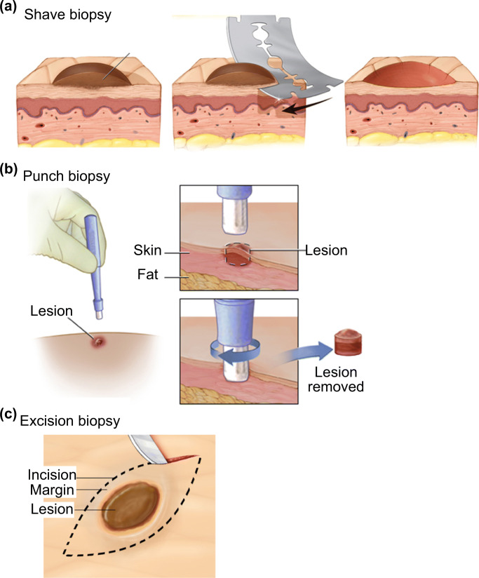
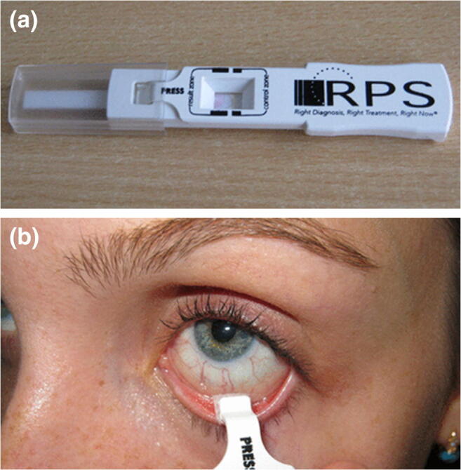
Conventional skin and blood sampling techniques for disease diagnosis, though effective, are often highly invasive and some even suffer from variations in analysis. With the improvements in molecular detection, the amount of starting sample quantity needed has significantly reduced in some diagnostic procedures, and this has led to an increased interest in microsampling techniques for disease biomarker detection. The miniaturization of sampling platforms driven by microsampling has the potential to shift disease diagnosis and monitoring closer to the point of care. The faster turnaround time for actionable results has improved patient care. The variations in sample quantification and analysis remain a challenge in the microsampling field. The future of microsampling looks promising. Emerging techniques are being clinically tested and monitored by regulatory bodies. This process is leading to safer and more reliable diagnostic platforms. This review discusses the advantages and disadvantages of current skin and blood microsampling techniques.

Keywords: Blood sampling; Microneedle; Microsampling; Minimally invasive; Point-of-care device; Skin biopsy.


Figures

Fig. 1
Fig. 1
Phlebotomy is one of the most commonly used conventional blood sampling techniques. The process consists of using an invasive needle and/or catheter to sample > 0.1–1 mL of blood for biomarker analysis. Picture retrieved from https://www.butlertech.org/event/phlebotomy-program-starts/
Fig. 2
Fig. 2
Dried blood spot (DBS) is the most common blood microsampling approach. It involves using a collection card to collect a sample of blood via finger-prick. Due to its low sample demand, it is often performed on infants (panel a). b) The volume of blood needs to be standardized in DBS for downstream analysis. Because of the strict requirement on sample volume, a technician is usually required to perform the technique. c) The hematocrit (HCT) level can lead to variations in analysis and can sometimes be distinguished by the color of sample. The HCT level of the top sample was 0.35 and the bottom was 0.5. d) Youhnovski et al. compared the precision and accuracy of DBS and PCDBS. PCDBS showed higher precision (%CV) and accuracy (%nominal). Pictures retrieved/adopted from http://www.ewbbu.com/mobile-health.html, Govender et al. , Wilhelm et al. and Youhnovski et al. . All figures are under a Creative Commons Attribution 2.0. Full terms at http://creativecommons.org/licenses/by/2.0
Fig. 3
Fig. 3
Plasma microsampling involves sampling whole blood and separating plasma out for analysis. a) The plasma collection card was designed to separate plasma from whole blood without centrifugation. b) The coefficients of variation (%CV) were similar for both the plasma extraction card and conventional liquid-liquid extraction. Reprinted (adapted) with permission from Kim et al. . Copyright 2018 American Chemical Society
Fig. 4
Fig. 4
Blood microsampling with volumetric absorptive microsampling (VAMS) and hemaPEN are emerging techniques for facilitating convenient and accurate sampling. a) VAMS sticks before (left) and after (right) sampling. b) Blood sample recovered from VAMS tip displayed less than 5% volumetric variation when compared to pipetting across the HCT range of 20 to 70%. c) The application of hemaPEN following finger-prick. Pictures retrieved from Denniff and Spooner and https://www.trajanscimed.com/pages/hemapen
Fig. 5
Fig. 5
Microprojection arrays, a solid microneedle-based device, was used to capture biomarkers in the skin of live mice as reported by Bhargav et al. a-c) The surface of microprojection arrays imaged with SEM. d) Surface modifications with EDC/NHS improved the capturing efficiency. The fluorescence intensities of EDC/NHS-treated MPAs showed an 18-fold increase. Pictures retrieved from Bhargav et al.
Fig. 6
Fig. 6
Hollow microneedles were combined with sensors for real-time biomarker monitoring. a) The dimension of hollow microneedle reported in Li et al. b) The absorption of blood led to color change in the reaction zones. The color change was used to determine the concentrations of glucose and cholesterol. c) The calibration curves for glucose and cholesterol measured by using the one-touch hollow microneedle device. Both measurements showed linear correlation coefficients (0.99 and 0.98). d) Schematic representation of the hollow microneedle array from Mohan et al. e) Real-time alcohol detection of 30 mM alcohol in artificial interstitial fluid (20.1 mg/mL BSA) for 100 min. The artificial interstitial fluid was located in a reservoir under a piece of excised mouse skin and was sampled by the microneedle array after penetrating the skin. Pictures retrieved from Li et al. , and Mohan et al.
Fig. 7
Fig. 7
Microneedle-based microbiopsies were used in skin and blood microsampling. a) A side-by-side comparison between conventional punch biopsy and the skin microbiopsy. b) The spring-loaded applicator used in microbiopsy application. c) After application, the microneedle captured pieces of skin tissue in the channel. d) Spatial detection of HPV by sampling cutaneous warts. Skin microbiopsy provided a more accurate spatial detection as demonstrated by DNA gel electrophoresis. e) Designs of skin (top) and absorbent microbiopsies (bottom). The main difference between the two designs was the middle absorbent layer. f) The absorbent microbiopsy was used to sample patients with Leishmaniasis in rural areas. g) The PCR data suggested the absorbent microbiopsy was able to detect Leishmaniasis more accurately than the finger-prick method. Pictures retrieved from Lin et al. , Tom et al. and Kirstein et al. . All figures are under a Creative Commons Attribution 2.0. Full terms at http://creativecommons.org/licenses/by/2.0
Fig. 8
Fig. 8
The microneedle patch reported by Samant et al. utilized absorbent microneedles to sample interstitial fluid from animal models. a Schematic of the microneedle patch. b The bottom view of the device, showing the absorbent paper was sandwiched by stainless steel covers. c Extraction of interstitial fluid from rat skin in vivo. Reproduced from Samant and Prausnitz with permission from the Royal Society of Chemistry
Fig. 9
Fig. 9
Skin biopsy is the current standard for skin conditions diagnosis. The three main types of skin biopsies are shave (a), punch (b), and excision (c) biopsy. All three types of skin biopsies are conducted bu a trained medical professional and the biopsy is sent for dermatopathology diagnosis in most cases. b The procedure for punch biopsy involves removing the suspicious location for analysis by a trained medical professional. Pictures adopted from https://www.mayoclinic.org/tests-procedures/skin-biopsy/about/pac-20384634 & https://myhealth.alberta.ca/Health/pages/conditions.aspx?hwid=hw234496
Fig. 10
Fig. 10
Tape stripping is a non-invasive technique for skin sampling. a The typical procedure of conventional tape stripping. 1) The penetration formulation was applied on the surface of the skin. 2) The formulation was spread across the sampling area. 3 & 4) The tape was applied and subsequently removed for downstream analysis. b The tape stripping biopsy kit from DermTech. The technique was performed by a technician at a clinic, and the tape was sent to a laboratory for analysis. c Performance of LINC00518 and/or PRAME preferentially expressed antigen in melanoma detection in the validation sets suggested a 91% sensitivity and 69% specificity. Both numbers were higher than using a dermoscopy alone. Pictures retrieved from Lademann et al. and Gerami et al.
Fig. 11
Fig. 11
Fractional skin harvesting (FSH) was designed for skin grafting. a Full-thickness skin columns harvested with a 19-gauge coring needle from the donor. (Cohen et al. 2014) Epidermis; (Graber et al. 2017) dermis including adnexal structures; (Global Point Of Care Diagnostics Market will reach USD 40.50 Billion by 2022: Zion Market Research n.d.) subcutaneous fat. Each mark on the ruler in the photograph spans 1 mm. b The recipient site i) before and ii) after the 8-week recovery time. i) Human skin columns were applied in random orientation to a full-thickness wound on the dorsal skin of a mouse; arrow highlights the epidermal head of one skin column. ii) The recipient site formed a ‘fishing net’ in the middle (arrow) after recovery. c The FSH resulted in samples with smaller surface area and mass than conventional split-thickness skin graft. Rσ = surface area ratio; Rm = mass ratio. Pictures retrieved/adopted from Tam et al. . All figures are under a Creative Commons Attribution 2.0. Full terms at http://creativecommons.org/licenses/by/2.0
Fig. 12
Fig. 12
Reverse iontophoresis was utilized in glucose monitoring. a In reverse iontophoresis, an electrical current is applied on skin to electro-osmotically drive interstitial fluid, which usually contains targeting analytes, through the epidermis to the skin surface for analysis or sample collection. b The GlucoWatch was one of the reversion iontophoresis-based diagnostic platforms for glucose level monitoring. The device was separated into two pieces, a watch (panel i) and an electrode assembly (panel ii). c GlucoWatch model GW2B accuracy test across a range of glucose concentrations. The medium relative absolute difference (RAD) value at the lowest glucose concentration (≤70 mg/dL) was the highest among all testing concentrations. Only 32% of the data collected met the ISO criteria (±20%). Pictures retrieved/adopted from Potts et al. and the Diabetes Research in Children Network (DirecNet) Study Group
Fig. 13
Fig. 13
The use of follicular pathway in bodily fluid sampling provided a calibration-free glucose monitoring platform. a Schematic illustration of the glucose preferential pathway (hair follicles). b A screen-printed array fixed onto a volunteer’s forearm. The array was connected to a potentiostat. c The platform enabled good tracking of the blood glucose as compared to commercial platform. Pictures retrieved from Lipani et al.
Fig. 14
Fig. 14
Nanopore biosensor for detecting analytes in bodily fluids. a Schematic illustration of the nanopore technique. The binding of analyte (glucose in this case) caused conformational change. The passing of proteins through the nanopore led to a change in current. b The glucose quantification with nanopore was similar to the commercial glucose meter in general. The asparagine measurement was less similar to the benchmarking technique HPLC but demonstrated a higher sensitivity in saliva sample. Pictures retrieved from Galenkamp et al. . All figures are under a Creative Commons Attribution 4.0. Full terms at http://creativecommons.org/licenses/by/4.0
Fig. 15
Fig. 15
Saliva sampling for disease diagnosis. Tao et al showed that oral rinse saliva sampling followed by lateral flow immunoassays (LFIA) were able to detect malaria infection in volunteers (a) and (b). PCR was used by Qureishi et al to detect Human Papilloma virus infection using saliva testing (c). mAb, monoclonal antibody; CI, confidense interval; DNA ISA, DNA in situ hybridisation; p16 IHC, p16 immunohistochemistry; PPV, positive predictive value; NPV, negative predictive value. b Table estimating the sensitivity of the lateral flow immunoassay as reported in Tao et al. . LFIA, lateral flow immunoassay. Pictures retrieved/adopted from Qureishi et al. and Tao et al. . All figures are under a Creative Commons Attribution 4.0. Full terms at http://creativecommons.org/licenses/by/4.0
Fig. 16
Fig. 16
Sweat sampling with a wearable device. a Schematic illustration of the wearable device as reported by Hauke et al. . b) In vivo test data and pharmacokinetic model curves for one of the testing subjects. Pictures adopted from Hauke et al. . All figures are under a Creative Commons Attribution 4.0. Full terms at http://creativecommons.org/licenses/by/4.0
Fig. 17
Fig. 17
AdenoPlus test for adenoviral conjunctivitis diagnosis. a AdenoPlus test includes both sampling and diagnosis. b The sample collector with the sampler. Pictures retrieved from Kam et al.
Fig. 18
Fig. 18
Centralized vs. decentralized diagnostic models. The centralized model involves a centralized laboratory in the diagnostic process and thus lengthens the turnaround time due to sample handling and delivery. In contrast, sample collection and analysis can be done at the same or nearby location under a decentralized model. Pictures retrieved from https://www.butlertech.org/event/phlebotomy-program-starts/ and http://www.johnstoncc.edu/continuing-education/allied-health/phlebotomy/index.aspx
Fig. 19
Fig. 19
Typical process vs. Theranos’ product development process. Theranos moved their diagnostic platform directly from R&D stage to the market. It skipped the peer review, clinical testing and government clearance procedure

References

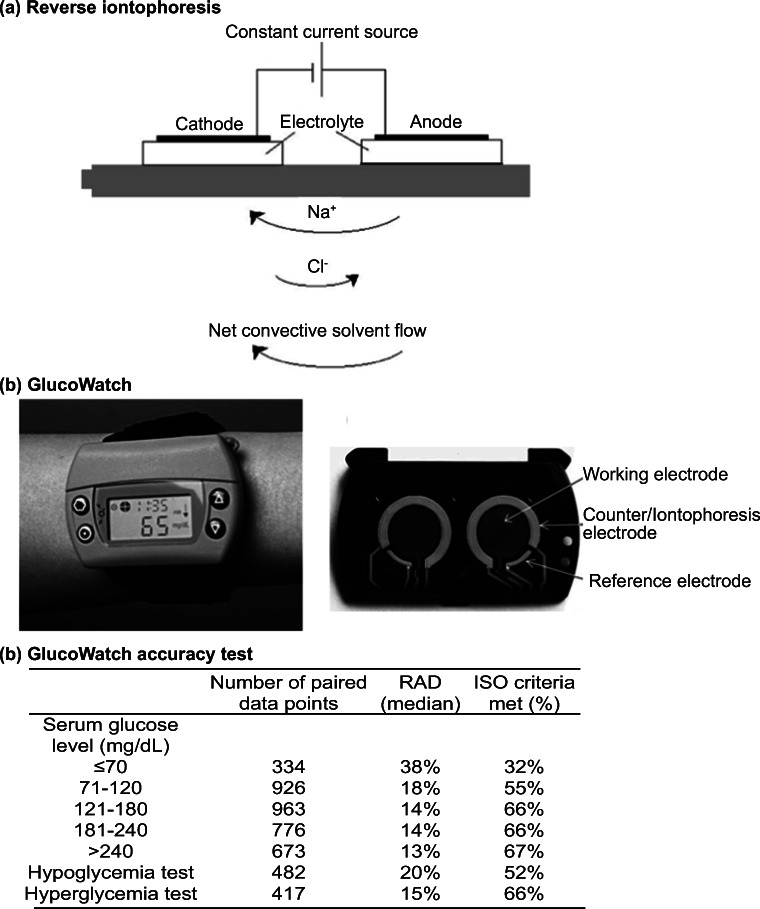
    1. Ackerman N, et al. Glucose monitoring via reverse iontophoresis. American Chemical Society, Polymer Preprints, Division of Polym. Chem. 1999;40:303–304.
    1. Alguire PC, Mathes BM. Skin biopsy techniques for the internist. J. Gen. Intern. Med. 1998;13:46–54. - PMC - PubMed
    1. Andrews E, et al. Concurrent human papillomavirus–associated tonsillar carcinoma in 2 couples. J. Infect. Dis. 2009;200:882–887. - PubMed
    1. Arima A, et al. Selective detections of single-viruses using solid-state nanopores. Sci. Rep. 2018;8:16305. - PMC - PubMed
    1. Asha SE, et al. Impact from point-of-care devices on emergency department patient processing times compared with central laboratory testing of blood samples: A randomised controlled trial and cost-effectiveness analysis. Emerg. Med. J. 2014;31:714–719. - PubMed

Publication types

LinkOut - more resources