Review
. 2019 Sep;20(9):e47928.
doi: 10.15252/embr.201947928. Epub 2019 Aug 14.

Metabolic implications of organelle-mitochondria communication

Affiliations
Review

Metabolic implications of organelle-mitochondria communication

Isabel Gordaliza-Alaguero et al. EMBO Rep. 2019 Sep.

Abstract

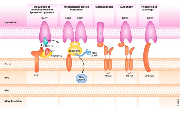
Cellular organelles are not static but show dynamism-a property that is likely relevant for their function. In addition, they interact with other organelles in a highly dynamic manner. In this review, we analyze the proteins involved in the interaction between mitochondria and other cellular organelles, especially the endoplasmic reticulum, lipid droplets, and lysosomes. Recent results indicate that, on one hand, metabolic alterations perturb the interaction between mitochondria and other organelles, and, on the other hand, that deficiency in proteins involved in the tethering between mitochondria and the ER or in specific functions of the interaction leads to metabolic alterations in a variety of tissues. The interaction between organelles is an emerging field that will permit to identify key proteins, to delineate novel modulation pathways, and to elucidate their implications in human disease.

Keywords: contact sites; diabetes; endoplasmic reticulum; insulin resistance; lipid droplets.


Conflict of interest statement

The authors declare that they have no conflict of interest.

The authors declare that they have no conflict of interest.

Figures

Figure 1
Figure 1. Contacts between mitochondria and other organelles
Mitochondria interact with other membranous compartments in the cell. Mitochondria interact with the Golgi apparatus; however, the identities of the proteins involved in this interaction have not been discovered yet. Mitochondria are also in contact with lysosomes, but the mediators of these contacts remain unknown. MFN2 in mitochondria interacts with melanosomes. ECI2 and TOM20 bridge the peroxisome to the mitochondria. Mitochondria are anchored to lipid droplets by the MFN2–PLIN1 interaction. Mitochondria–ER contacts harbor a singular architecture and are hubs for several cellular processes such as Ca2+ signaling and lipid trafficking (see further details in Figs 2 and 3).
Figure 2
Figure 2. The architecture of mitochondria–ER contact sites: tethering complexes
Mitochondria are bridged to the ER by several protein complexes. In the ER, VAPB or MOSPD2 bind to PTPIP51 in mitochondria. IP3R in the ER is anchored to VDAC in the OMM by the cytosolic protein GRP75. MFN2 is present both at the ER and in the OMM. From the ER, MFN2 interacts with either MFN1 or MFN2 in the mitochondria. BAP31 in the ER partners up with FIS1 in the mitochondria. BiP in the ER, WASF3 in the cytosol, and ATAD3A in the IMM have been suggested to form a complex that tethers both organelles.
Figure 3
Figure 3. Cellular functions at mitochondria–ER contact sites
The main processes that take place at the MAM are as follows: phospholipid trafficking, mitochondrial dynamics, Ca2+ signaling, unfolded protein response (UPR), apoptosis initiation, and autophagosome formation. MAMs are hubs for phospholipid exchange between the ER and mitochondria. Mitochondria take phosphatidylserine from the ER, which is supplied with phosphatidylethanolamine by mitochondria. Mitochondrial dynamics processes are regulated at the interface between the ER and mitochondria. The mitochondrial fission effector DRP1 is recruited by MFF and MiD49/51 to the mitochondrial surface, and it interacts with STX17 in the ER membrane. The ER wraps around the mitochondrion, which is finally excised into two daughter mitochondria after mtDNA replication. Mitochondrial fusion is promoted by TCHP binding to MFN2. This interaction separates MFN2 tethers and promotes the fusogenic function of mitochondrion‐bound MFN2. The ER is the cellular Ca2+ reservoir. IP3R, GRP75, and VDAC form a Ca2+ channeling complex that allows Ca2+ flux from the ER to mitochondria. MCU transports the intermembrane Ca2+ to the mitochondrial matrix. The UPR is regulated at the interface between mitochondria and the ER. One of the key regulators is MFN2, which inhibits the UPR by interacting with PERK. It is not known whether PERK interacts with MFN2 in mitochondria, MFN2 in the ER or with both. MAMs are also involved in the initiation of apoptosis. Sustained pro‐apoptotic stimuli lead to BCL2 sequestration by the reticulum proteins BAP31 and CDIP in order to initiate apoptotic signaling cascades. Autophagosomes arise from mitochondria–ER contact sites. Proteins involved in autophagosome formation are recruited to these locations. STX17 in the ER attracts ATG14L and the PI3K complex. The mitochondrial component involved in this process is still not known.
Figure 4
Figure 4. Metabolic impact of alterations in proteins participating in mitochondria–ER contacts
(A) GRP75, MFN2, BAP31, or CypD depletion leads to deficient insulin signaling and glucose intolerance. The lack of GRP75, MFN2, BAP31, or CypD at mitochondria–ER contact sites causes deficient insulin signaling and glucose intolerance, probably through a mechanism that involves ERUPR or mtUPR. (B) IP3R1, VDAC1, FUNDC1, or PACS2 ablation results in enhanced insulin signaling and improved glucose tolerance. The lack of IP3R1, VDAC1, FUNDC1, or PACS2 at mitochondria–ER contact sites potentiates insulin signaling and improves glucose tolerance. The mechanism by which FUNCD1 or PACS2 causes these effects is mediated by the release of FGF21. As a result of decreased mitochondrial Ca2+ accumulation, IP3R1 or VDAC1 ablation may result in enhanced insulin signaling and improved glucose tolerance. (C) Mitochondria–ER contacts response to protein ablation. The deletion of proteins that participate in the MAM results in the activation of signaling pathways that either enhance or impair insulin sensitivity and glucose tolerance. It has been proposed that these signaling pathways are related to UPR, FGF21, and an adaptive mitochondrial response that may lead to an improved or to a worsened response to insulin and glucose handling. (D) ORP8 or ATAD3A deficiency causes lipid metabolism alterations. Various alterations in lipid metabolism are observed upon ORP8 and ATAD3A ablation. ORP8 deficiency causes an increase in circulating HDL, cholesterol, triglycerides, and phospholipids. On the other hand, a lack of ATAD3A results in impaired cholesterol and lipid metabolism, reduced cholesterol esters, and decreased steroidogenesis.
Figure 5
Figure 5. The architecture of mitochondria–LD contact sites
Mitochondria establish contacts with lipid droplets (LDs). Although these contacts are poorly studied, several proteins have been found to participate in them. MFN2 in mitochondria interacts with the LD protein PLIN1. Mitochondrial ASCL1 has been found to form a complex with SNAP23 and VAMP4, both present on the LD surface. Moreover, PLIN5 has been found both on the surface of LDs and in the OMM. It is known that PLIN5 interacts with ATGL; however, the protein complex through which PLIN5 anchors LDs to mitochondria is still uncharacterized.
Figure 6
Figure 6. Mitochondria–lysosome interface
Several processes take place at the contacts between mitochondria and lysosomes: (A) Regulation of mitochondrial and lysosomal dynamics by RAB7, TBC1D15, and FIS1 coordination; (B) mitochondrial protein translation in ribosomes anchored to the endosomal surface by interaction with RAB7 and RAB5 in close proximity to mitochondria; (C) melanogenesis in melanosomes that interact with mitochondria through MFN2; (D) autophagosome to lysosome fusion, which is supported by MFN2–RAB7 interaction; and (E) possible phospholipid exchange through VPS13A interaction with RAB7.

References

    1. Valm AM, Cohen S, Legant WR, Melunis J, Hershberg U, Wait E, Cohen AR, Davidson MW, Betzig E, Lippincott‐Schwartz J (2017) Applying systems‐level spectral imaging and analysis to reveal the organelle interactome. Nature 546: 162–167 - PMC - PubMed
    1. Benador IY, Veliova M, Mahdaviani K, Petcherski A, Wikstrom JD, Assali EA, Acín‐Pérez R, Shum M, Oliveira MF, Cinti S et al (2018) Mitochondria bound to lipid droplets have unique bioenergetics, composition, and dynamics that support lipid droplet expansion. Cell Metab 27: 869–885.e6 - PMC - PubMed
    1. Boutant M, Kulkarni SS, Joffraud M, Ratajczak J, Valera‐Alberni M, Combe R, Zorzano A, Cantó C (2017) Mfn2 is critical for brown adipose tissue thermogenic function. EMBO J 36: 1543–1558 - PMC - PubMed
    1. Dolman NJ, Gerasimenko JV, Gerasimenko OV, Voronina SG, Petersen OH, Tepikin AV (2005) Stable Golgi‐mitochondria complexes and formation of Golgi Ca2+ gradients in pancreatic acinar cells. J Biol Chem 280: 15794–15799 - PubMed
    1. Marsh BJ, Mastronarde DN, Buttle KF, Howell KE, McIntosh JR (2001) Organellar relationships in the Golgi region of the pancreatic beta cell line, HIT‐T15, visualized by high resolution electron tomography. Proc Natl Acad Sci USA 98: 2399–2406 - PMC - PubMed

Publication types