Comparative Study
. 2019 Oct 1;317(4):H765-H776.
doi: 10.1152/ajpheart.00247.2019. Epub 2019 Aug 16.

Exosomes derived from induced vascular progenitor cells promote angiogenesis in vitro and in an in vivo rat hindlimb ischemia model

Affiliations
Comparative Study

Exosomes derived from induced vascular progenitor cells promote angiogenesis in vitro and in an in vivo rat hindlimb ischemia model

Takerra K Johnson et al. Am J Physiol Heart Circ Physiol. .

Abstract

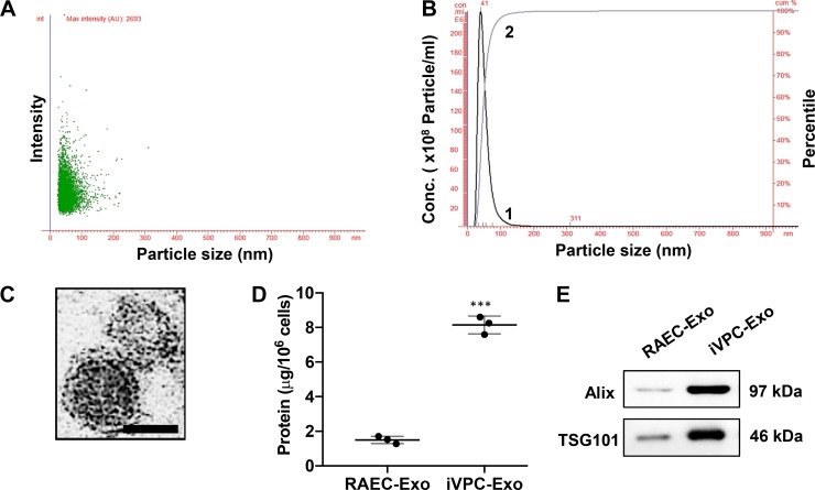
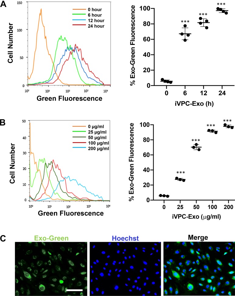
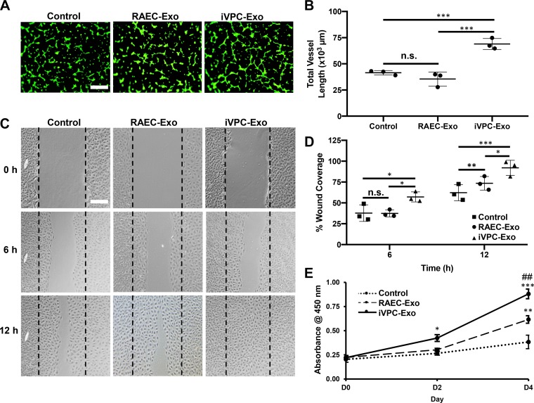
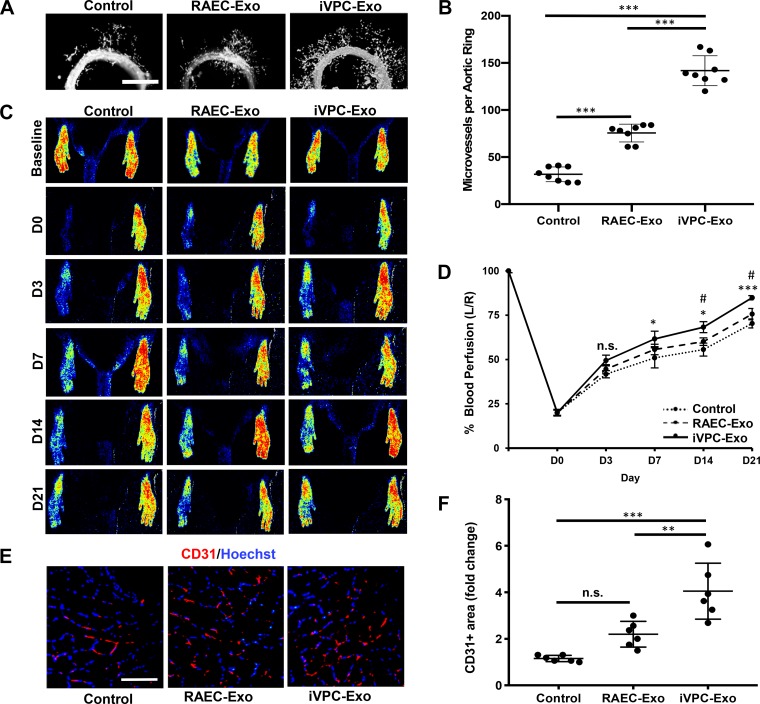
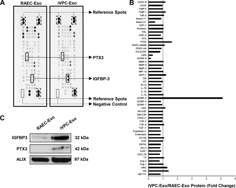
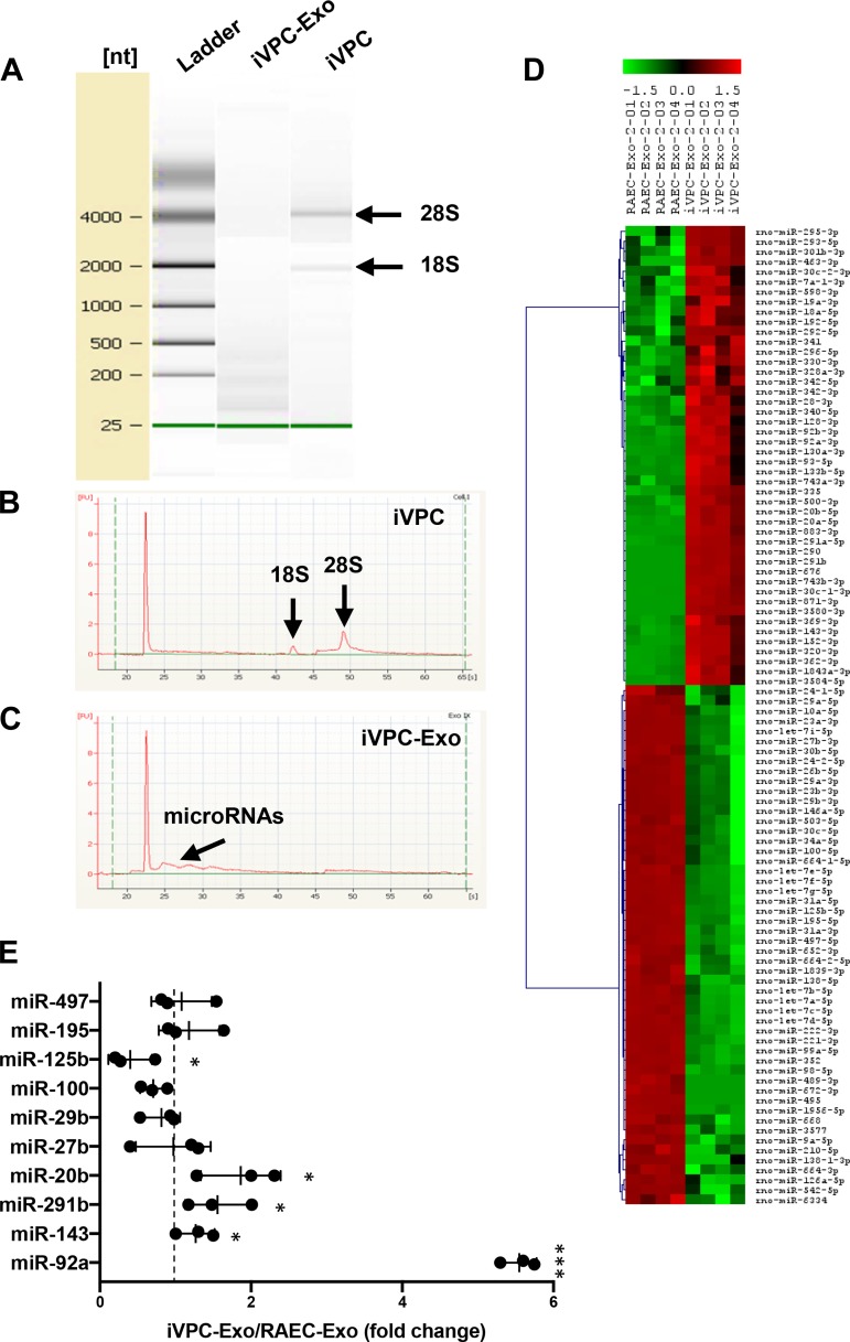
Induced vascular progenitor cells (iVPCs) were created as an ideal cell type for regenerative medicine and have been reported to positively promote collateral blood flow and improve cardiac function in a rat model of myocardial ischemia. Exosomes have emerged as a novel biomedicine that mimics the function of the donor cells. We investigated the angiogenic activity of exosomes from iPVCs (iVPC-Exo) as a cell-free therapeutic approach for ischemia. Exosomes from iVPCs and rat aortic endothelial cells (RAECs) were isolated using a combination of ultrafiltration and size-exclusion chromatography. Nanoparticle tracking analysis revealed that exosome isolates fell within the exosomal diameter (<150 nm). These exosomes contained known markers Alix and TSG101, and their morphology was validated using transmission electron microscopy. When compared with RAECs, iVPCs significantly increased the secretion of exosomes. Cardiac microvascular endothelial cells and aortic ring explants were pretreated with RAEC-Exo or iVPC-Exo, and basal medium was used as a control. iVPC-Exo exerted an in vitro angiogenic effect on the proliferation, tube formation, and migration of endothelial cells and stimulated microvessel sprouting in an ex vivo aortic ring assay. Additionally, iVPC-Exo increased blood perfusion in a hindlimb ischemia model. Proangiogenic proteins (pentraxin-3 and insulin-like growth factor-binding protein-3) and microRNAs (-143-3p, -291b, and -20b-5p) were found to be enriched in iVPC-Exo, which may mediate iVPC-Exo induced vascular growth. Our findings demonstrate that treatment with iVPC-Exo promotes angiogenesis in vitro, ex vivo, and in vivo. Collectively, these findings indicate a novel cell-free approach for therapeutic angiogenesis.NEW & NOTEWORTHY The results of this work demonstrate exosomes as a novel physiological mechanism by which induced vascular progenitor cells exert their angiogenic effect. Moreover, angiogenic cargo of proteins and microRNAs may define the biological contributors in activating endothelial cells to form a new capillary plexus for ischemic vascular diseases.

Keywords: angiogenesis; endothelial cell; exosomes; microRNA; progenitor cell.


Conflict of interest statement

No conflicts of interest, financial or otherwise, are declared by the authors.

Figures

Fig. 1.
Fig. 1.
Characterization of iVPC-Exo. iVPCs were incubated in culture medium containing 1% exosome-free fetal bovine serum for 24 h. Medium was collected and subjected to exosome isolation. Isolated exosomes were examined using NTA to determine exosomal size and concentration. Scatter plot graphs of exosomes demonstrating the particle size vs. light intensity of iVPC-Exo (A) and the distribution of particle size vs. concentration of iVPC-Exo (B) are shown. Curve 1 illustrates the relationship between particle number and distribution of particle size (concentration/mL; left, y-axis). Curve 2 describes the correlation between the cumulative percentage distribution of particles (percentile; right, y-axis) and particle size (x-axis). Morphology of iVPC-Exo was visualized under transmission electron microscopy (scale bar = 50 nm; C). Protein amount in exosomes from the same number of RAECs or iVPCs was compared (D) (***P < 0.001 vs. RAEC-Exo). Exosomal markers, Alix and TSG101, in RAEC-Exo and iVPC-Exo were determined via Western blot analysis (E). Each lane represented an exosomal lysate collected from 2.5 × 10⁶ cells. For A, B, D, and E: n = 3. iVPC, induced vascular progenitor cell; iVPC-Exo, iVPC-secreted exosomes; NTA, nanoparticle tracking analysis; RAEC-Exo, rat aortic endothelial cell-secreted exosomes.
Fig. 2.
Fig. 2.
Uptake of iVPC-Exo into CMVECs. CMVECs were incubated with Exo-Green labeled iVPC-Exo to measure the efficiency of uptake. A: flow cytometry histogram of CMVECs after incubation with 200 µg/mL of labeled iVPC-Exo for 0 h, 6 h, 12 h, or 24 h (left). Quantification of time-course assay result from flow cytometry analysis (right) (***P < 0.001 vs. 0 h; n = 4). B: flow cytometry histogram of CMVECs after incubation with 0, 25, 50, 100, or 200 µg/mL of labeled iVPC-Exo for 24 h (left). Quantification of dose-response assay result from flow cytometry analysis (right) (***P < 0.001 vs. 0 µg/mL). C: representative images demonstrated iVPC-Exo uptake into CMVECs after incubation with 200 µg/mL of iVPC-Exo for 24 h using fluorescent microscopy. Images displayed Exo-Green (green) for labeled exosomes in iVPCs, Hoechst 33342 (blue) staining of nuclei, and merged image, respectively (scale bar = 100 µm). For B and C: n = 3. CMVEC, cardiac microvascular endothelial cells; iVPC, induced vascular progenitor cell; iVPC-Exo, iVPC-secreted exosomes.
Fig. 3.
Fig. 3.
iVPC-Exo enhance tube formation, migration, and proliferation of CMVECs. CMVECs were left untreated (control) or pretreated with 200 µg/mL of RAEC-Exo or iVPC-Exo for 24 h and subjected to the following angiogenic assay in vitro. A and B: cells were seeded on top of Matrigel and used for tube formation assay and stained with calcein AM (green) for visualization, and representative images were acquired using fluorescent microscopy. Tube formation was quantified by vessel length using NIH AngioTool software (***P < 0.001). C and D: representative images of the scratch wound healing assay acquired at time points 0 h, 6 h, and 12 h using a bright-field microscope. Percentage of wound coverage was quantified by calculating area of gap using ImageJ software (*P < 0.05, **P < 0.01, and ***P < 0.001). E: CMVEC proliferation was measured by using CCK-8 analysis (*P < 0.05 iVPC-Exo vs. control on day 2, **P < 0.01 RAEC-Exo vs. control on day 4, ***P < 0.001 iVPC-Exo vs. control on day 4, and ##P < 0.01 iVPC-Exo vs. RAEC-Exo on day 4). For A and C: scale bar = 100 µm. For AE: n = 3. CMVEC, cardiac microvascular endothelial cells; D, day; iVPC-Exo, induced vascular progenitor cell-secreted exosomes; n.s., not significant; RAEC-Exo, rat aortic endothelial cell-secreted exosomes.
Fig. 4.
Fig. 4.
iVPC-Exo promotes microvessel sprouting from aortic ring and recovery of blood perfusion in rat ischemic hindlimb. A and B: aortic ring explants were left untreated (control) or treated with 200 µg/mL RAEC-Exo or iVPC-Exo (n = 8). A: images were taken using fluorescent microscopy to visualize microvessel sprouts emerging from aortic rings after 5 days of treatment. B: number of microvessels per ring was then quantified (***P < 0.001). C–F: ligated limbs were left untreated (control) or treated with 30 µg RAEC-Exo or iVPC-Exo (n = 10). C: blood perfusion in both hind limbs was assessed before ligation (baseline) and on day 0, 3, 7, 14, and 21 postligation by using laser speckle imaging. D: perfusion was measured, and the percentage of perfusion was calculated measuring left limb/right limb (L/R) (*P < 0.05 iVPC-Exo vs. control on the same day, ***P < 0.001 iVPC-Exo vs. control on day 21, #P < 0.05 iVPC-Exo vs. RAEC-Exo on the same day, n.s. = not significant). E and F: sections of the gastrocnemius muscle on the ligated side were subjected to immunohistochemistry analysis for CD31, an endothelial cell marker, and counterstained with Hoechst 33342 (scale bar = 200 µm). E: representative images after staining of CD31-positive cells in the gastrocnemius were taken at day 21 postligation. F: quantification of the CD31-positive area was shown (**P < 0.05, ***P < 0.001, n.s. = not significant). D, day; iVPC-Exo, induced vascular progenitor cell-secreted exosomes; RAEC-Exo, rat aortic endothelial cell-secreted exosomes.
Fig. 5.
Fig. 5.
Identification of angiopeptides in RAEC-Exo and iVPC-Exo. A and B: a total of 53 angiogenesis-related proteins in RAEC-Exo and iVPC-Exo were examined using an angiogenesis antibody array (n = 2). A: when compared with RAEC-Exo, insulin-like growth factor-binding protein-3 (IGFBP3) and pentraxin-3 (PTX3) in iVPC-Exo are most increased and labeled (two dots represent one angiopeptide in duplicate). Positive (reference spots) and negative membrane controls are also labeled. B: signal intensities were quantified using ImageJ densitometry analysis. Level of protein from RAEC-Exo was set to 1 (dashed line). C: IGFBP3 and PTX3 were then validated using Western blot analysis. Exosomal marker Alix was used as a loading control. n = 3. iVPC-Exo, induced vascular progenitor cell-secreted exosomes; RAEC-Exo, rat aortic endothelial cell-secreted exosomes.
Fig. 6.
Fig. 6.
RNA analysis and microRNA profiling of RAEC-Exo and iVPC-Exo. A–C: an equal amount of RNA from RAEC-Exo and iVPC-Exo was analyzed using a Bioanalyzer. A: electrophoretic separation of RNA from iVPC and iVPC-Exo. RNA 6000 ladder standard marked six RNA fragments ranging in size from 0.25 to 4 kb. Bands of 18S and 28S ribosomal RNA units are indicated. B: iVPC electropherogram indicates peaks that correspond to ribosomal RNA. C: iVPC-Exo electropherogram shows possible microRNAs. D: exosomal microRNA profiling was performed (n = 2). The microRNAs with different levels between RAEC-Exo and iVPC-Exo (P < 0.01) was shown in a heat map. Hierarchical clustering displayed on the axis to the left demonstrates microRNA cluster relationships. E: angiogenic-related microRNAs (angiomiR) in D were validated using RT-PCR. Level of microRNA in RAEC-Exo was set to 1 (dashed line). U6 was used as an internal control (*P < 0.05, ***P < 0.001 iVPC-Exo vs. RAEC-Exo). n = 3. iVPC, induced vascular progenitor cell; iVPC-Exo, iVPC-secreted exosomes; miR, microRNA; RAEC-Exo, rat aortic endothelial cell-secreted exosomes.

References

    1. Annex BH. Therapeutic angiogenesis for critical limb ischaemia. Nat Rev Cardiol 10: 387–396, 2013. doi:10.1038/nrcardio.2013.70. - DOI - PubMed
    1. Baker M, Robinson SD, Lechertier T, Barber PR, Tavora B, D’Amico G, Jones DT, Vojnovic B, Hodivala-Dilke K. Use of the mouse aortic ring assay to study angiogenesis. Nat Protoc 7: 89–104, 2011. doi:10.1038/nprot.2011.435. - DOI - PubMed
    1. Barile L, Lionetti V, Cervio E, Matteucci M, Gherghiceanu M, Popescu LM, Torre T, Siclari F, Moccetti T, Vassalli G. Extracellular vesicles from human cardiac progenitor cells inhibit cardiomyocyte apoptosis and improve cardiac function after myocardial infarction. Cardiovasc Res 103: 530–541, 2014. doi:10.1093/cvr/cvu167. - DOI - PubMed
    1. Belair DG, Whisler JA, Valdez J, Velazquez J, Molenda JA, Vickerman V, Lewis R, Daigh C, Hansen TD, Mann DA, Thomson JA, Griffith LG, Kamm RD, Schwartz MP, Murphy WL. Human vascular tissue models formed from human induced pluripotent stem cell derived endothelial cells. Stem Cell Rev Rep 11: 511–525, 2015. doi:10.1007/s12015-014-9549-5. - DOI - PMC - PubMed
    1. Beltrami C, Besnier M, Shantikumar S, Shearn AIU, Rajakaruna C, Laftah A, Sessa F, Spinetti G, Petretto E, Angelini GD, Emanueli C. Human pericardial fluid contains exosomes enriched with cardiovascular-expressed microRNAs and promotes therapeutic angiogenesis. Mol Ther 25: 679–693, 2017. doi:10.1016/j.ymthe.2016.12.022. - DOI - PMC - PubMed

MeSH terms

LinkOut - more resources