Review
. 2019 Nov 1;317(5):H891-H922.
doi: 10.1152/ajpheart.00259.2019. Epub 2019 Aug 16.

Guidelines for evaluating myocardial cell death

Affiliations
Review

Guidelines for evaluating myocardial cell death

Paras K Mishra et al. Am J Physiol Heart Circ Physiol. .

Erratum in

  • Corrigendum.
    [No authors listed] [No authors listed] Am J Physiol Heart Circ Physiol. 2019 Dec 1;317(6):H1390. doi: 10.1152/ajpheart.zh4-2975-corr.2019. Am J Physiol Heart Circ Physiol. 2019. PMID: 31797698 Free PMC article. No abstract available.

Abstract

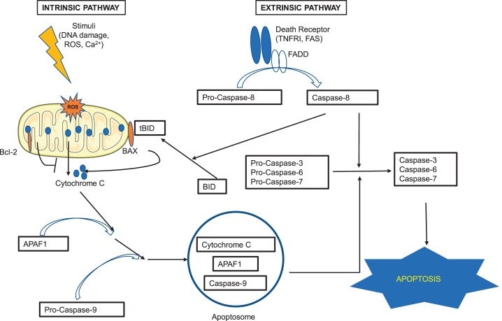
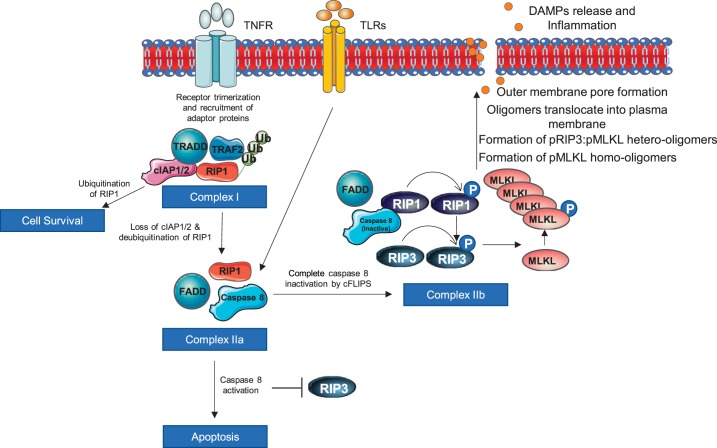
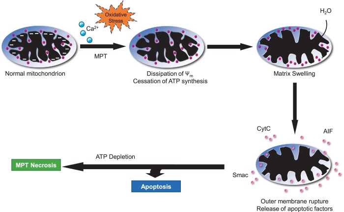
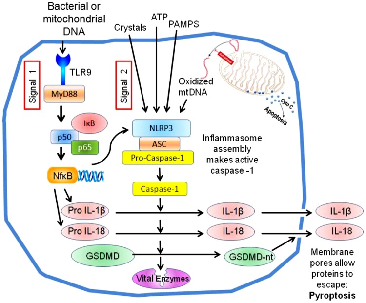
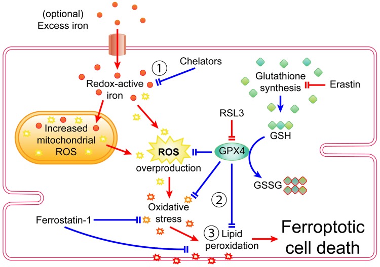
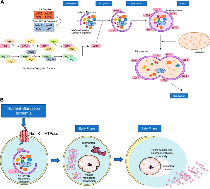
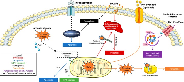
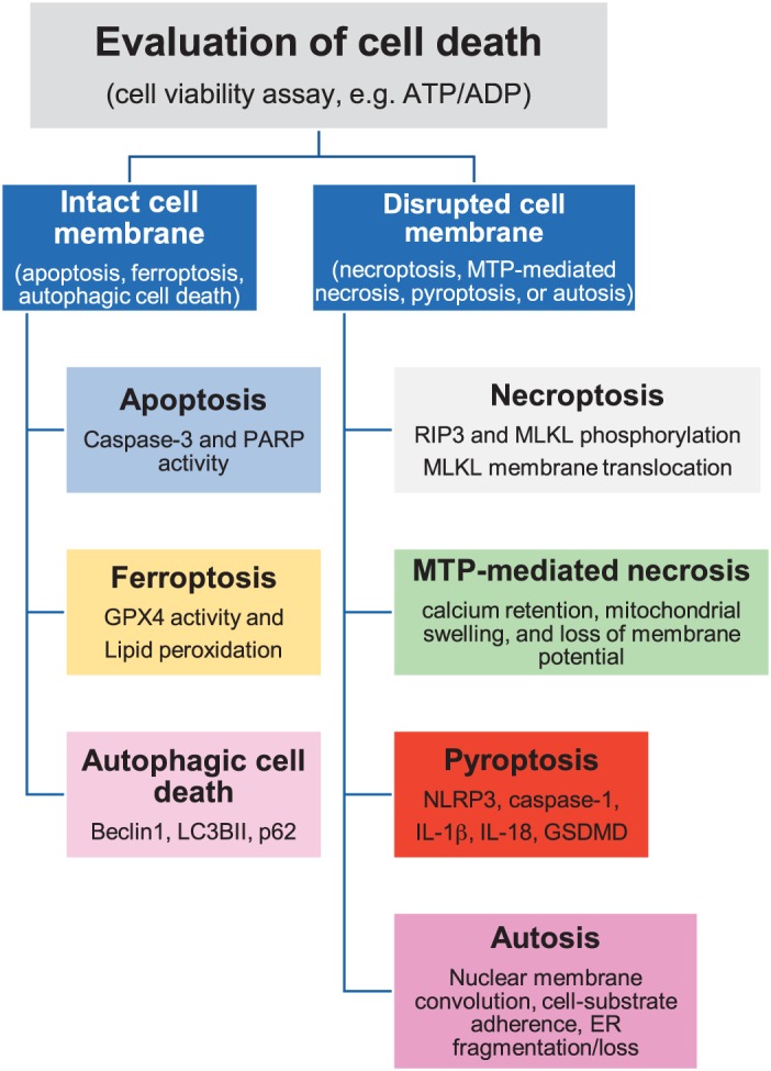
Cell death is a fundamental process in cardiac pathologies. Recent studies have revealed multiple forms of cell death, and several of them have been demonstrated to underlie adverse cardiac remodeling and heart failure. With the expansion in the area of myocardial cell death and increasing concerns over rigor and reproducibility, it is important and timely to set a guideline for the best practices of evaluating myocardial cell death. There are six major forms of regulated cell death observed in cardiac pathologies, namely apoptosis, necroptosis, mitochondrial-mediated necrosis, pyroptosis, ferroptosis, and autophagic cell death. In this article, we describe the best methods to identify, measure, and evaluate these modes of myocardial cell death. In addition, we discuss the limitations of currently practiced myocardial cell death mechanisms.

Keywords: apoptosis; autophagic cell death; cardiovascular disease; ferroptosis; heart; mitochondrial-mediated necrosis; necroptosis; pyroptosis.


Conflict of interest statement

A. Abbate has served as a consultant to Astra-Zeneca, Janssen, Merck, Novartis, Olatec and Serpin pharma.

Figures

Fig. 1.
Fig. 1.
Apoptosis pathway. There are 2 major pathways that lead to apoptosis: extrinsic and intrinsic. The extrinsic pathway is triggered when ligands bind to the death receptors (such as tumor necrosis factor receptor I and Fas) on the surface of the cell. The binding induces conformational changes of the receptors that, with the help of the adaptor protein Fas-associated protein with death domain (FADD), activate pro-caspase-8 to caspase-8. The activated caspase-8 then activates pro-caspases-3,-6, and -7, the activation of which eventually leads to apoptosis. The intrinsic pathway is mitochondria dependent and happens in response to insults such as DNA damage, oxidative stress, and high calcium concentration. Activation of pro-apoptotic proteins (such as BAX) neutralizes the effects of the antiapoptotic proteins of the Bcl-2 family and leads to the release of apoptogenic factor cytochrome c from the mitochondria. Cytochrome c binds to APAF1 (apoptosis protease activating factor-1), and together they bind to and activate pro-caspase-9. The complex of cytochrome c, APAF1, and activated caspase-9 forms apoptosome, which also activates pro-caspases-3, -6, and -7, and leads to apoptosis. The cross-talk between the extrinsic and intrinsic pathways is through BH3-interacting domain (BID). When BID is cleaved by activated caspase-8, it is activated to the truncated form of BID (tBID) and translocates to mitochondria, promoting cytochrome c release. See Refs. and .
Fig. 2.
Fig. 2.
Necroptosis pathway. TNF receptors induce formation of complex I. If receptor-interacting protein kinase (RIP)1 remains ubiquitinated, the cell survives. Complex I forms complex II by deubiquitination of RIP1, and it is mediated by loss of cellular inhibitor of apoptosis 1/2 (cIAP1/2). Caspase 8 activation in complex II inhibits RIP3 and promotes apoptosis, whereas caspase inactivation promotes necroptosis by phosphorylating RIP1, which in turn phosphorylates RIP3. Phosphorylated RIP3 causes mixed-lineage kinase domain-like (MLKL) phosphorylation that triggers MLKL oligomerization and subsequently translocation to the cell membrane, where it makes pore and release damage-associated molecular patterns (DAMPs).
Fig. 3.
Fig. 3.
Mitochondrial permeability transition (MPT)-mediated necrosis. Because of calcium overload and/or extremely high oxidative stress, the mitochondrial membrane becomes permeable and results in dissipation of mitochondrial membrane potential, leading to cessation of ATP synthesis. Increased permeability also causes influx of cytoplasmic water molecules, which causes matrix swelling that ultimately results in mitochondrial membrane rupture and release of apoptotic factors. In the presence of ATP, it leads to apoptosis. However, depletion of ATP causes MPT necrosis.
Fig. 4.
Fig. 4.
The canonical NACHT, LRR, and PYD domains-containing protein 3 (NLRP3) inflammasome pathway of pyroptosis. A number of substances generated during ischemia-reperfusion (termed signal 2) trigger the assembly of the NLRP3 inflammasome, which results in activation of the cysteine-aspartic protease caspase-1. Simultaneous activation of Toll-like receptors (signal 1) increases the expression of pro-interleukins and NLRP3 via NF-κB. Caspase-1 activates pro-interleukins and releases the NH2-terminal domain of gasdermin D, which makes large pores in the plasma membrane. Those pores allow the proinflammatory interleukins to be secreted, and the loss of membrane integrity also kills the cell (pyroptosis). Caspase 1 is also reported to attack many other proteins in the cell, including some vital enzymes like GAPDH, which could also contribute to its toxicity.
Fig. 5.
Fig. 5.
Signaling pathway of ferroptosis in cardiomyocytes. The defining characteristics of ferroptosis are 1) iron dependency 2), disturbances in glutathione (GSH) and glutathione peroxidase 4 (GPX4)-mediated redox balance, and 3) lipid peroxidation. Pointed arrowheads indicate transport, products, or activation. Blunt arrowheads indicate inhibition. Red arrows indicate ferroptotic activity or features that promote ferroptosis. Blue arrows indicate processes that inhibit ferroptosis. GSSG, glutathione disulfate; ROS, reactive oxygen species; RSL3, Ras-selective lethal 3.
Fig. 6.
Fig. 6.
Autophagic cell death pathway and autosis. A: steps of adaptive autophagy/autophagic cell death and proteins involved in different steps. B: factors inducing autosis mediated by autophagy machinery and changes in the cell morphology during early and late phases of autosis. LC3, microtubule-associated proteins 1A/1B light chain 3; RER, rough endoplasmic reticulum.
Fig. 7.
Fig. 7.
Interactions between 6 forms of myocardial cell death. The nature of the signals in the myocardium determines the cell death pathway a cell undergoes. However, oxidative damage to mitochondria can trigger multiple cell death pathways. Different colors denote different forms of cell death. DAMPs, damage-associated molecular patterns; LC3, microtubule-associated proteins 1A/1B light chain 3; MPT, Mitochondrial permeability transition; ROS, reactive oxygen species.
Fig. 8.
Fig. 8.
Evaluation of myocardial cell death. Different steps to distinguish and identify cell death mechanisms in the heart. GPX4, glutathione peroxidase 4; GSDMD, gasdermine D; LC3BII microtubule-associated proteins 1A/1B light chain 3BII; MLKL, mixed-lineage kinase domain-like; MTP, mitochondrial permeability transition pore; NLPR3, NACHT, LRR, and PYD domains-containing protein 3; PARP, poly-ADP ribose polymerase; RIP3, receptor-interacting protein kinase 3.

References

    1. Abbate A, De Falco M, Morales C, Gelpi RJ, Prisco M, De Luca A, Palleiro J, Fedele V, Feroce F, Baldi F, Vetrovec GW, Baldi A. Electron microscopy characterization of cardiomyocyte apoptosis in ischemic heart disease. Int J Cardiol 114: 118–120, 2007. doi:10.1016/j.ijcard.2005.11.025. - DOI - PubMed
    1. Abramov AY, Duchen MR. Measurements of threshold of mitochondrial permeability transition pore opening in intact and permeabilized cells by flash photolysis of caged calcium. Methods Mol Biol 793: 299–309, 2011. doi:10.1007/978-1-61779-328-8_19. - DOI - PubMed
    1. Adameova A, Goncalvesova E, Szobi A, Dhalla NS. Necroptotic cell death in failing heart: relevance and proposed mechanisms. Heart Fail Rev 21: 213–221, 2016. doi:10.1007/s10741-016-9537-8. - DOI - PubMed
    1. Adameova A, Hrdlicka J, Szobi A, Farkasova V, Kopaskova K, Murarikova M, Neckar J, Kolar F, Ravingerova T, Dhalla NS. Evidence of necroptosis in hearts subjected to various forms of ischemic insults. Can J Physiol Pharmacol 95: 1163–1169, 2017. doi:10.1139/cjpp-2016-0609. - DOI - PubMed
    1. Adedoyin O, Boddu R, Traylor A, Lever JM, Bolisetty S, George JF, Agarwal A. Heme oxygenase-1 mitigates ferroptosis in renal proximal tubule cells. Am J Physiol Renal Physiol 314: F702–F714, 2018. doi:10.1152/ajprenal.00044.2017. - DOI - PMC - PubMed

MeSH terms

Substances

LinkOut - more resources