. 2019 Oct;37(7):534-544.
doi: 10.1002/cbf.3430. Epub 2019 Aug 16.

The CTIP-mediated repair of TNF-α-induced DNA double-strand break was impaired by miR-130b in cervical cancer cell

Affiliations

The CTIP-mediated repair of TNF-α-induced DNA double-strand break was impaired by miR-130b in cervical cancer cell

Lei Yang et al. Cell Biochem Funct. 2019 Oct.

Abstract

Chemotherapeutic drugs that induce DNA damage have the potential to kill cancer cells, but DNA repair protects cells from damage-induced cell death. Thus, eliminating DNA repair is a potential approach to overcome cell drug resistance. In this study, we observed that the gene expression of C-terminal binding protein interacting protein (CTIP) was promoted by TNF-α stimulation and prevented TNF-α-induced double-strand breaks (DSBs) in the genomes of cervical cancer cells. The putative miR-130b targeted site within 3' untranslated region (UTR) of CTIP mRNA was identified through in silico analysis and confirmed based on experimental data. By targeting the CTIP gene, miR-130b caused the accumulation of DSBs and accelerated cell apoptosis in combination with poly ADP ribose polymerase (PARP) inhibitors. Additionally, overexpression of the CTIP gene elevated cancer cell viability by promoting proliferation while miR-130b antagonized CTIP-stimulated cell reproduction. Consequently, miR-130b destruction of DNA repair should be employed as a strategy to treat cervical cancer. SIGNIFICANCE OF THE STUDY: Cervical cancer threatens the health of women all over the world. In this study, we observed that miR-130b was able to cause the accumulation of DNA double-strand breaks through suppressing the gene expression of C-terminal binding protein interacting protein and to accelerate cell apoptosis by preventing DNA damage repairs in cervical cancer cells. As far as we know, the impact of miR-130b on the DNA double-strand break repair and on the cell apoptosis induced by the destruction of DNA repair in cervical cancer cells was firstly documented. It is reasonable to believe that miR-130b destruction of DNA repair may be employed as a strategy to treat cervical cancer in the future.

Keywords: C-terminal binding protein interacting protein; apoptosis; double-strand break; miR-130b; proliferation.


Figures

Figure 1
Figure 1
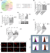
TNF‐α‐stimulated expressions of CTIP gene prevented cancer cells from TNF‐α‐induced DNA double‐strand breaks. A, There were significant augments in the levels of CTIP mRNAs transcribed in both HeLa and Siha cells. B, The expression levels of CTIP proteins increased obviously in both of HeLa and Siha cells. The graphic data obtained in Western blot assays were provided below the statistical analyses of the graphic data. Circles indicated the relative levels of CTIP mRNAs and CTIP proteins in HeLa cells and squares represented the levels in Siha cells at various time points when compared with the corresponding levels in target cells ahead of TNF‐α stimulations. C, The means of DNA Oliver tail moments in target cells treated with TNF‐α were apparently higher than the means in the cells with vehicle control. Circles represented the DNA Oliver tail moments in target cells transfected with pcDNA3.1 vectors, and triangles indicated the tail moments in the cells with pcDNA3.1::CTIP vectors. The solid signs and the hollow ones were respectively utilized to present the Oliver tail moments in HeLa and those in Siha cells. The representative graphic data were offered at the bottom of the statistical analyses of the graphic data. D, The mean levels of phosphorylated H2AX proteins in target cells that were exposed to TNF‐α increased when compared with the mean levels in the cells to vehicle control. The blank columns represented the relative levels of γ‐H2AX proteins in HeLa, and the shadow ones indicated the levels in Siha cells. The cells transfected with pcDNA3.1::CTIP and their control vectors were cultured in fresh media over 24‐hour time periods prior to TNF‐α stimulations. The representative graphic data were put on the right of the statistical analyses of the graphic data. Data were described as means ± SE (n = 3). The bars indicated the deviations from means. * P < .05. ** P < .005. *** P < .001. N.S., non‐significant. CTIP, C‐terminal binding protein interacting protein
Figure 2
Figure 2
The inhibitor roles of miR‐130bs on the repairs of TNF‐α double‐strand breaks were rescued by the overexpressed genes of CTIP. A, A putative miR‐130b targeted site exists in the 3′ UTR of CTIP mRNA. The DNA sequences of wild type or mutated 3′ UTRs that were not shown in the figure were presented as the dotted lines. The base pairs between the sequences of miR‐130b and those of the wild type or the mutated 3′ UTR of CTIP mRNA were showed as vertical lines. The mutated points in the wild type of 3′ UTR of CTIP mRNA were exhibited as the words in red. B, The expression levels of green fluorescence proteins as reporters were downregulated by miR‐130bs based on the base pairing between the seed sequences of miR‐130bs and the regions complementary to the seed sequences within the wild‐type 3′ UTR of CTIP mRNAs. The relative fluorescence intensities of green fluorescence protein were determined at 24‐hour post‐transfection. The blank columns represented the relative fluorescence intensities of reporter proteins in HeLa, and the shadow ones represented the levels in Siha cells. C, miR‐130bs caused the reductions in the contents of CTIP mRNAs in HeLa and Siha cells. The blank columns represented the relative levels of CTIP mRNAs in HeLa, and the shadow ones indicated the levels in Siha cells. D, miR‐130b decreased the mounts of CTIP proteins in HeLa and Siha cells. The graphic data acquired in Western blot assays located at the bottom of the statistical analyses of the graphic data. The blank columns showed the relative levels of CTIP proteins in HeLa, and the shadow ones exhibited the levels in Siha cells. E, The increments in Oliver tail moments caused by miR‐130bs were restored by the enhanced gene expression of CTIP in HeLa and Siha cells. The solid and the hollow circles were individually exploited to indicate the DNA Oliver tail moments in HeLa and that in Siha cells. F, The elevations in the mean levels of γ‐H2AX proteins induced by miR‐130b transfections were reversed by the overexpression of CTIP gene in HeLa and Siha cells. All the transfected target cells were cultured in fresh medium over 24‐hour time periods prior to TNF‐α stimulations. The blank columns represented the relative levels of γ‐H2AX proteins in HeLa, and the shadow ones represented the levels in Siha cells. The representative graphic data were provided below the statistical analyses of the graphic data. Data were described as means ± SE (n = 3). The bars indicated the deviations from means. * P < .05. ** P < .005. *** P < .001. N.S., non‐significant. CTIP, C‐terminal binding protein interacting protein; UTR, untranslated region
Figure 3
Figure 3
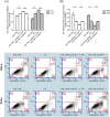
The miR‐130b‐induced cell apoptosis was relieved by the increased expression of CTIP gene in the presence of TNF‐α and PARP inhibitor. A, miR‐130b mimics decreased viabilities of HeLa and Siha cells incubated with TNF‐α agents along with PARP inhibitors, while pcDNA3.1::CTIP vectors elevated the viabilities of miR‐130b transfected target cells. The blank columns represented the relative levels of cell viabilities in HeLa, and the shadow ones indicated the levels in Siha cells. All the transfected cells were cultured in the fresh media over 24‐hour time periods and then incubated in the media containing TNF‐α and AZD2461 for another 24 hours ahead of CCK‐8 assays performed. B, miR‐130b mimics increased apoptosis rates of HeLa and Siha cells stimulated with TNF‐α agents coupled with PARP inhibitors while the enhanced expression of CTIP gene lowed the rates of target cells transfected with miR‐130b mimics. All the transfected cells were cultured in the fresh media over 48‐hour time periods and then incubated in the media containing TNF‐α and AZD2461 for another 24 hours prior to apoptosis assays performed. The graphic data presenting the apoptosis rates of tumour cells were provided below the statistical analyses of the graphic data, and the oligonucleotides used in the transfection assays were showed above the graphic data. The blank columns represented the relative levels of viabilities or apoptosis rates of HeLa cells, and the shadow ones represented the levels or rates of Siha cells. Data were described as means ± SE (n = 3). The bars indicated the deviations from means. * P < .05. ** P < .005. *** P < 0.001. N.S., non‐significant. CTIP, C‐terminal binding protein interacting protein; PARP, poly ADP ribose polymerase
Figure 4
Figure 4
The positive roles of expressed CTIP genes in the cell proliferations were antagonized by miR‐130bs. A, The overexpressions of CTIP genes augmented viabilities of HeLa and Siha cells, while the transfections of miR‐130b mimics downregulated the viabilities of target cells bearing the pcDNA3.1::CTIP vectors. The blank columns represented the relative levels of cell viabilities in HeLa, and the shadow ones indicated the levels in Siha cells. B, The elevations in the gene expression of CTIP promoted cancer cells to enter S phases from G1 phases, while miR‐130b mimics decreased the ratios between the percentages of S and G1 phases in cell cycles. The blank columns indicated the ratios between the percentages of S and G1 phases in cell cycles of HeLa and Siha cells. The representative figures indicating the distributions of cell cycle phases in target cells located under the statistical analyses of the graphic data. Data were described as means ± SE (n = 3). The bars indicated the deviations from means. * P < .05. ** P < .005. *** P < .001. N.S., non‐significant; CTIP, C‐terminal binding protein interacting protein

Similar articles

Cited by

References

    1. Thapa N, Maharjan M, Xiong Y, et al. Impact of cervical cancer on quality of life of women in Hubei, China. Sci Rep. 2018;8(1):11993. - PMC - PubMed
    1. Benard VB, Thomas CC, King J, Massetti GM, Doria‐Rose VP, Saraiya M. Vital signs: cervical cancer incidence, mortality, and screening—United States, 2007‐2012. MMWR. 2014;63:1004‐1009. - PMC - PubMed
    1. Aimagambetova G, Azizan A. Epidemiology of HPV infection and HPV‐related cancers in Kazakhstan: a review. Asian Pac J Cancer Prev. 2018;19(5):1175‐1180. - PMC - PubMed
    1. Hoeijmakers JH. DNA damage, aging and cancer. N Engl J Med. 2009;361(15):1475‐1485. - PubMed
    1. De Bont R, van Larebeke N. Endogenous DNA damage in humans: a review of quantitative data. Mutagenesis. 2004;19(3):169‐185. - PubMed