. 2019 Aug 16;38(1):358.
doi: 10.1186/s13046-019-1370-1.

miR-504 suppresses mesenchymal phenotype of glioblastoma by directly targeting the FZD7-mediated Wnt-β-catenin pathway

Affiliations

miR-504 suppresses mesenchymal phenotype of glioblastoma by directly targeting the FZD7-mediated Wnt-β-catenin pathway

Qiang Liu et al. J Exp Clin Cancer Res. .

Abstract

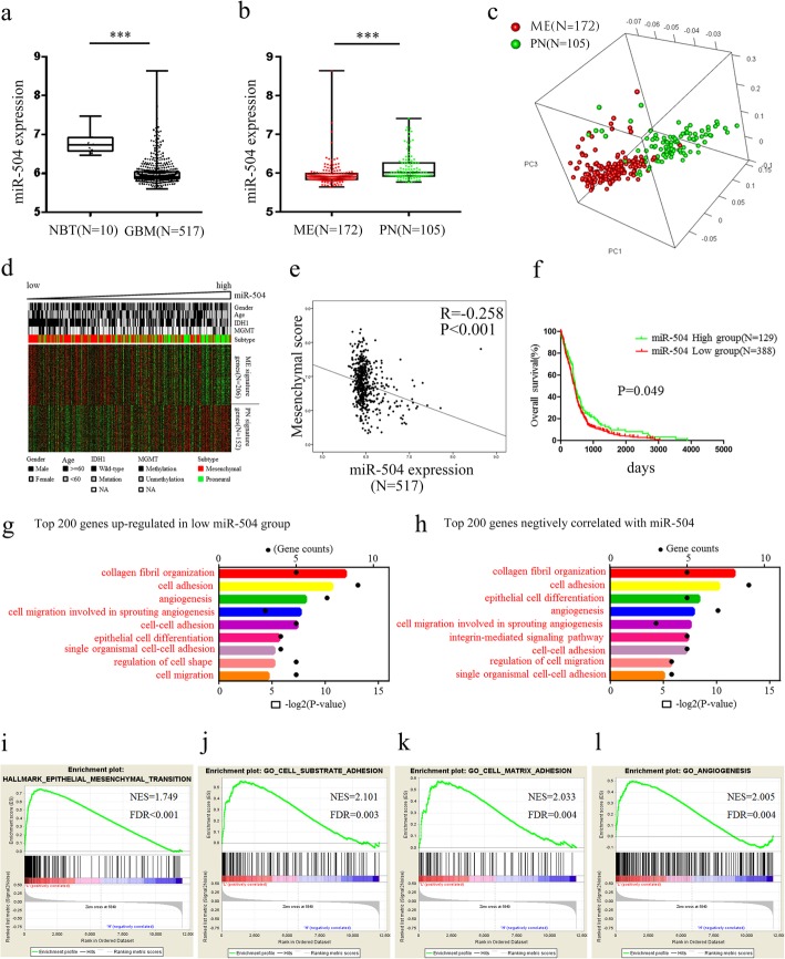
Background: MicroRNAs (miRNAs) play crucial roles in tumor initiation and development. Previously, we indicated that miR-504 is downregulated and suppresses tumor proliferation in glioblastoma (GBM). However, the regulation and relevant mechanism of miR-504 in GBM mesenchymal (ME) transition remain unclear.

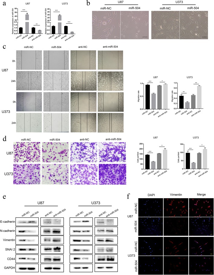
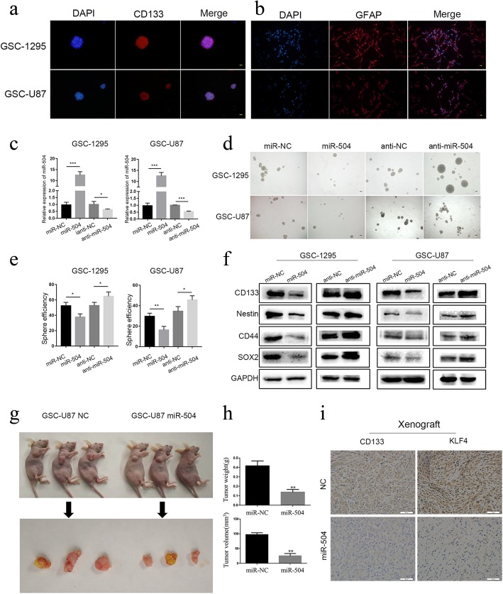
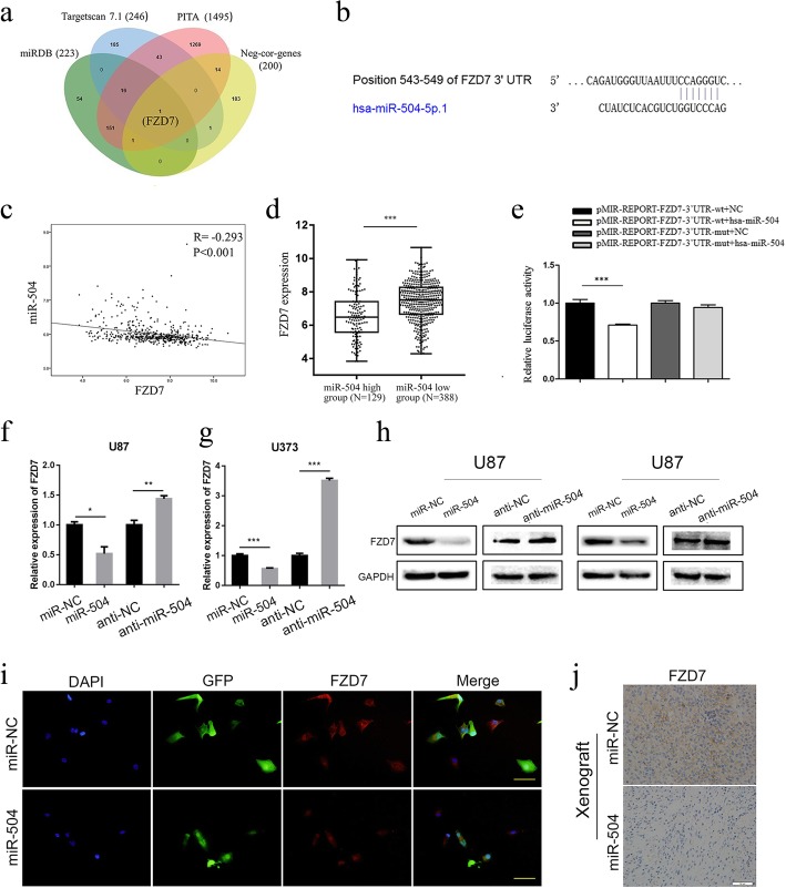
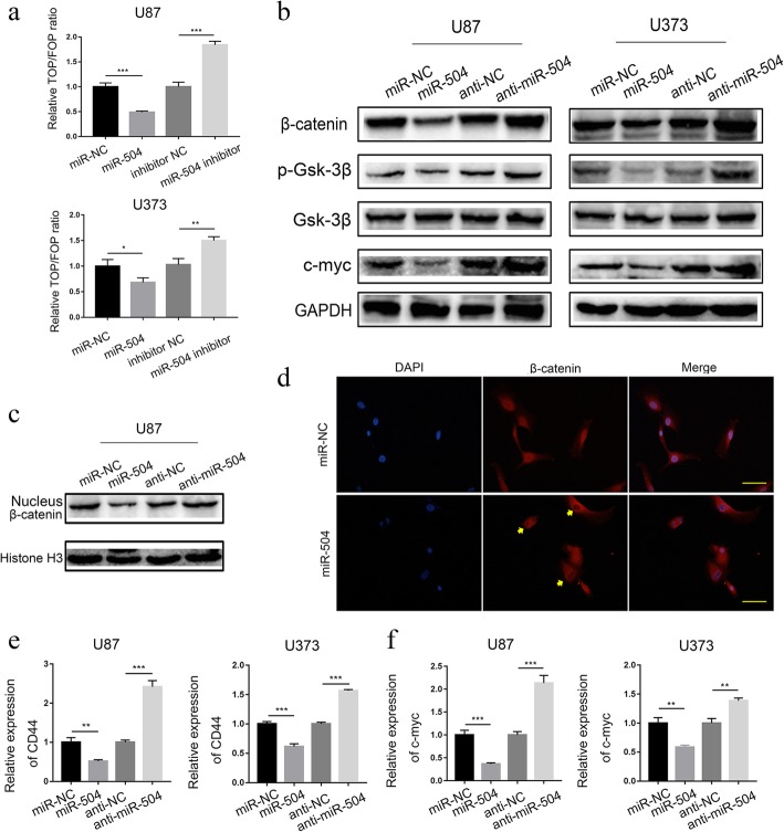
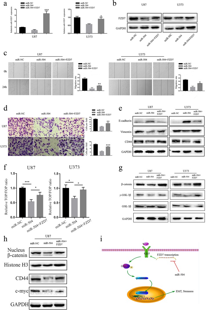
Methods: Transcriptome and clinical data were obtained from The Cancer Genome Atlas (TCGA) database. The potential functions of miR-504 were predicted using gene ontology analysis. GBM cell migration and invasion were examined using wound healing and Transwell assays. Epithelial-mesenchymal transition (EMT) progression in GBM cell lines was detected with immunofluorescence and western blotting. The stemness activity of glioma stem-like cells (GSCs) was assessed by sphere formation assay and tumor xenograft model. miR-504 binding to the FZD7 (frizzled class receptor 7) 3' untranslated region (3'UTR) was validated using dual luciferase reporter assay. TOP/FOP Flash assays were conducted to determine the effects of miR-504 on Wnt/β-catenin signaling.

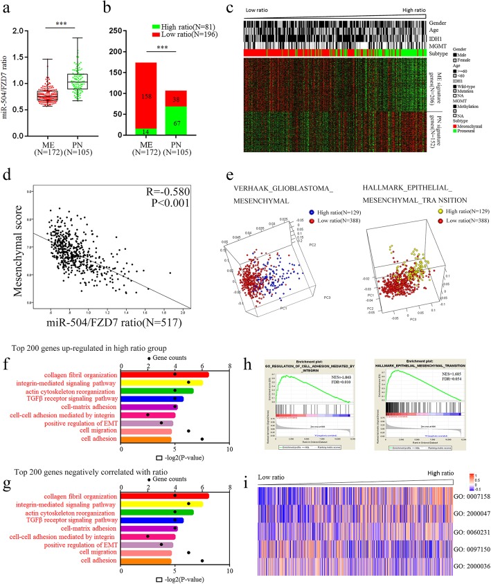
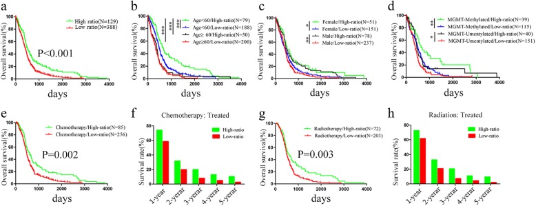
Results: Analysis of TCGA transcriptomic data showed that low miR-504 expression correlated with ME subtype transition and poor survival in patients with GBM. Functional experiments showed that miR-504 overexpression suppressed malignant behaviors of GBM cells, such as migration, invasion, EMT, and stemness activity. Furthermore, miR-504 was a negative regulator of the Wnt-β-catenin pathway by directly repressing FZD7 expression, and FZD7 overexpression reversed the EMT inhibition caused by miR-504. Moreover, the low miR-504/FZD7 expression ratio was a ME subtype marker and could serve as a significant prognostic indicator and predict the clinical outcome of chemotherapy and radiotherapy for patients with GBM in TCGA dataset.

Conclusions: Our results suggest that miR-504 suppresses the aggressive biological processes associated with the ME phenotype of GBM and could be a potential candidate for therapeutic applications in these malignant brain tumors.

Keywords: EMT; FZD7; GBM; Mesenchymal phenotype; Wnt–β-catenin pathway; miR-504.


Conflict of interest statement

The authors declare that they have no competing interests.

Figures

Fig. 1
Fig. 1
Downregulation of miR-504 in ME subtype GBM. a Analysis of TCGA data of miR-504 expression levels in NBT (n = 10) and GBM (n = 517). b Analysis of TCGA data of miR-504 expression levels in ME subtype GBM (n = 172) and PN subtype GBM (n = 105). c PCA based on the miR-504–related genes shows a distinct distribution pattern between patients with the ME and the PN subtype. d The expression pattern of ME and PN signature genes corresponding to miR-504 expression. e Pearson correlation analysis between miR-504 and ME score in TCGA. f Kaplan–Meier analysis of overall survival of patients with GBM from TCGA database according to miR-504 expression level. g GO analysis of the top 200 differentially upregulated genes in miR-504 low-expression tumors. h GO analysis of top 200 genes negatively related to miR-504. il GSEA highlighting positive association of decreased miR-504 expression levels with EMT, cell adhesion, and angiogenesis. ***P < 0.001
Fig. 2
Fig. 2
miR-504 suppresses the ME properties and EMT of GBM cells. a qRT-PCR detection of miR-504 expression in U87 and U373 cells transfected with miR-504 overexpression and inhibition vectors. a Representative images of GBM cell morphological changes after miR-504 transfection (scale bars = 100 μm). (c, d) Representative results of wound healing assays (c) and Transwell assays (d) showing the effect of miR-504 overexpression and inhibition on the migration and invasive abilities of GBM cells (scale bars = 100 μm). e Western blot analysis of the levels of representative epithelial and ME markers. f Immunofluorescence examination of ME marker expression in U87 and U373 cells transfected with miR-504 or miR-NC (red, vimentin; blue, DAPI; scale bars = 100 μm). Data are the mean ± SD of three independent experiments. *P < 0.05,**P < 0.01, ***P < 0.001
Fig. 3
Fig. 3
miR-504 attenuates GSC stemness activity. a Immunofluorescence showing tumor spheres comprised of CD133-positive GSCs (red, CD133; blue, DAPI; scale bars = 100 μm). b GSCs differentiated into GFAP-positive cells after serum induction (red, GFAP; blue, DAPI; scale bars = 100 μm). c qRT-PCR detection of miR-504 expression in GSC-U87 and GSC-1295 cells transfected with miR-504 overexpression and inhibition vectors. d Representative images of tumor spheres formed after 14-days culture (scale bars = 500 μm). e Western blot analysis of the levels of the representative stem cell markers. fh miR-504 reduced GSC-U87 cell tumorigenicity in vivo. i Immunohistochemistry for CD133 and KLF4 in xenograft sections (scale bar = 50 μm). Data are the mean ± SD of three independent experiments. *P < 0.05, **P < 0.01, ***P < 0.001
Fig. 4
Fig. 4
miR-504 targets the FZD7 3′UTR directly. a Venn diagram identifying the overlap between predicted genes and miR-504–negatively related genes. b The predicted target site of FZD7 3′UTR binding to miR-504. c Pearson correlation analysis of miR-504 and FZD7 in TCGA. d TCGA data analysis of FZD7 expression levels between miR-504 high-expression tumors (n = 129) and miR-504 low-expression tumors (n = 388). e Measurement of firefly/Renilla luciferase activity in HEK-293 T cells co-transfected with wild-type-FZD7–3′-UTR or mutant-type-FZD7–3′-UTR reporters and NC mimic or miR-504 mimic. f, g qRT-PCR determination of FZD7 mRNA levels in U87 and U373 cells after transfection with miR-504, miR-NC, anti-NC, or anti-miR-504. h Western blot analysis of FZD7 protein levels in U87 and U373 cells after transfection with miR-504, miR-NC, anti-NC or anti-miR-504 . i Immunofluorescence examination of FZD7 expression in U87 cells transfected with miR-504 or miR-NC (red, FZD7; green, GFP; blue, DAPI; scale bars = 100 μm). j Immunohistochemistry of FZD7 in xenograft sections (scale bar = 50 μm). Data are the mean ± SD of three independent experiments. *P < 0.05, **P < 0.01, ***P < 0.001
Fig. 5
Fig. 5
miR-504 regulates Wnt–β-catenin signaling activation. a TOP/FOPFlash reporter activity assay detection of relative Wnt signaling activity in U87 and U373 cells. b Western blot assessment of the effects of miR-504 on β-catenin, p-GSK3β, GSK3β, and c-MYC protein levels in U87 and U373 cells. c, d Western blot (c) and immunofluorescence (d) assay detection of nuclear accumulation of β-catenin in U87 cells. e, f qRT-PCR analysis of the relative mRNA expression of downstream target genes of the Wnt–β-catenin pathway, i.e., CD44 (e) and c-MYC (f), in U87 and U373 cells. Data are the mean ± SD of three independent experiments. *P < 0.05, **P < 0.01, ***P < 0.001
Fig. 6
Fig. 6
miR-504 inhibits GBM cell ME transition by suppressing the FZD7-mediated Wnt–β-catenin pathway. a, b qRT-PCR (a) and western blot (b) of FZD7 expression in miR-504–transfected U87 and U373 cells after FZD7 transfection. c, d Wound healing assay (c) and Transwell assay (d) evaluation of the restorative effect of FZD7 on miR-504–induced inhibition of migration and invasion in U87 and U373 cells (scale bar = 100 μm). e Western blot detection of EMT markers in U87 and U373 cells co-transfected with miR-504 and FZD7. f TOP/FOPFlash reporter activity detection of relative Wnt signaling activity in U87 and U373 cells. g Western blot detection of β-catenin, p-GSK3β, and GSK3β expression levels in miR-504 and FZD7 co-transfected cells. h Western blot assessment of the effects of FZD7 on nuclear β-catenin, c-MYC, and CD44 protein levels in U87 cells. i The diagram illustrated that miR-504 decreased the EMT and stemness via inhibiting the FZD7 mediated Wnt/β-catenin signaling pathway activity. Data are the mean ± SD of three independent experiments. *P < 0.05, **P < 0.01, ***P < 0.001
Fig. 7
Fig. 7
Identification of the miR-504/FZD7 ratio as a ME subtype marker in GBM. a Distribution of the miR-504/FZD7 ratio between ME subtype GBM (n = 172) and PN subtype GBM (n = 105) in TCGA. b Chi-square analysis of the relationship between the miR-504/FZD7 ratio and molecular subtype. c The expression patterns of ME and PN signature genes corresponding to the miR-504/FZD7 ratio. d Pearson correlation analysis of the miR-504/FZD7 ratio and ME score in TCGA. e PCA based on ME signature genes and EMT signature genes showed distinct distribution patterns between GBM of high- and low-ratio groups. f GO analysis of top 200 genes negatively related to the miR-504/FZD7 ratio. g GO analysis of top 200 differentially upregulated genes in low–miR-504/FZD7 ratio tumors in TCGA. h GSEA comparison of ME phenotype between high- and low–miR-504/FZD7 ratio cases. i Clustering of GSVA scores and analyses of biological processes associated with the miR-504/FZD7 ratio in GBM according to TCGA dataset (GO:0007158, neuron cell–cell adhesion; GO:2000047, regulation of cell–cell adhesion mediated by cadherin; GO:0060231, mesenchymal to epithelial transition; GO:0097150, neuronal stem cell population maintenance; GO:2000036, regulation of stem cell population maintenance). ***P < 0.001
Fig. 8
Fig. 8
The miR-504/FZD7 ratio is a prognostic indicator in GBM. a Kaplan–Meier assessment of the association between the miR-504/FZD7 ratio and overall survival of patients with GBM in TCGA. bd Prognostic significance of the miR-504/FZD7 ratio in cohorts stratified by age (b), sex (c), and MGMT promoter status (d). e Kaplan–Meier analysis of survival of chemotherapy-treated patients with GBM based on the miR-504/FZD7 ratio. f Comparison of 1–5-year survival rates between high and low miR-504/FZD7 ratio groups in chemotherapy-treated patients with GBM. g Kaplan–Meier analysis of overall survival according to the miR-504/FZD7 ratio of standard radiotherapy–treated patients with GBM from TCGA. h Comparison of 1–5-year survival rates between high and low miR-504/FZD7 ratio groups in standard radiotherapy–treated patients with GBM. *P < 0.05, **P < 0.01, ***P < 0.001

Similar articles

Cited by

References

    1. Siegel R, Miller K, Jemal A. Cancer statistics, 2018. CA Cancer J Clin. 2018;68(1):7–30. doi: 10.3322/caac.21442. - DOI - PubMed
    1. Brandsma D, Stalpers L, Taal W, Sminia P, van den Bent M. Clinical features, mechanisms, and management of pseudoprogression in malignant gliomas. Lancet Oncol. 2008;9(5):453–461. doi: 10.1016/S1470-2045(08)70125-6. - DOI - PubMed
    1. Wang Y, Jiang T. Understanding high grade glioma: molecular mechanism, therapy and comprehensive management. Cancer Lett. 2013;331(2):139–146. doi: 10.1016/j.canlet.2012.12.024. - DOI - PubMed
    1. Phillips H, Kharbanda S, Chen R, Forrest W, Soriano R, Wu T, et al. Molecular subclasses of high-grade glioma predict prognosis, delineate a pattern of disease progression, and resemble stages in neurogenesis. Cancer Cell. 2006;9(3):157–173. doi: 10.1016/j.ccr.2006.02.019. - DOI - PubMed
    1. Verhaak R, Hoadley K, Purdom E, Wang V, Qi Y, Wilkerson M, et al. Integrated genomic analysis identifies clinically relevant subtypes of glioblastoma characterized by abnormalities in PDGFRA, IDH1, EGFR, and NF1. Cancer Cell. 2010;17(1):98–110. doi: 10.1016/j.ccr.2009.12.020. - DOI - PMC - PubMed

MeSH terms