. 2019 Sep 3;116(36):17951-17956.
doi: 10.1073/pnas.1905943116. Epub 2019 Aug 16.

CD8αα homodimers function as a coreceptor for KIR3DL1

Affiliations

CD8αα homodimers function as a coreceptor for KIR3DL1

Jie Geng et al. Proc Natl Acad Sci U S A. .

Abstract

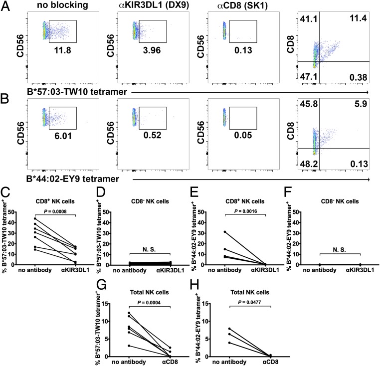
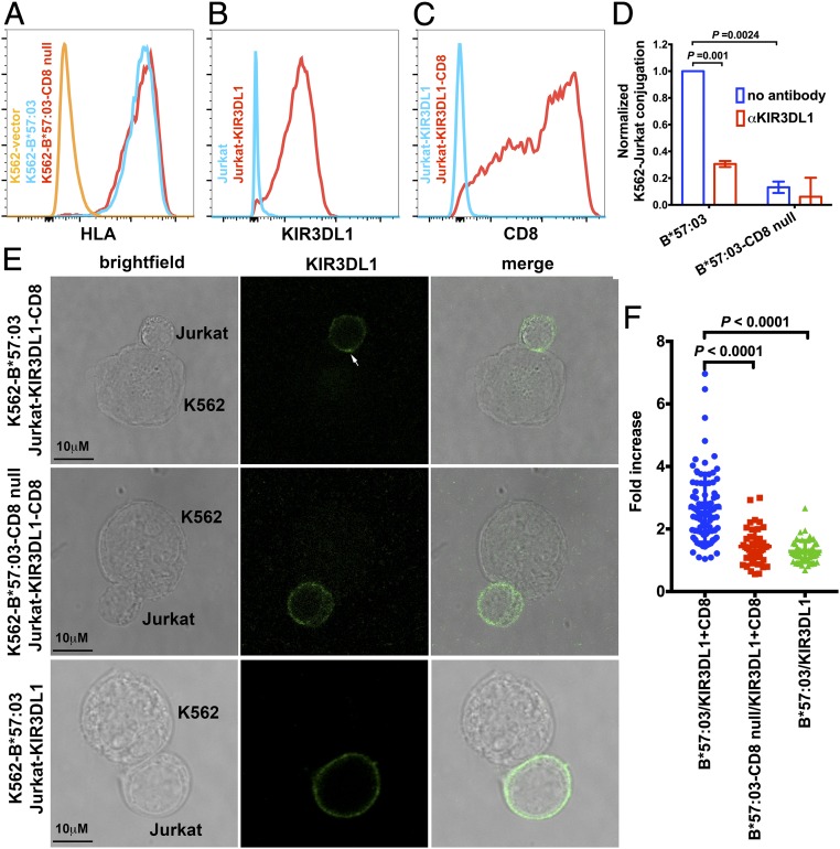
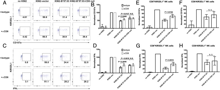
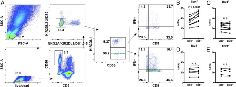
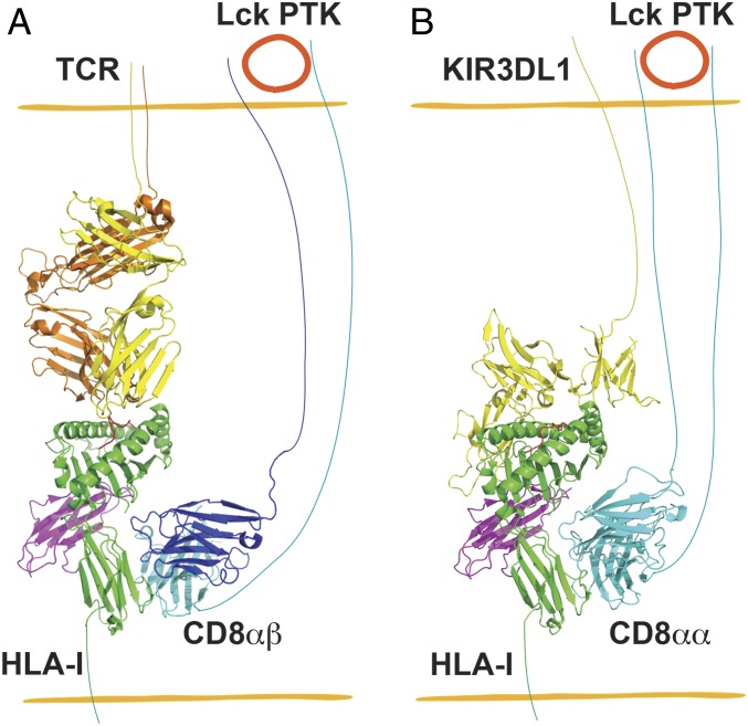
Cluster of differentiation 8 (CD8) is a cell surface glycoprotein, which is expressed as 2 forms, αα homodimer or αβ heterodimer. Peptide-loaded major histocompatibility complex class I (pMHC-I) molecules are major ligands for both forms of CD8. CD8αβ is a coreceptor for the T cell receptor (TCR) and binds to the same cognate pMHC-I as the TCR, thus enabling or augmenting T cell responses. The function of CD8αα homodimers is largely unknown. While CD8αβ heterodimer is expressed exclusively on CD8+ T cells, the CD8αα homodimer is present in subsets of T cells and human natural killer (NK) cells. Here, we report that the CD8αα homodimer functions as a coreceptor for KIR3DL1, an inhibitory receptor of NK cells that is specific for certain MHC-I allotypes. CD8αα enhances binding of pMHC-I to KIR3DL1, increases KIR3DL1 clustering at the immunological synapse, and augments KIR3DL1-mediated inhibition of NK cell activation. Additionally, interactions between pMHC-I and CD8αα homodimers regulate KIR3DL1+ NK cell education. Together, these findings reveal another dimension to the modulation of NK cell activity.

Keywords: CD8αα; KIR3DL1; NK cell activation; coreceptor.


Conflict of interest statement

The authors declare no conflict of interest.

Figures

Fig. 1.
Fig. 1.
CD8 augments specific HLA-B staining to KIR3DL1. NK cells (CD3CD56+ PBMCs) were stained with B*57:03-TW10 (A) or B*44:02-EY9 (B) tetramers (column I). Tetramer staining was blocked by anti-KIR3DL1 (column II) or anti-CD8 (column III) antibodies. There is a correlation of tetramer staining with CD8 expression level on NK cells (column IV). Representative staining data from one of 7 donors for B*57:03 and 4 donors for B*44:02 are shown in A and B. Tetramer staining data with additional donors are shown in SI Appendix, Fig. S3. (CH) Compiled tetramer staining data are shown. N.S., not significant.
Fig. 2.
Fig. 2.
CD8 facilitates cell conjugation induced by KIR3DL1–Bw4 interactions and KIR3DL1 clustering induced by HLA-Bw4. (A) Surface expression levels of B*57:03 or B*57:03-CD8null on the surface of K562 cells were assessed with flow cytometry after staining with the HLA-I–specific antibody W6/32. KIR3DL1 (B) and CD8 (C) expression on the surface of Jurkat cells was confirmed by flow cytometry. (D) Cell conjugation between Jurkat-KIR3DL1-CD8 cells and K562 cells expressing no HLA-I, B*57:03, or B*57:03-CD8null was tested by flow cytometry. K562 cells were labeled with CFSE. Cell conjugation was either blocked with anti-KIR3DL1 or medium. CFSE and CD8 double-positive cells are quantified as conjugates. Cell conjugation was normalized to that between Jurkat-KIR3DL1-CD8 cells and K562-B*57:03 cells after background correction (based on conjugates between Jurkat-KIR3DL1-CD8 cells and K562 cells). Data before normalization are shown in SI Appendix, Table S1. Conjugation between Jurkat-KIR3DL1-CD8 cells and K562-B*57:03 cells is partially blocked by anti-KIR3DL1. K562-B*57:03 cells are more efficient than K562-B*57:03-CD8null cells in forming conjugates with Jurkat-KIR3DL1-CD8 cells (n = 3 replicates). (E and F) Jurkat-KIR3DL1-CD8 cells were incubated with K562 cells expressing B*57:03 or B*57:03-CD8null and then fixed and stained with anti-KIR3DL1 before analysis by confocal microscopy. As another control, Jurkat-KIR3DL1 cells incubated with K562-B*57:03 were also assessed. The arrowhead in E indicates clustering at the interface between the Jurkat and K562 cells. The intensity of staining of the Jurkat cells at cell–cell interfaces was compared with that measured at a noncontact area. (F) Data are plotted as the fold increase in intensity above the noncontact area background. The results from 5 independent experiments for K562-B*57:03/Jurkat-KIR3DL1-CD8 and 2 independent experiments for K562-B*57:03-CD8null/Jurkat-KIR3DL1-CD8 and K562-B*57:03/Jurkat-KIR3DL1 with a total of >40 conjugates per condition are shown. Data are shown as mean ± SD. Statistical analyses were performed with paired Student’s t tests using GraphPad Prism version 7.
Fig. 3.
Fig. 3.
CD8 enhances the inhibitory ability of HLA-Bw4 toward KIR3DL1+ NK cell activation. NK cells (CD3CD56+ PBMCs) were activated by incubation with K562 cells or K562 cells expressing exogenous B*57:03 or B*57:03-CD8null. KIR3DL1+ NK cell activation was determined by testing CD107a (A and B) and IFN-γ (C and D) induction. A and C are representative data, while B and D are compiled data (n = 4). Cell activation was normalized to the NK cell + K562-vec condition after background correction (based on untreated NK cells). vec, empty vector. Data before normalization are shown in SI Appendix, Tables S2 and S3. Anti-CD8 antibody can partially rescue B*57:03-mediated KIR3DL1+ NK cell inhibition. B*57:03-CD8null showed reduced inhibition of KIR3DL1+ NK cell activation, and anti-CD8 antibody has no significant effect on B*57:03-CD8null-induced inhibition of KIR3DL1+ NK cell activation. (EH) B*57:03 showed stronger inhibition of NK cell activation than B*57:03-CD8null only in CD8+, but not CD8, KIR3DL1+ NK cells. Statistical analyses were performed with paired Student’s t tests. N.S., not significant.
Fig. 4.
Fig. 4.
CD8 modulates NK cell education by facilitating HLA-Bw4 binding to KIR3DL1. (A) Gating strategy for selective detection of KIR3DL1+ and KIR3DL1 NK cells. Live lymphocytes were first gated, from which CD3CD56+ cells were selected. For selective gating, CD3CD56+ NK cells negative for KIR2DL1/L2/L3/S1/S2/S3/S4/S5 and NKG2A were selected. These cells were further gated to identify KIR3DL1+ and KIR3DL1 cells. Selectively gated KIR3DL1+ and KIR3DL1 NK cells were then examined for IFN-γ expression. Functional gates were set on KIR3DL1+ or KIR3DL1 NK cells in unstimulated PBMCs. The NK cell activation rate was characterized by the fraction of activated cells among all cells within the CD8+ or CD8 gate. (B and C) In HLA-Bw4+ donors, KIR3DL1+ (i.e., KIR3DL1+others) NK cells (B), but not KIR3DL1 (i.e., KIRNKG2A) NK cells (C), showed CD8-dependent activation by K562 cells (n = 8 donors). (D and E) In HLA-Bw4 donors, neither KIR3DL1+ (D) nor KIR3DL1 (E) NK cells showed CD8 dependency (n = 5 donors). Statistical analyses were performed with paired Student’s t tests. N.S., not significant.
Fig. 5.
Fig. 5.
Schematic description of the parallels of CD8 coreceptor function in T cells and NK cells. (A) CD8αβ heterodimers are coreceptors for CD8+ T cell binding to target cells displaying the appropriate pHLA-I. The protein tyrosine kinase (PTK) Lck associates with the CD8αβ cytoplasmic tail, triggering enhanced T cell signaling. (B) Similar to A, CD8αα interacts with the nonpolymorphic region of HLA-I and enhances HLA-I engagement to KIR3DL1 of NK cells and the inhibitory function of KIR3DL1. In A, the TCR–HLA-A*02:01 complex structure (Protein Data Bank [PDB] ID code 5c0a) (47) was superimposed onto H2-Dd-CD8αβ (PDB ID code 3dmm) (48) by aligning Cαs of HLA-A*02:01 and H2-Dd, followed by deletion of H2-Dd. Similarly, in B, the KIR3DL1–HLA-B*57:01 complex structure (PDB ID code 3vh8) (15) was superimposed onto HLA-A*02:01-CD8αα complex (PDB ID code 1akj) (30) by aligning Cαs of HLA-B*57:01 and HLA-A*02:01, and HLA-A*02:01 was then deleted. The CD8αα and KIR3DL1 binding sites on HLA-I are predicted to be nonoverlapping and synergistic for enhancing KIR function.

References

    1. Cole D. K., Gao G. F., CD8: Adhesion molecule, co-receptor and immuno-modulator. Cell. Mol. Immunol. 1, 81–88 (2004). - PubMed
    1. Purbhoo M. A., et al. , The human CD8 coreceptor effects cytotoxic T cell activation and antigen sensitivity primarily by mediating complete phosphorylation of the T cell receptor ζ chain. J. Biol. Chem. 276, 32786–32792 (2001). - PubMed
    1. Garcia K. C., et al. , CD8 enhances formation of stable T-cell receptor/MHC class I molecule complexes. Nature 384, 577–581 (1996). - PubMed
    1. Huang J., Edwards L. J., Evavold B. D., Zhu C., Kinetics of MHC-CD8 interaction at the T cell membrane. J. Immunol. 179, 7653–7662 (2007). - PubMed
    1. Hayday A., Theodoridis E., Ramsburg E., Shires J., Intraepithelial lymphocytes: Exploring the third way in immunology. Nat. Immunol. 2, 997–1003 (2001). - PubMed

Publication types