. 2019 Dec 17;21(12):1565-1577.
doi: 10.1093/neuonc/noz139.

Integrated clinical, histopathological, and molecular data analysis of 190 central nervous system germ cell tumors from the iGCT Consortium

Affiliations

Integrated clinical, histopathological, and molecular data analysis of 190 central nervous system germ cell tumors from the iGCT Consortium

Hirokazu Takami et al. Neuro Oncol. .

Abstract

Background: We integrated clinical, histopathological, and molecular data of central nervous system germ cell tumors to provide insights into their management.

Methods: Data from the Intracranial Germ Cell Tumor Genome Analysis (iGCT) Consortium were reviewed. A total of 190 cases were classified as primary germ cell tumors (GCTs) based on central pathological reviews.

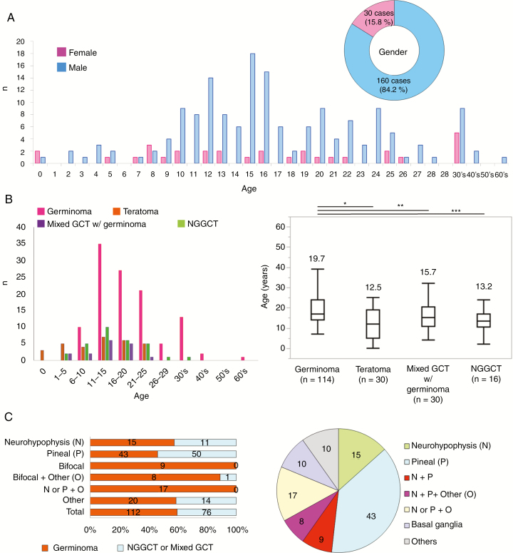
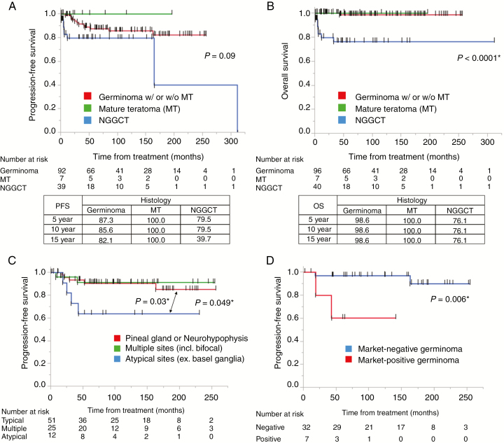
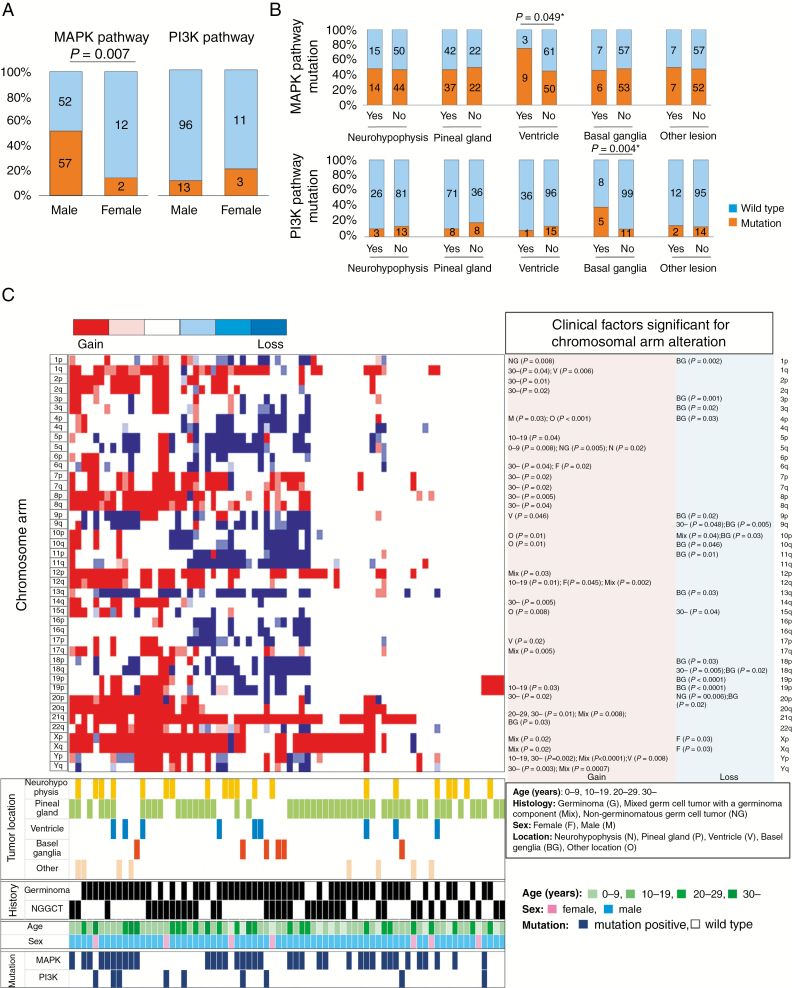
Results: All but one of the cases that were bifocal (neurohypophysis and pineal glands) and cases with multiple lesions including neurohypophysis or pineal gland were germinomas (34 of 35). Age was significantly higher in patients with germinoma than other histologies. Comparison between tumor marker and histopathological diagnoses showed that 18.2% of histopathologically diagnosed germinomas were marker positive and 6.1% of non-germinomatous GCTs were marker negative, suggesting a limitation in the utility of markers or histopathology alone using small specimens for diagnosis. Comparison between local and central histopathological diagnoses revealed a discordance of 12.7%. Discordance was significantly less frequent in biopsy cases, implying difficulty in detecting all histopathological components of heterogeneous GCTs. Germinomas at the typical sites (neurohypophysis or pineal gland) showed a better progression-free survival than those at atypical sites (P = 0.03). A molecular clinical association study revealed frequent mitogen-activated protein kinase (MAPK) pathway mutations in males (51.4% vs 14.3%, P = 0.007), and phosphatidylinositol-3 kinase/mammalian target of rapamycin (PI3K/mTOR) pathway mutations in basal ganglia cases (P = 0.004). Basal ganglia cases also had frequent chromosomal losses. Some chromosomal aberrations (2q, 8q gain, 5q, 9p/q, 13q, 15q loss) showed potential prognostic significance.

Conclusions: The in-depth findings of this study regarding clinical and molecular heterogeneity will increase our understanding of the pathogenesis of this enigmatic tumor.

Keywords: germ cell tumor; pathological diagnosis; prognosis, tumor marker; rare disease.


Figures

Fig. 1
Fig. 1
(A) Age distribution of GCT patients is shown in a bar graph (blue, male; pink, female). Mean and median ages were 17.5 and 16 years. The pie chart shows the male to female ratio (approximately 5:1). (B) Left: The distribution of histological subtypes according to age. Of note, patients <6 years (n = 12) had mostly teratomas and yolk sac tumors (n = 10, 83.3%). Patients ≥30 years (n = 17) mostly had germinomas (n = 16, 94.1%). Right: Germinomas (19.7 ± 9.0) occurred in significantly older patients than did teratomas (12.5 ± 7.7, *P < 0.0001), mixed germinomas (15.7 ± 6.7, **P = 0.03), or non-germinomatous GCTs (13.2 ± 5.8, ***P = 0.004). (C) Left: The distribution of histology according to tumor locations. The majority of bifocal or other multifocal tumors (34/35 cases, 97.1%) were germinomas. About half of cases with solitary lesions were germinoma. Half were non-germinoma (NGGCT) or mixed GCTs with a germinoma component (mixed GCTs). Right: The pie chart shows the distribution of tumor locations in germinoma cases. Bifocal germinomas constitute 15.0% of total germinomas.
Fig. 2
Fig. 2
(A, B) Kaplan–Meier curves showing PFS and OS of GCT patients. Tables in the lower panel show 5-, 10-, and 15-year PFS and OS values. PFS of germinomas and NGGCTs were not statistically different, although all 7 cases of mature teratoma did not show recurrence. OS of these 3 histological classifications were markedly statistically significant. (C) Kaplan–Meier curves of PFS for germinomas at different locations show that those at a typical single site (neurohypophysis or pineal gland) (n = 51) had significantly longer PFS than those at atypical sites (n = 12) (P = 0.03). (D) Kaplan–Meier curves of PFS for marker-positive germinoma and marker-negative germinoma. Marker-positive germinoma cases showed significantly worse PFS than marker-negative germinoma cases (P = 0.006).
Fig. 3
Fig. 3
(A) Percent presence of MAPK pathway and PI3K/mTOR pathway mutations is shown partitioned by sex. MAPK pathway mutations were significantly more frequent in male cases. There was no statistical difference in frequency of PI3K/mTOR pathway mutations (female: 21.4%, vs male: 11.9%, P = 0.32). (B) MAPK pathway mutations (top row) and PI3K/mTOR pathway mutations (bottom row) were analyzed for association with tumor locations. MAPK pathway mutations were more common in cases with ventricular lesions, whereas PI3K/mTOR pathway mutations were more common in cases with basal ganglia lesions. (C) Distribution of chromosome aberrations in 74 cases was described in a heatmap, with clinical and molecular (mutation) information at the bottom. Two major clusters were created based on chromosomal instability data, one with abundant alterations (left) and one with scant alterations (right). Of note, cases with basal ganglia lesions, cases in older patients, and cases with PI3K/mTOR mutations obviously preferentially aggregated in the left cluster with abundant chromosomal aberrations. Detailed multivariate analyses of clinical factors significantly associated with each chromosomal arm aberration are listed to the right of the heatmap.

References

    1. Shibui S. Report of brain tumor registry of Japan (2001–2004). Neurol Med Chir. 2014;54(suppl. 1) :1–102.
    1. Louis D, Ohgaki H, Wiestler O, Cavenee W.. WHO Classification of Tumours of the Central Nervous System. Revised 4th ed. Lyon, France: International Agency for Research on Cancer; 2016.
    1. Calaminus G, Frappaz D, Kortmann RD, et al. . Outcome of patients with intracranial non-germinomatous germ cell tumors-lessons from the SIOP-CNS-GCT-96 trial. Neuro Oncol. 2017;19(12):1661–1672. - PMC - PubMed
    1. Al-Hussaini M, Sultan I, Abuirmileh N, Jaradat I, Qaddoumi I. Pineal gland tumors: experience from the SEER database. J Neurooncol. 2009;94(3):351–358. - PMC - PubMed
    1. Goldman S, Bouffet E, Fisher PG, et al. . Phase II trial assessing the ability of neoadjuvant chemotherapy with or without second-look surgery to eliminate measurable disease for nongerminomatous germ cell tumors: a Children’s Oncology Group Study. J Clin Oncol. 2015;33(22):2464–2471. - PMC - PubMed

Publication types

MeSH terms

Substances