. 2019 Dec;12(2-3):77-94.
doi: 10.1007/s12307-019-00229-x. Epub 2019 Aug 16.

Kinins in Glioblastoma Microenvironment

Affiliations

Kinins in Glioblastoma Microenvironment

Mona N Oliveira et al. Cancer Microenviron. 2019 Dec.

Abstract

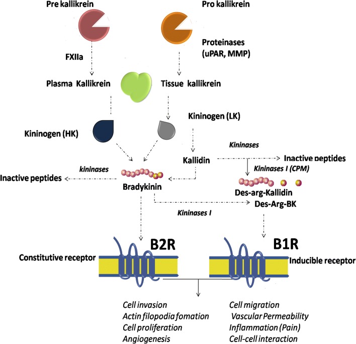
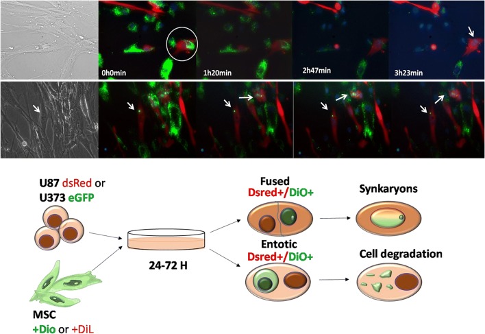
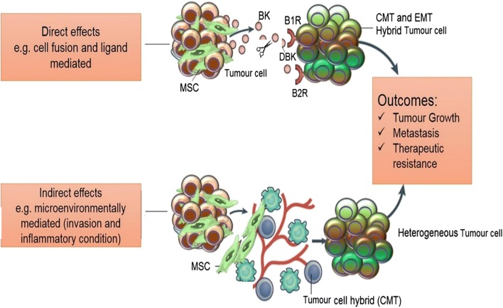
Tumour progression involves interactions among various cancer cell clones, including the cancer stem cell subpopulation and exogenous cellular components, termed cancer stromal cells. The latter include a plethora of tumour infiltrating immunocompetent cells, among which are also immuno-modulatory mesenchymal stem cells, which by vigorous migration to growing tumours and susequent transdifferentiation into various types of tumour-residing stromal cells, may either inhibit or support tumour progression. In the light of the scarce therapeutic options existing for the most malignant brain tumour glioblastoma, mesenchymal stem cells may represent a promising novel tool for cell therapy, e.g. drug delivery vectors. Here, we review the increasing number of reports on mutual interactions between mesenchymal stem cells and glioblastoma cells in their microenvironment. We particularly point out two novel aspects: the different responses of cancer cells to their microenvironmental cues, and to the signalling by kinin receptors that complement the immuno-modulating cytokine-signalling networks. Inflammatory glioblastoma microenvironment is characterised by increasing expression of kinin receptors during progressive glioma malignancy, thus making kinin signalling and kinins themselves rather important in this context. In general, their role in tumour microenvironment has not been explored so far. In addition, kinins also regulate blood brain barrier-related drug transfer as well as brain tumour angiogenesis. These studies support the on-going research on kinin antagonists as candidates in the development of anti-invasive agents for adjuvant glioblastoma therapy.

Keywords: Co-culture; Glioma; Kinin receptors; Mesenchymal stem cells; Microenvironment; Tumour heterogeneity.


Conflict of interest statement

The authors declare that they have no conflict of interest.

Figures

Fig. 1
Fig. 1
Zebrafish embryo as an animal model to study MSC-GBM cell interactions. The interactions between MSCs and U373 GBM cell line increase the invasion of U373 cells in zebrafish embryo brain (a) as described by Breznik et al. [26]. Image taken at higher magnification shows MSC-GBM mixed tumours with invasive U373 cells. MSCs follow U373 cells into the zebrafish brain parenchyma (b). Scale bars: 250 μm (A); 50 μm (B). Image courtesy of Miloš Vittori
Fig. 2
Fig. 2
Schematic diagram of the kallikrein-kinin system (KKS) metabolic cascade. Plasma- and kallikrein-related peptidases are secreted as zymogens and become activated by a cascade of pro-peptide proteolytic cleavages. Kallikreins cleave serum and tissue high molecular-weight (HK) and low molecular-weight (LK) kininogens to generate bradykinin (BK) and kallidin (Lys-BK) which, in turn, are cleaved either by kininase (originating inactive peptides), or arginine aminopeptidyl carboxy-peptidases (M and N), originating their des-Arg metabolites. Kinins are rapidly inactivated, mainly by angiotensin converting enzyme (ACE, kininase II). Des-Arg-Bradykinin (DBK) and Bradykinin bind to BR1 and BR2 receptors, respectively, thus inducing signalling pathways involved in the regulation of several (patho)physiological processes, such as blood pressure variations, inflammation and cancer
Fig. 3
Fig. 3
Heterotypic cell fusion and cell cannibalism. Time-lapse frame imaging of BM-MSC DiO/U87 dsRED co-culture showing entosis, cell fusion. (a) Demonstration of the entosis-like process on BM-MSC DiO (green) and U87dsRed cells (red), marked with a circle, followed by a cell fusion event, after 3 h, which is marked with an arrow (nuclear stained in blue). (b) Time-lapse depicting the nuclear fusion process. Fused nuclei exhibit a combined green staining marked by arrows. Scale bar: 50 μm. (c) Illustration of GBM cell-cell interaction with BM-MSC after co-culture, showing fused cells and the cellular degradation process. Original data published in Oliveira et al. [21]
Fig. 4
Fig. 4
Summary of the proposed role of bradykinins in MSC-GBM cross-talk. Depiction of the heterotypic cell-cell interaction between GBM cells and MSCs in complex tumour microenvironment. This interaction is regulated by bradykinin and Des-Arg–bradykinin binding to their respective receptors in direct (above) and indirect (bellow) binding conditions. Direct binding occurs when tumour cells secret BK which, in turn, is converted to its metabolite. Indirect binding is mediated by the tumour microenvironment- cytokines and tumour cell hybrids influence MSC behaviour and lead to the formation of the cancer mesenchymal transition cells that make up the heterogenous tumour

References

    1. Louis DN, Perry A, Reifenberger G, von Deimling A, Figarella-Branger D, Cavenee WK, Ohgaki H, Wiestler OD, Kleihues P, Ellison DW. The 2016 World Health Organization classification of tumors of the central nervous system: a summary. Acta Neuropathol. 2016;131:803–820. doi: 10.1007/s00401-016-1545-1. - DOI - PubMed
    1. Philips A, Henshaw DL, Lamburn G, O’Carroll MJ. Brain tumours: rise in glioblastoma multiforme incidence in England 1995-2015 suggests an adverse environmental or lifestyle factor. J Environ Public Health. 2018;2018:7910754–7910710. doi: 10.1155/2018/7910754. - DOI - PMC - PubMed
    1. Bao S, Wu Q, McLendon RE, et al. Glioma stem cells promote radioresistance by preferential activation of the DNA damage response. Nature. 2006;444:756–760. doi: 10.1038/nature05236. - DOI - PubMed
    1. Wood MD, Reis GF, Reuss DE, Phillips JJ. Protein analysis of glioblastoma primary and posttreatment pairs suggests a mesenchymal shift at recurrence. J Neuropathol Exp Neurol. 2016;75:925–935. doi: 10.1093/jnen/nlw068. - DOI - PMC - PubMed
    1. Wang Q, Hu X, Muller F, et al. Tumor evolution of glioma intrinsic gene expression subtype associates with immunological changes in the microenvironment. Cancer Cell. 2017;32:42–56. doi: 10.1016/j.ccell.2017.06.003. - DOI - PMC - PubMed

LinkOut - more resources