. 2019 Sep:26:101295.
doi: 10.1016/j.redox.2019.101295. Epub 2019 Aug 8.

Vitamin D receptor activation regulates microglia polarization and oxidative stress in spontaneously hypertensive rats and angiotensin II-exposed microglial cells: Role of renin-angiotensin system

Affiliations

Vitamin D receptor activation regulates microglia polarization and oxidative stress in spontaneously hypertensive rats and angiotensin II-exposed microglial cells: Role of renin-angiotensin system

Changmeng Cui et al. Redox Biol. 2019 Sep.

Abstract

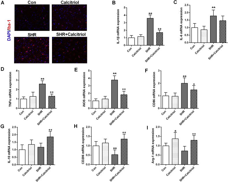
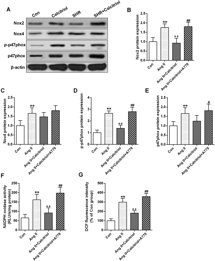
Hypertension is one of the major predisposing factors for neurodegenerative disease characterized with activated renin-angiotensin system (RAS) in both periphery and brain. Vitamin D (VitD) is recently recognized as a pleiotropic hormone with strong neuroprotective properties. While multiple lines of evidence suggest that VitD can act on RAS, the evidence concerning the crosstalk between VitD and RAS in the brain is limited. Therefore, this study aims to evaluate whether VitD can modulate brain RAS to trigger neuroprotective actions in the brain of spontaneously hypertensive rats (SHR). Our data showed that calcitriol treatment induced VDR expression and inhibited neural death in the prefrontal cortex of SHR. Sustained calcitriol administration also inhibited microglia M1 polarization, but enhanced M2 polarization, accompanied with decreased expression of proinflammatory cytokines. We then further explored the potential mechanisms and showed that SHR exhibited overactivated classical RAS with increased expression of angiotensin II (Ang II) receptor type 1 (AT1), angiotensin converting enzyme (ACE) and Ang II production, whereas the counteracting arm of traditional RAS, ACE2/Ang(1-7)/MasR, was impaired in the SHR brain. Calcitriol nonsignificantly suppressed AT1 and ACE but markedly reduced Ang II formation. Intriguingly, calcitriol exerted pronouncedly impact on ACE2/Ang(1-7)/MasR axis with enhanced expression of ACE2, MasR and Ang(1-7) generation. Meanwhile, calcitriol ameliorated the overactivation of NADPH-oxidase (Nox), the downstream of RAS, in SHR, and also mitigated oxidative stress. In microglial (BV2) cells, we further found that calcitriol induced ACE2 and MasR with no significant impact on ACE and AT1. In accordance, calcitriol also attenuated Ang II-induced Nox activation and ROS production, and shifted the microglia polarization from M1 to M2 phenotype. However, co-treatment with A779, a specific MasR antagonist, abrogated the antioxidant and neuroimmune modulating actions of VitD. These findings strongly indicate the involvement of ACE2/Ang(1-7)/MasR pathway in the neuroprotective mechanisms of VitD in the hypertensive brain.

Keywords: ACE2/Ang(1–7)/MasR axis; Neuroinflammation; Oxidative stress; Renin-angiotensin system; Vitamin D.


Figures

Image 1
Graphical abstract
Fig. 1
Fig. 1
Neuroprotective effects of calcitriol on hypertensive brain. Representative images of immunofluorescence assays of VDR (A) and VDR Western blot analysis (B) in the prefrontal cortex. (C) Representative images of Tunel and Nissl staining. Statistical graphs of Tunel (D) and Nissl (E) staining. 100 ng/kg calcitriol was administrated daily for 6 weeks in both control (Con) and SHR animals. Data are means ± SD (n = 7). **p < 0.01 compared to control group. ++p < 0.01 compared to SHR group.
Fig. 2
Fig. 2
Anti-inflammatory effects of calcitriol on hypertensive brain. Representative images of immunofluorescence assays of Iba-1 (A). Expression of proinflammatory M1 mediators, IL-1β (B), IL-6 (C), TNFα (D), iNOS (E) and CD86 (F), and immunoregulatory M2 mediators, IL-10 (G), CD206 (H) and Arg-1 (I). 100 ng/kg calcitriol was administrated daily for 6 weeks in both control (Con) and SHR animals. Data are means ± SD (n = 7). *p < 0.05, **p < 0.01 compared to control group. +p < 0.05, ++p < 0.01 compared to SHR group.
Fig. 3
Fig. 3
The impacts of calcitriol on RAS in the hypertensive brain. Protein expression or concentrations of ACE (A), Ang II (B), AT1 (C), ACE2 (D), Ang (1–7) (E) and MasR (F). 100 ng/kg calcitriol was administrated daily for 6 weeks in both control (Con) and SHR animals. Data are means ± SD (n = 7). *p < 0.05, **p < 0.01 compared to control group. ++p < 0.01 compared to SHR group.
Fig. 4
Fig. 4
The impacts of calcitriol on protein expression and activity of Nox in the hypertensive brain. Representative Western blot (A) and statistical graphs of protein expression of Nox2 (B), p-p47phox (C), p47phox (D) and Nox4 (E), and total Nox activity (F). 100 ng/kg calcitriol was administrated daily for 6 weeks in both control (Con) and SHR animals. Data are means ± SD (n = 7). **p < 0.01 compared to control group. +p < 0.05, ++p < 0.01 compared to SHR group.
Fig. 5
Fig. 5
Anti-oxidative effects of calcitriol in the hypertensive brain. Representative images (A) and statistical graph (B) of immunofluorescence assays of DHE. Concentrations of lipid peroxidation product, MDA (C) and activity of the antioxidant enzymes, SOD (D) and CAT (E). 100 ng/kg calcitriol was administrated daily for 6 weeks in both control (Con) and SHR animals. Data are means ± SD (n = 7). **p < 0.01 compared to control group. +p < 0.05, ++p < 0.01 compared to SHR group.
Fig. 6
Fig. 6
The impacts of calcitriol treatment on VDR activation and RAS in microglia BV2 cells. Representative western blots (A) and statistical graphs of protein expression of VDR (B), AT1 (C), ACE (D), ACE2 (E) and MasR (F). 1  μM calcitriol was pre-treated for 30min before exposure to 100 nM Ang II for 24 h. Data are means ± SD (n = 6). **p < 0.01 compared to control group. ++p < 0.01 compared to SHR group.
Fig. 7
Fig. 7
The impacts of calcitriol treatment on Nox signaling and cellular superoxide generation in microglia BV2 cells. Representative western blots (A) and statistical graphs of protein expression of Nox2 (B), Nox4 (C), p-p47phox (D) and p47phox (E). NADPH oxidase activity (F) and ROS generation (DCF fluorescence intensity) (G). 1  μM calcitriol was pre-treated for 30min before exposure to 100 nM Ang II for 24 h. 1 μM A779 was added simultaneously with Ang II to block MasR. Data are means ± SD (n = 6). **p < 0.01 compared to control group. ++p < 0.01 compared to Ang II treated group. ##p < 0.05, ##p < 0.01 compared to Ang II + Calcitriol treated group.
Fig. 8
Fig. 8
The impacts of calcitriol treatment on microglial polarization in BV2 cells. Expression of proinflammatory M1 mediators, IL-1β (A), IL-6 (B), TNFα (C), CD86 (D) and iNOS (E), and immunoregulatory M2 mediators, Arg-1 (F) and CD206 (G). Representative images of immunofluorescence assays of CD86 (red) and CD206 (green) (H). 1  μM calcitriol was pre-treated for 30min before exposure to 100 nM Ang II for 24 h. 1 μM A779 was added simultaneously with Ang II to block MasR. Data are means ± SD (n = 6). *p < 0.05, **p < 0.01 compared to control group. ++p < 0.01 compared to Ang II treated group. ##p < 0.01 compared to Ang II + Calcitriol treated group. (For interpretation of the references to colour in this figure legend, the reader is referred to the Web version of this article.)
Supplementary Fig. 1
The impacts of calcitriol treatment on body weight growth and blood pressure. Body weight was measured weekly (A). Systolic blood pressure (B) and heart rate (C) were measured at the end of the experiment. 100ng/kg calcitriol was administrated daily for 6 weeks in both control (Con) and SHR animals. Data are means ± SD (n=10). **p < 0.01 compared to control group; +p < 0.05 compared to SHR group
Supplementary Fig. 2
VDR/AT1 and VDR/MasR positive cells in prefrontal cortex. Double immunofluorescence labeling for VDR (red) and AT1 (green) (A) or VDR (red) and MasR (green) (B), respectively

References

    1. Arnold A.C., Gallagher P.E., Diz D.I. Brain renin-angiotensin system in the nexus of hypertension and aging. Hypertens. Res. : Off. J. Jpn. Soc. Hypertens. 2013;36(1):5–13. - PMC - PubMed
    1. Farag E., Sessler D.I., Ebrahim Z., Kurz A., Morgan J., Ahuja S., Maheshwari K., John Doyle D. The renin angiotensin system and the brain: new developments. J. Clin. Neurosci. : Off. J. Neurosurg. Soc. Australas. 2017;46:1–8. - PubMed
    1. Labandeira-Garcia J.L., Rodriguez-Perez A.I., Garrido-Gil P., Rodriguez-Pallares J., Lanciego J.L., Guerra M.J. Brain renin-angiotensin system and microglial polarization: implications for aging and neurodegeneration. Front. Aging Neurosci. 2017;9:129. - PMC - PubMed
    1. Gironacci M.M., Cerniello F.M., Longo Carbajosa N.A., Goldstein J., Cerrato B.D. Protective axis of the renin-angiotensin system in the brain. Clin. Sci. 2014;127(5):295–306. - PubMed
    1. Brocca M.E., Pietranera L., Meyer M., Lima A., Roig P., de Kloet E.R., De Nicola A.F. Mineralocorticoid receptor associates with pro-inflammatory bias in the hippocampus of spontaneously hypertensive rats. J. Neuroendocrinol. 2017;29(7) - PubMed

Publication types

MeSH terms