. 2019 Aug;18(2):990-996.
doi: 10.3892/ol.2019.10446. Epub 2019 Jun 7.

Primary osteosarcoma in elderly patients: A report of three cases

Affiliations

Primary osteosarcoma in elderly patients: A report of three cases

Qiang Xu et al. Oncol Lett. 2019 Aug.

Abstract

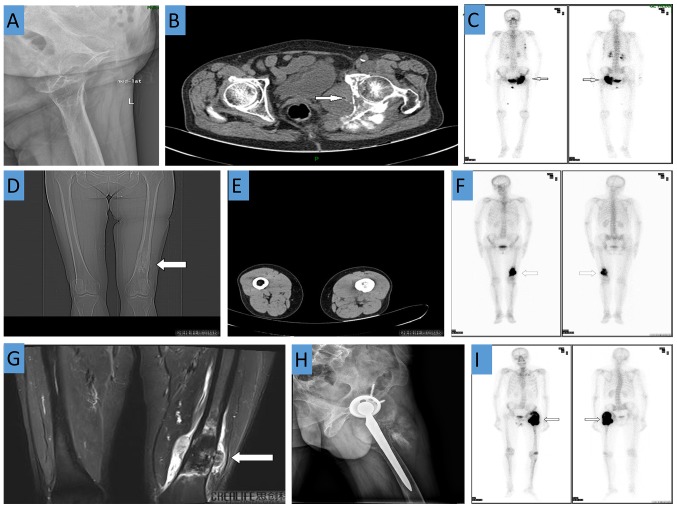
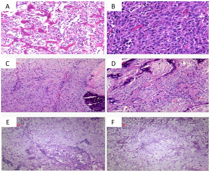
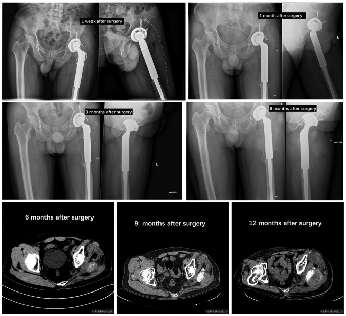
Osteosarcoma is the most common type of primary malignant bone tumor in children and young adults. However, primary osteosarcoma in elderly patients is rare. The present study reports 3 cases of advanced osteosarcoma in elderly patients. The pathological findings in all 3 cases confirmed the diagnosis of primary osteosarcoma. Notably, each patient received different treatment options. Chemoradiotherapy was recommended in case 1 due to the age of the patient. However, the patient requested to be discharged and was lost to follow-up. Conversely, in case 2, the 62-year-old female patient underwent systemic chemotherapy, but no surgical treatment, and in case 3, the 51-year-old male patient underwent complete tumor resection and received systemic chemotherapy for late tumor recurrence. Early diagnosis of osteosarcoma in elderly patients is difficult, and misdiagnosis or a missed diagnosis is common. In clinical practice, bone tumors in elderly patients should be investigated carefully. Imaging examinations are essential for diagnosis, and biopsy is required for confirmation. However, the efficacy of chemotherapy for elderly patients with primary osteosarcoma remains uncertain. Collectively, due to the small number of reports of osteosarcoma in the elderly population, the 3 cases in the present study raise awareness of this rare condition.

Keywords: chemoradiotherapy; elderly patients; imaging; osteosarcoma; pathology; primary malignant bone tumor; surgery.


Figures

Figure 1.
Figure 1.
Case 1: (A) X-ray revealed bony changes in the upper left suprapubic branch, ischium and acetabulum; (B) CT demonstrated irregular bone destruction of the left acetabulum and ischium and adjacent soft tissue swelling; (C) A whole body bone scan revealed increased bone metabolism in the left acetabulum, ischium, femoral head, left fourth and fifth ribs and T8 vertebra. Case 2: CT demonstrates a soft tissue mass shadow and a bony mass shadow attached to the surface of the bone at the distal end of the left femur. (D) Coronal view. (E) Transverse view. (F) Increased bone metabolism at the distal end of the left femur. (G) MRI of the distal femur shows irregular bone destruction with an abnormal signal shadow. Case 3: (H) X-ray shows that after left artificial hip joint replacement, a soft tissue mass shadow is visible in the upper left femur; (I) A whole body bone scan revealed increased bone metabolism in the left upper femur.
Figure 2.
Figure 2.
Post-operative histological observations. (A and B) Case 1, diffuse distribution of tumor cells between the trabecular bone and tumor cells in fibrous tissue and muscle tissue are distributed in patches and reveal infiltrating growth (A, HE staining; magnification, ×100; B, HE staining; magnification, ×200). (C and D) Case 2, spindle cells in the tissue proliferating in bundles or weave patterns, adherent peripheral osteoblasts, and calcification were observed (HE staining; magnification, ×100). (E and F) Case 3, a large number of irregular bone-like structures are arranged in trabecular or reticulated patterns, and damaged bone tissue and immature cartilage (HE staining; magnification, ×100). HE, hematoxylin and eosin.
Figure 3.
Figure 3.
X-ray or CT imaging data at patient follow-up in case 3.

References

    1. Kansara M, Teng MW, Smyth MJ, Thomas DM. Translational biology of osteosarcoma. Nat Rev Cancer. 2014;14:722–735. doi: 10.1038/nrc3838. - DOI - PubMed
    1. Poletajew S, Fus L, Wasiutynski A. Current concepts on pathogenesis and biology of metastatic osteosarcoma tumors. Ortop Traumatol Rehabil. 2011;13:537–545. doi: 10.5604/15093492.971038. (In English, Polish) - DOI - PubMed
    1. Centrella M, Horowitz MC, Wozney JM, McCarthy TL. Transforming growth factor-beta gene family members and bone. Endocr Rev. 1994;15:27–39. doi: 10.1210/edrv-15-1-27. - DOI - PubMed
    1. Bismar H, Klopinger T, Schuster EM, Balbach S, Diel I, Ziegler R, Pfeilschifter J. Transforming growth factor beta (TGF-beta) levels in the conditioncd mcdia of human bone cclls relationship to donor age bone volvme and cconcentration of TGF-beta in human bone matrix in vivo. Bone. 1999;24:565–569. doi: 10.1016/S8756-3282(99)00082-4. - DOI - PubMed
    1. Savage SA, Mirabello L. Using epidemiology and genomics to understand osteosarcoma etiology. Sarcoma. 2011;2011:548151. doi: 10.1155/2011/548151. - DOI - PMC - PubMed