Review
. 2019 Jul 2;4(7):430-444.
doi: 10.1302/2058-5241.4.180076. eCollection 2019 Jul.

Lisfranc fracture-dislocations: current management

Affiliations
Review

Lisfranc fracture-dislocations: current management

Inmaculada Moracia-Ochagavía et al. EFORT Open Rev. .

Abstract

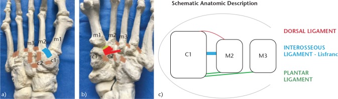
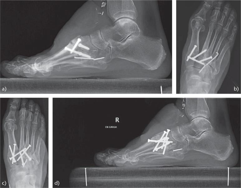
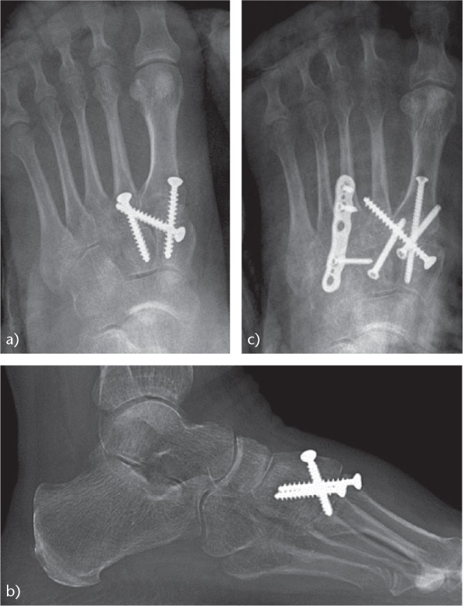
It is essential to know and understand the anatomy of the tarsometatarsal (TMT) joint (Lisfranc joint) to achieve a correct diagnosis and proper treatment of the injuries that occur at that level.Up to 20% of Lisfranc fracture-dislocations go unnoticed or are diagnosed late, especially low-energy injuries or purely ligamentous injuries. Severe sequelae such as post-traumatic osteoarthritis and foot deformities can create serious disability.We must be attentive to the clinical and radiological signs of an injury to the Lisfranc joint and expand the study with weight-bearing radiographs or computed tomography (CT) scans.Only in stable lesions and in those without displacement is conservative treatment indicated, along with immobilisation and initial avoidance of weight-bearing.Through surgical treatment we seek to achieve two objectives: optimal anatomical reduction, a factor that directly influences the results; and the stability of the first, second and third cuneiform-metatarsal joints.There are three main controversies regarding the surgical treatment of Lisfranc injuries: osteosynthesis versus primary arthrodesis; transarticular screws versus dorsal plates; and the most appropriate surgical approach.The surgical treatment we prefer is open reduction and internal fixation (ORIF) with transarticular screws or with dorsal plates in cases of comminution of metatarsals or cuneiform bones. Cite this article: EFORT Open Rev 2019;4:430-444. DOI: 10.1302/2058-5241.4.180076.

Keywords: Lisfranc fracture-dislocation; Lisfranc joint; diagnosis; results; tarsometatarsal joint; treatment.


Conflict of interest statement

ICMJE Conflict of interest statement: The author declares no conflict of interest relevant to this work.

Figures

Fig. 1
Fig. 1
Mechanism of indirect injury in fracture-dislocations of the Lisfranc joint [tarsometatarsal (TMT)] joint: longitudinal force with the foot in plantar flexion.
Fig. 2
Fig. 2
Anatomy of the TMT joint: (a) Dorsal view. In blue, dorsal TMT ligament first cuneiform to second metatarsal (c1-m2). In brown, inter-metatarsal ligaments, which do not exist between the first and second metatarsals (m1-m2). (b) Plantar view. In red, plantar TMT ligament; in green, interosseous ligament (ligament of Lisfranc), exclusive between the first cuneiform and the second metatarsal (c1-m2). (c) Schematic anatomic description.
Fig. 3
Fig. 3
Open fracture of the Lisfranc and Chopart joints produced in a traffic accident (high-energy mechanism).
Fig. 4
Fig. 4
Radiological study of a lesion of the Lisfranc joint: (a) Anteroposterior (AP) radiograph. Note the discontinuity of the medial cortex of the second metatarsal (m2) with the medial cortical of the second cuneiform (c2) (yellow and red lines). (b) ‘Fleck sign’, fracture-avulsion of the Lisfranc ligament (circle). (c) Internal oblique radiograph, showing continuity of the medial cortex of the cuboid and the medial cortex of the fourth metatarsal (m4) (red line). (d) Lateral radiograph showing dorsal dislocation of the metatarsals (red lines).
Fig. 5
Fig. 5
Study of the Lisfranc joint by means of CT scan: (a) CT scan allows an accurate description of subtle lesions of the TMT joint. (b) Comminution of the cuneiforms and bases of the metatarsals. Increased space between the first and second metatarsals, and fracture-avulsion of the Lisfranc ligament (‘fleck sign’).
Fig. 6
Fig. 6
The 1986 Myerson classification for Lisfranc fracture-dislocations.
Fig. 7
Fig. 7
Plantar ecchymosis is a pathognomonic sign of Lisfranc injury.
Fig. 8
Fig. 8
Treatment protocol recommended by us for fracture-dislocations of the Lisfranc joint.
Fig. 9
Fig. 9
Position that we usually use on the surgical table to facilitate the placement of the osteosynthesis material.
Fig. 10
Fig. 10
Intra-operative images: (a) note the separation between the first and second metatarsals (black arrow) that causes instability due to rupture of the Lisfranc ligament complex (black line). (b) Reduction and closure of the first intermetatarsal space.
Fig. 11
Fig. 11
Osteosynthesis of a Lisfranc lesion: (a) comminuted fracture of the base of the second metatarsal; (b) the first inter-metatarsal space was reduced with a Lisfranc screw and fixed with a dorsal plate on the second cuneiform-metatarsal joint.
Fig. 12
Fig. 12
Surgical treatment of Lisfranc lesion: (a) comminuted fracture of the second, third and fourth metatarsal bases. Although there was no clear increase in inter-metatarsal space, there was ligamentous instability. (b) Post-operative anteroposterior (AP) projection. ORIF of the first column was performed and stabilisation of the second and third rays with a Lisfranc screw and dorsal plates. Osteosynthesis of the base of the fourth metatarsal was also performed. The joint between the fourth and fifth metatarsals and the cuboid were not fixed, given that they are articulations of adaptation to the ground and must have mobility. (c) Post-operative lateral projection.
Fig. 13
Fig. 13
Painful post-traumatic OA after a non-anatomical reduction of a Lisfranc injury. Arthrodesis of the Lisfranc joint was performed with complete relief of symptoms: (a) Lateral view before the arthrodesis; (b) AP radiograph before the arthrodesis; (c) AP view after the arthrodesis; (d) lateral radiograph after the arthrodesis.
Fig. 14
Fig. 14
Another case of post-traumatic OA of the Lisfranc joint due to a non-anatomical reduction associated with instability of Lisfranc joint: (a) AP view before the arthrodesis; (b) lateral radiograph before the arthrodesis; (c) radiograph after the arthrodesis. The result was satisfactory.

References

    1. Cassebaum WH. Lisfranc fracture-dislocations. Clin Orthop Relat Res 1963;30(30):116–129. - PubMed
    1. Desmond EA, Chou LB. Current concepts review: Lisfranc injuries. Foot Ankle Int 2006;27(8):653–660. - PubMed
    1. Myerson MS, Cerrato R. Current management of tarsometatarsal injuries in the athlete. Instr Course Lect 2009;58:583–594. - PubMed
    1. Stavlas P, Roberts CS, Xypnitos FN, Giannoudis PV. The role of reduction and internal fixation of Lisfranc fracture-dislocations: a systematic review of the literature. Int Orthop 2010;34(8):1083–1091. - PMC - PubMed
    1. Benirschke SK, Meinberg E, Anderson SA, Jones CB, Cole PA. Fractures and dislocations of the midfoot: Lisfranc and Chopart injuries. J Bone Joint Surg [Am] 2012;94(14):1325–1337. - PubMed

LinkOut - more resources