. 2020 Jan;1460(1):43-56.
doi: 10.1111/nyas.14208. Epub 2019 Aug 18.

Progranulin promotes diabetic fracture healing in mice with type 1 diabetes

Affiliations

Progranulin promotes diabetic fracture healing in mice with type 1 diabetes

Jianlu Wei et al. Ann N Y Acad Sci. 2020 Jan.

Abstract

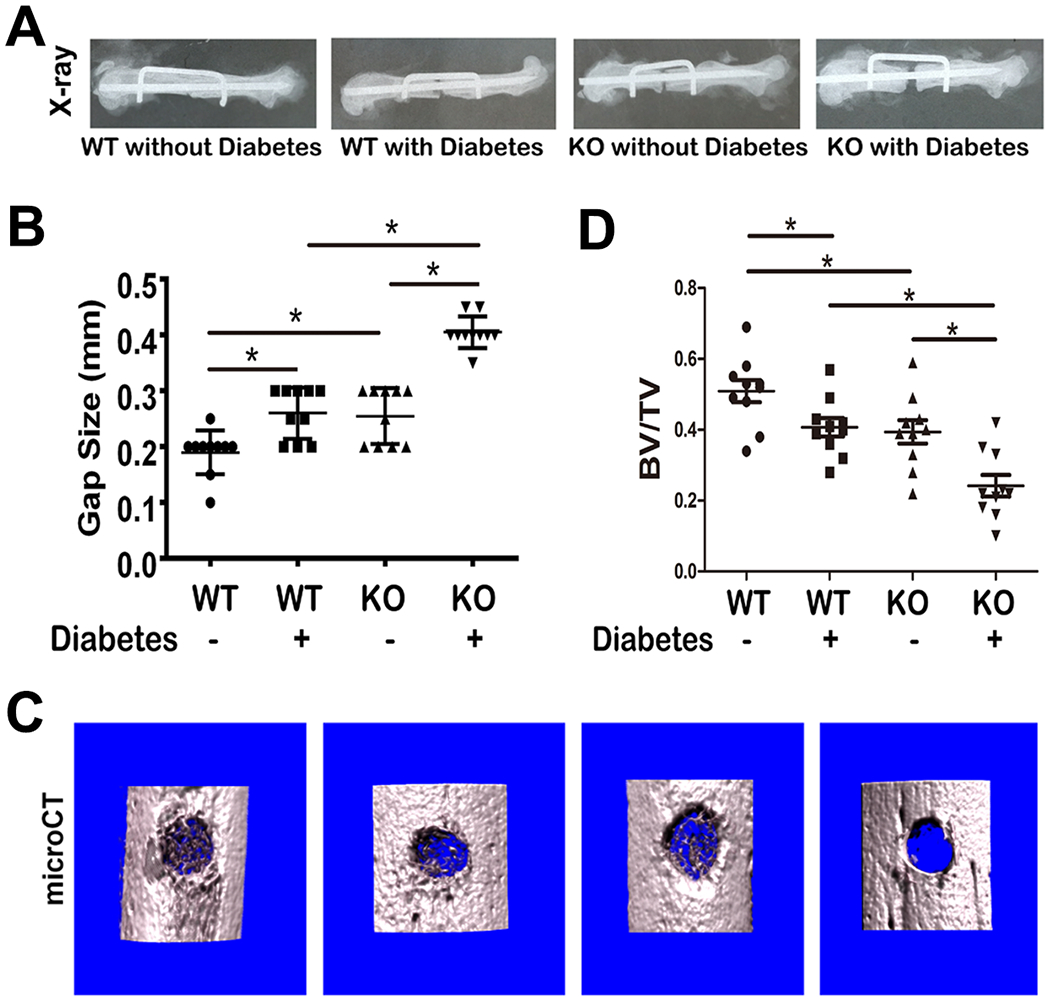
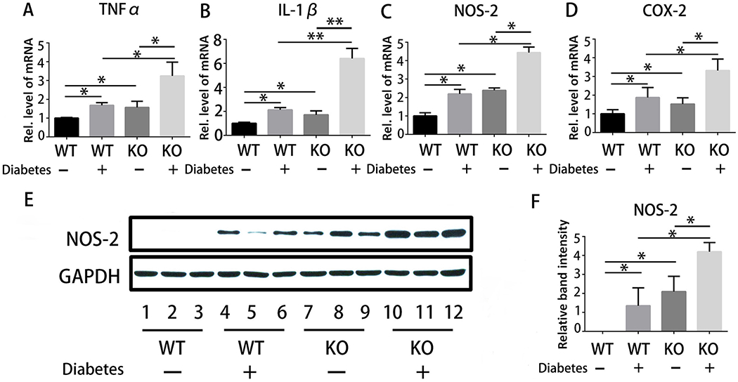
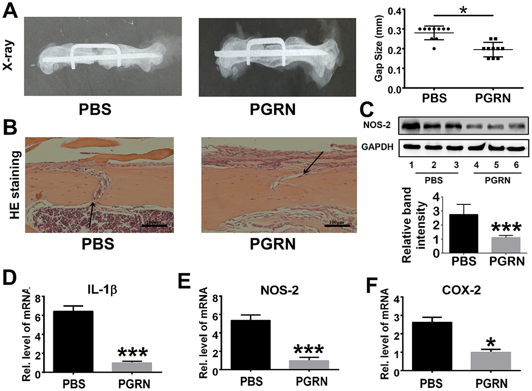
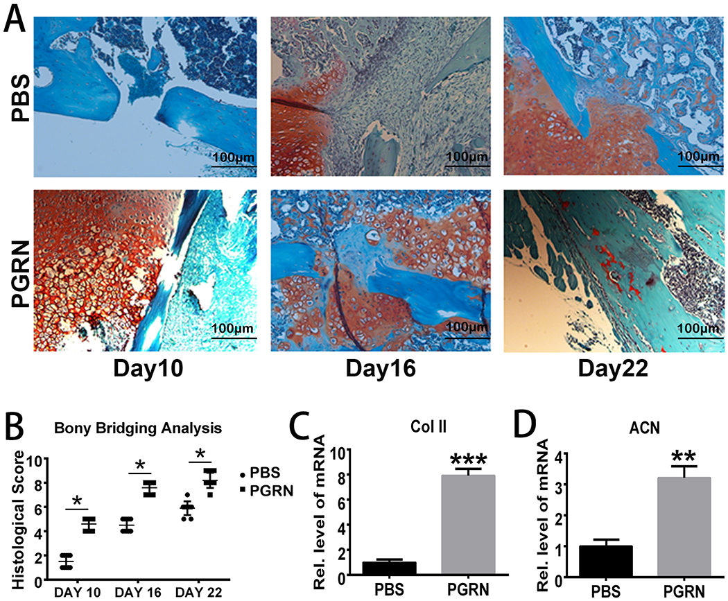
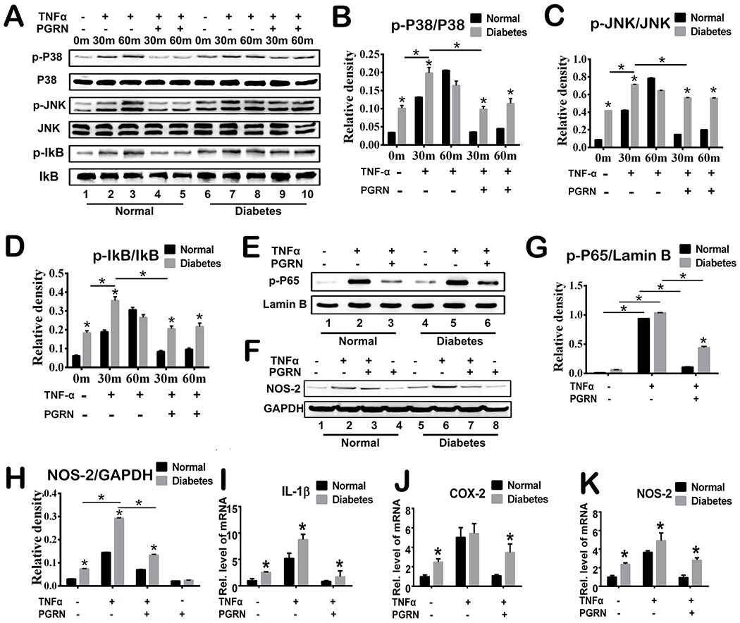
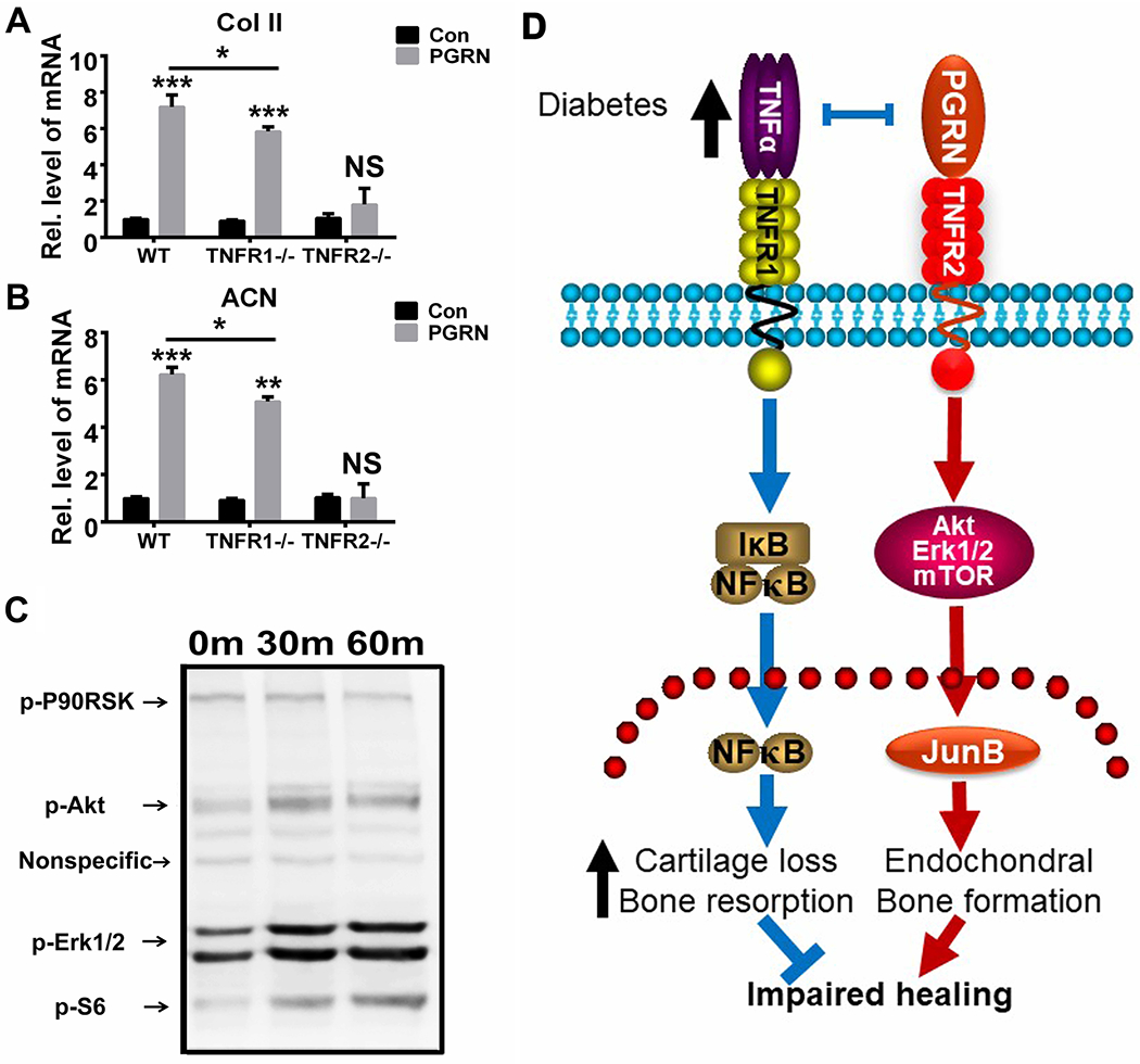
Type 1 diabetes mellitus (T1DM) is an autoimmune disease characterized by insulin deficiency, and patients with diabetes have an increased risk of bone fracture and significantly impaired fracture healing. Proinflammatory cytokine tumor necrosis factor-alpha is significantly upregulated in diabetic fractures and is believed to underlie delayed fracture healing commonly observed in diabetes. Our previous genetic screen for the binding partners of progranulin (PGRN), a growth factor-like molecule that induces chondrogenesis, led to the identification of tumor necrosis factor receptors (TNFRs) as the PGRN-binding receptors. In this study, we employed several in vivo models to ascertain whether PGRN has therapeutic effects in diabetic fracture healing. Here, we report that deletion of PGRN significantly delayed bone fracture healing and aggravated inflammation in the fracture models of mice with T1DM. In contrast, recombinant PGRN effectively promoted diabetic fracture healing by inhibiting inflammation and enhancing chondrogenesis. In addition, both TNFR1 proinflammatory and TNFR2 anti-inflammatory signaling pathways are involved in PGRN-stimulated diabetic fracture healing. Collectively, these findings illuminate a novel understanding concerning the role of PGRN in diabetic fracture healing and may have an application in the development of novel therapeutic intervention strategies for diabetic and other types of impaired fracture healing.

Keywords: TNFR1; TNFR2; impaired fracture healing; progranulin; type 1 diabetes.


Conflict of interest statement

Conflict of interest

We herein declare that we have no conflict of interest.

Figures

Fig.1
Fig.1. Deletion of PGRN further delays diabetic bone fracture healing.
(A) Bone healing of WT with or without diabetes, and PGRN−/− mice with or without diabetes, as indicated, after 4 weeks of establishment of femoral segmental bone defect model, assayed by X-ray. (B) Residual gap size based on radiology results in panel A. (C) Representative microCT image of drill-hole model. (D) Quantification of BV/TV, based on microCT. Each group contained 10 mice. Student’s t-test was used for the statistical analysis.
Fig.2
Fig.2. Deletion of PGRN accelerates inflammation in diabetic fracture healing.
(A-D) Real-time PCR assay for transcriptional levels of pro-inflammatory cytokines, including TNFα, IL-1β, COX-2 and NOS-2, in the drill-hole model; n=10. (E) Expression of NOS-2 in WT without diabetes, WT with diabetes, PGRN KO without diabetes and PGRN KO with diabetes in the drill-hole model, assayed by immunoblotting. (F) Quantitative analysis of immunoblotting in (E), assayed by ImageJ program. Student’s t-test analysis was used for the statistical analysis. The values are the mean ±SD . *p < 0.05 vs. control group.
Fig.3
Fig.3. Recombinant PGRN promotes diabetic bone fracture healing by inhibiting inflammation.
(A) Recombinant PGRN significantly enhanced diabetic bone regeneration in WT mice subjected to the femoral segmental bone defect model, assayed by X-ray. Residual gap size was quantified based on radiology results. (B) HE staining of PBS- and PGRN-treated diabetic drill-hole model. Black arrows indicate bone gap. (C) Expression and quantification of NOS-2 in callus of PBS-/PGRN-treated diabetic drill-hole model mice assayed by Western blot and ImageJ program. (D-F) Transcriptional levels of IL-1β, NOS-2 and COX-2 from the callus of drill-hole model. Each group contained 10 mice. Student’s t-test was used for the statistical analysis. The values are the mean ±SD. *p < 0.05 and ***p < 0.001 vs. control group.
Fig.4
Fig.4. Recombinant PGRN promotes diabetic bone fracture healing by accelerating chondrogenesis.
(A) Safranin O staining of Bonnarens and Einhorn model in diabetic bone fracture healing at various time points. (B) Quantification of bony bridging analysis based on the Safranin O staining. Each group contained 10 mice. Two way ANOVA was used for the statistical analysis. (C-D) Transcriptional levels of Collagen II (Col II) and aggrecan (ACN) in PBS-/PGRN- treated diabetic callus on Day 10. Student’s t-test was used for the statistical analysis. The values are the mean ±SD. **p < 0.01 and ***p < 0.001 vs. control group.
Fig.5
Fig.5. PGRN promotes diabetic fracture healing by inhibiting TNFα-mediated catabolic responses.
(A) Primary mouse bone marrow cells were incubated with or without TNFα (10ng/ml) in the presence or absence of PGRN (200 ng/ml) for 48 hours, and phosphorylation and expression of the indicated signaling molecules were determined by Western Blotting. (B-D) Relative band density of p-P38/P38, p-JNK/JNK and p-IkB/IkB based on Western Blotting. (E) Phosphorylation and expression of p-65 was determined by Western Blotting assay. Lamin B is employed as loading control. (F) Expression of NOS-2 and GAPDH (serving as a loading control) was determined by Western Blotting assay. (G-H) Relative band density of p-P65/lamin B and NOS-2 based on Western Blotting. (I-K) Primary mouse bone marrow cells were cultured without or with TNFα in absence or presence of PGRN for 6 hours. Transcriptional levels of IL-1β, COX-2 and NOS-2 were determined by Real-time PCR assay. Two-way ANOVA was used for the statistical analysis. The values are the mean ±SD. *p < 0.05 vs. control group.
Fig.6
Fig.6. PGRN promotes chondrogenesis through TNFR2-Akt/Erk1/2/mTOR signaling.
(A&B) Primary mouse bone marrow cells of WT, TNFR1−/− and TNFR2−/− mouse were cultured with or without PGRN (200 ng/ml) for 6 hours, transcriptional levels of type II collagen (Col II) and aggrecan (ACN) were determined by Real-time PCR assay. The values are the mean ±SD. **p < 0.01 and ***p < 0.001 vs. control group. Two-way ANOVA was used for the statistical analysis. (C) Primary mouse bone marrow cells of WT mice were incubated in presence of PGRN, and phosphorylation of the indicated signaling molecules were determined by cocktail scanning. (D) A proposed model for the role of PGRN in diabetic bone fracture healing process.

References

    1. Shaw JE, Sicree RA, Zimmet PZ. 2010. Global estimates of the prevalence of diabetes for 2010 and 2030. Diabetes research and clinical practice 87:4–14 - PubMed
    1. Sellmeyer DE, Civitelli R, Hofbauer LC, Khosla S, Lecka-Czernik B, Schwartz AV. 2016. Skeletal Metabolism, Fracture Risk, and Fracture Outcomes in Type 1 and Type 2 Diabetes. Diabetes 65:1757–66 - PMC - PubMed
    1. Ferrari SL, Abrahamsen B, Napoli N, Akesson K, Chandran M, et al. 2018. Diagnosis and management of bone fragility in diabetes: an emerging challenge. Osteoporos Int 29:2585–96 - PMC - PubMed
    1. Strotmeyer ES, Cauley JA. 2007. Diabetes mellitus, bone mineral density, and fracture risk. Current opinion in endocrinology, diabetes, and obesity 14:429–35 - PubMed
    1. Vestergaard P 2007. Discrepancies in bone mineral density and fracture risk in patients with type 1 and type 2 diabetes—a meta-analysis. Osteoporosis International 18:427–44 - PubMed

Publication types

MeSH terms

Substances