. 2019 Dec;39(12):2379-2391.
doi: 10.1177/0271678X19870583. Epub 2019 Aug 18.

cGMP-dependent protein kinase I in vascular smooth muscle cells improves ischemic stroke outcome in mice

Affiliations

cGMP-dependent protein kinase I in vascular smooth muscle cells improves ischemic stroke outcome in mice

Maria Shvedova et al. J Cereb Blood Flow Metab. 2019 Dec.

Abstract

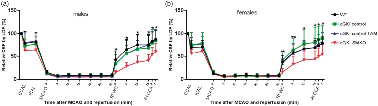
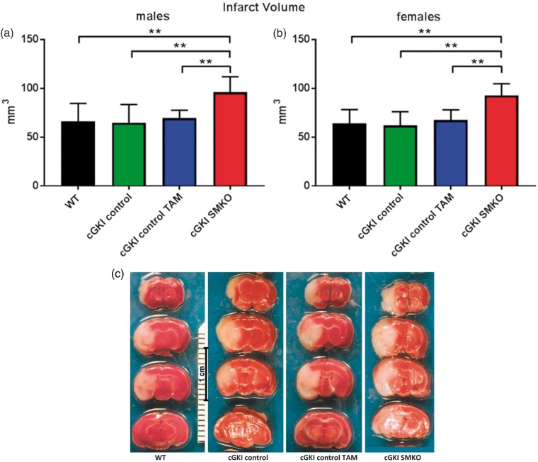
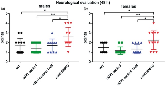
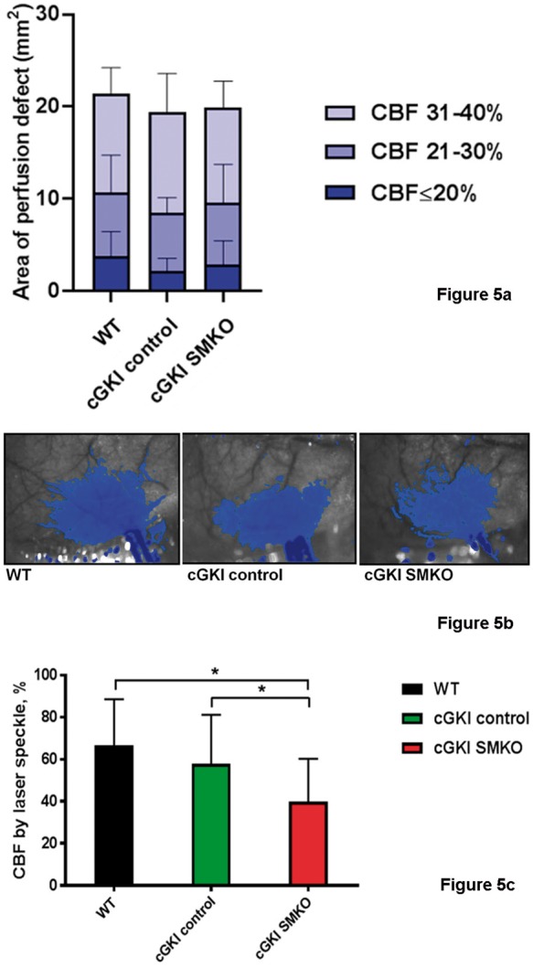
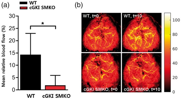
Recent works highlight the therapeutic potential of targeting cyclic guanosine monophosphate (cGMP)-dependent pathways in the context of brain ischemia/reperfusion injury (IRI). Although cGMP-dependent protein kinase I (cGKI) has emerged as a key mediator of the protective effects of nitric oxide (NO) and cGMP, the mechanisms by which cGKI attenuates IRI remain poorly understood. We used a novel, conditional cGKI knockout mouse model to study its role in cerebral IRI. We assessed neurological deficit, infarct volume, and cerebral perfusion in tamoxifen-inducible vascular smooth muscle cell-specific cGKI knockout mice and control animals. Stroke experiments revealed greater cerebral infarct volume in smooth muscle cell specific cGKI knockout mice (males: 96 ± 16 mm3; females: 93 ± 12 mm3, mean±SD) than in all control groups: wild type (males: 66 ± 19; females: 64 ± 14), cGKI control (males: 65 ± 18; females: 62 ± 14), cGKI control with tamoxifen (males: 70 ± 8; females: 68 ± 10). Our results identify, for the first time, a protective role of cGKI in vascular smooth muscle cells during ischemic stroke injury. Moreover, this protective effect of cGKI was found to be independent of gender and was mediated via improved reperfusion. These results suggest that cGKI in vascular smooth muscle cells should be targeted by therapies designed to protect brain tissue against ischemic stroke.

Keywords: Cerebral blood flow; cGMP-dependent protein kinase I; ischemia–reperfusion injury; middle cerebral artery occlusion-reperfusion model; stroke.


Figures

Figure 1.
Figure 1.
(a) Cerebrovascular expression of cGKI in cGKI SMKO and control mice. Posterior communicating arteries exhibit cGKI immunoreactivity in cGKI control and WT mouse in vascular smooth muscle cells (left panels, red color), which are identified by their location within the vessel wall and their reaction with an anti smoothelin antibody in subsequent brain sections (right panels, red color). However, cGKI expression was not detected in smooth muscle cells of cGKI SMKO mice; the anti-cGKI antibody reactivity in smooth muscle cells of these vessels is similar to that detected in cells in sections exposed to non-immune IgG (lower panel). Immunostaining of the SCO shows cGKI immunoreactivity in cGKI SMKO mice, demonstrating the smooth muscle cell-specificity of the cGKI knockout in the mice. Counterstaining was performed with hematoxylin. Original magnification, × 60; the bar is 40 µm-long. (b) Ablation of cGKI in aorta of cGKI SMKO mice. Representative Western blot demonstrating control levels of cGKI in aortae of cGKI control TAM mice and lower levels of cGKI in cGKI SMKO mice.
Figure 2.
Figure 2.
Cerebral blood flow by LDF (%): common carotid artery ligation (CCAL), internal carotid artery ligation (ICAL), middle cerebral artery occlusion (MCAO), partial reperfusion through the Willis circle (Rf WC), the reperfusion through the ipsilateral common carotid artery (Rf CCA) (*p < 0.05; **p < 0.01, WT, cGKI control, and cGKI control TAM mice vs. cGKI SMKO mice).
Figure 3.
Figure 3.
Greater infarct volume in cGKI SMKO mice as compared to all control groups in both males and females subgroups. (a, b) analysis of infarct volumes at 48 h after reperfusion in male and female subgroups (**p < 0.001). (c) TTC staining of representative brain coronal sections at 48 h of reperfusion after 30 min filament MCAO.
Figure 4.
Figure 4.
Greater neurological deficit in cGKI SMKO mice than in control mice in both males and females subgroups (*p < 0.05, **p < 0.01).
Figure 5.
Figure 5.
(a) The area of perfusion defect measured using laser speckle flowmetry. The number of pixels displaying three different severities of intraischemic CBF reduction was quantified using a thresholding paradigm and then converted to area (mm2) by the known scale factor. There was no statistically significant difference among the groups for any of the CBF segments. (b) Representative laser speckle flowmetry images in WT, cGKI control and cGKI SMKO mice 30 min after distal MCAO. Superimposed blue pixels indicate regions with ≤40% residual CBF. Imaging field dimensions are 6 × 8 mm. Imaging field is positioned over the right hemisphere. The long blue towards the bottom of each image is the clip artifact. (c) CBF after 5 min of reperfusion by laser speckle. After reperfusion by removing the microvascular clip occluding the distal middle cerebral artery, CBF was restored to a higher level in WT (n = 8) and cGKI control (n = 9) than cGKI SMKO (n = 9) mice (*p < 0.05).
Figure 6.
Figure 6.
NO-dependent cerebrovascular responses in WT and cGKI SMKO mice. (a) MAHMA NONOate administered intravenously (0.1 mg/kg) increased the mean relative cerebral blood flow (%) in WT mice but the mean relative cerebral blood flow of cGKI SMKO mice was significantly lower than in WT mice. (b) Representative relative blood flow images before (t=0 ) and 10 min (t=10) after injection of MAHMA NONOate (0.1 mg/kg, i.v.) in WT and cGKI SMKO mice. These panels represent relative blood flow images. For each mouse, the max blood flow at t=0 was scaled to 100% and the same scaling factor was applied to the blood flow map at t=10 min after injection of the NO donor. Values are mean ± SD. *p < 0.05 for comparison between WT and cGKI SMKO mice (n = 4 for each group).

References

    1. Francis SH, Corbin JD. Cyclic nucleotide-dependent protein kinases: intracellular receptors for cAMP and cGMP Action. Crit Rev Clin Lab Sci 1999; 36: 275–328. - PubMed
    1. Zhang R, Wang L, Zhang L, et al. Nitric oxide enhances angiogenesis via the synthesis of vascular endothelial growth factor and cGMP after stroke in the rat. Circ Res 2003; 92: 308–313. - PubMed
    1. Chen X, Wang N, Liu Y, et al. Yonkenafil: a novel phosphodiesterase type 5 inhibitor induces neuronal network potentiation by a cGMP-dependent Nogo-R axis in acute experimental stroke. Exp Neurol 2014; 261: 267–277. - PubMed
    1. Pfeifer A, Klatt P, Massberg S, et al. Defective smooth muscle regulation in cGMP kinase I-deficient mice. EMBO J 1998; 17: 3045–3051. - PMC - PubMed
    1. Pilz RB, Broderick KE. Role of cyclic GMP in gene regulation. Front Biosci 2005; 10: 1239–1268. - PubMed

Publication types

MeSH terms

Substances

LinkOut - more resources