. 2020 Jan-Feb;15(1-2):134-144.
doi: 10.1080/15592294.2019.1656156. Epub 2019 Aug 26.

Impaired T cell proliferation by ex vivo BET-inhibition impedes adoptive immunotherapy in a murine melanoma model

Affiliations

Impaired T cell proliferation by ex vivo BET-inhibition impedes adoptive immunotherapy in a murine melanoma model

Jonathan Chee et al. Epigenetics. 2020 Jan-Feb.

Abstract

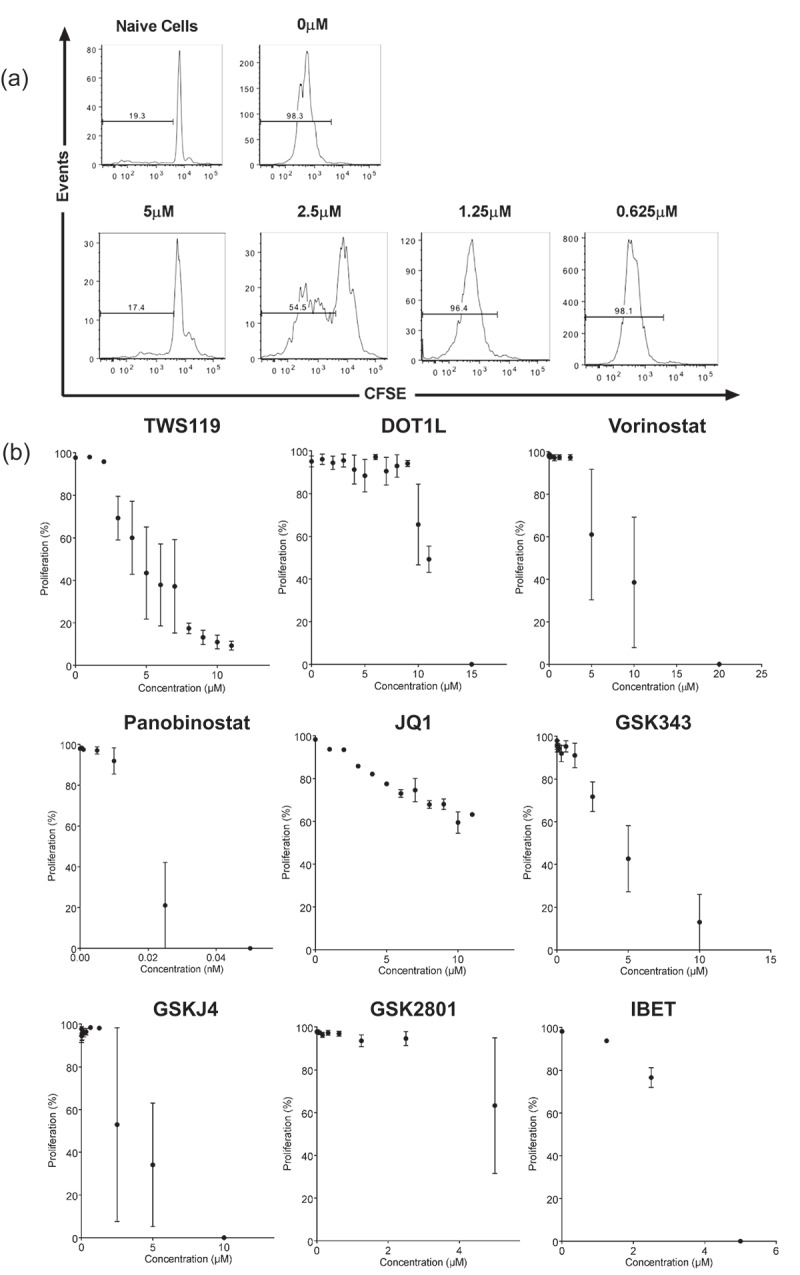
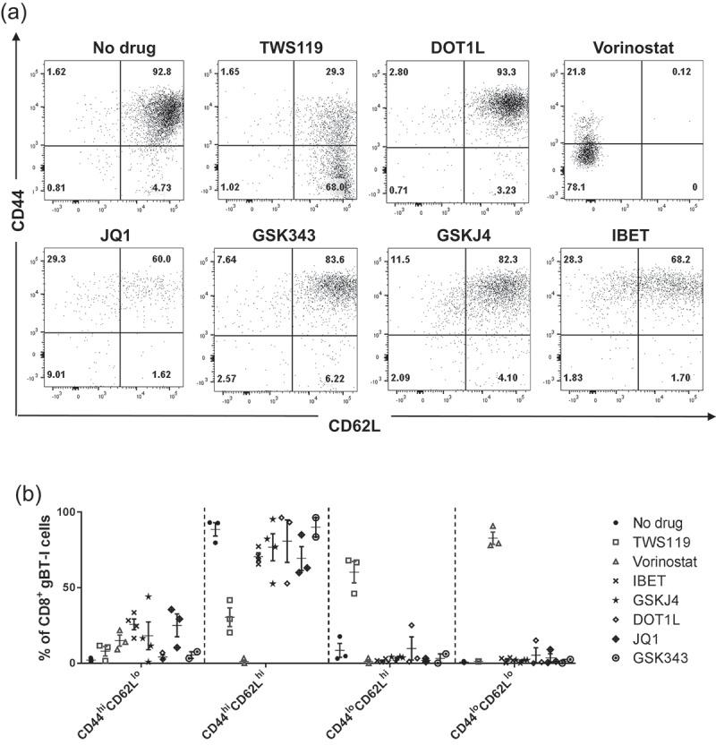
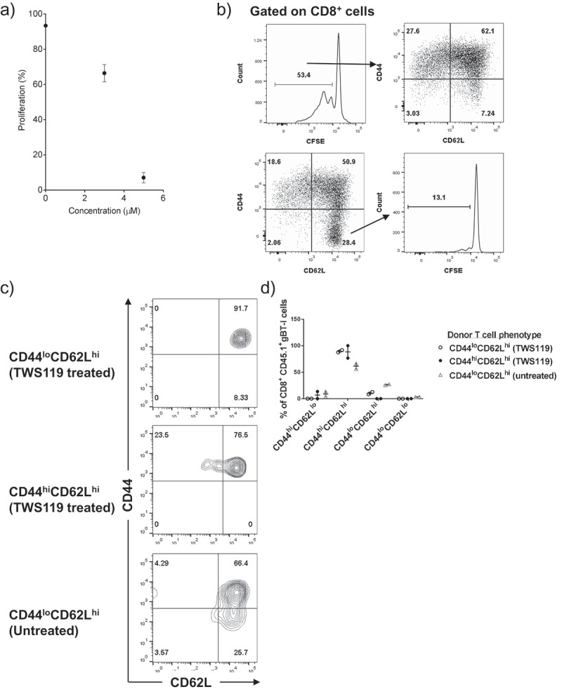
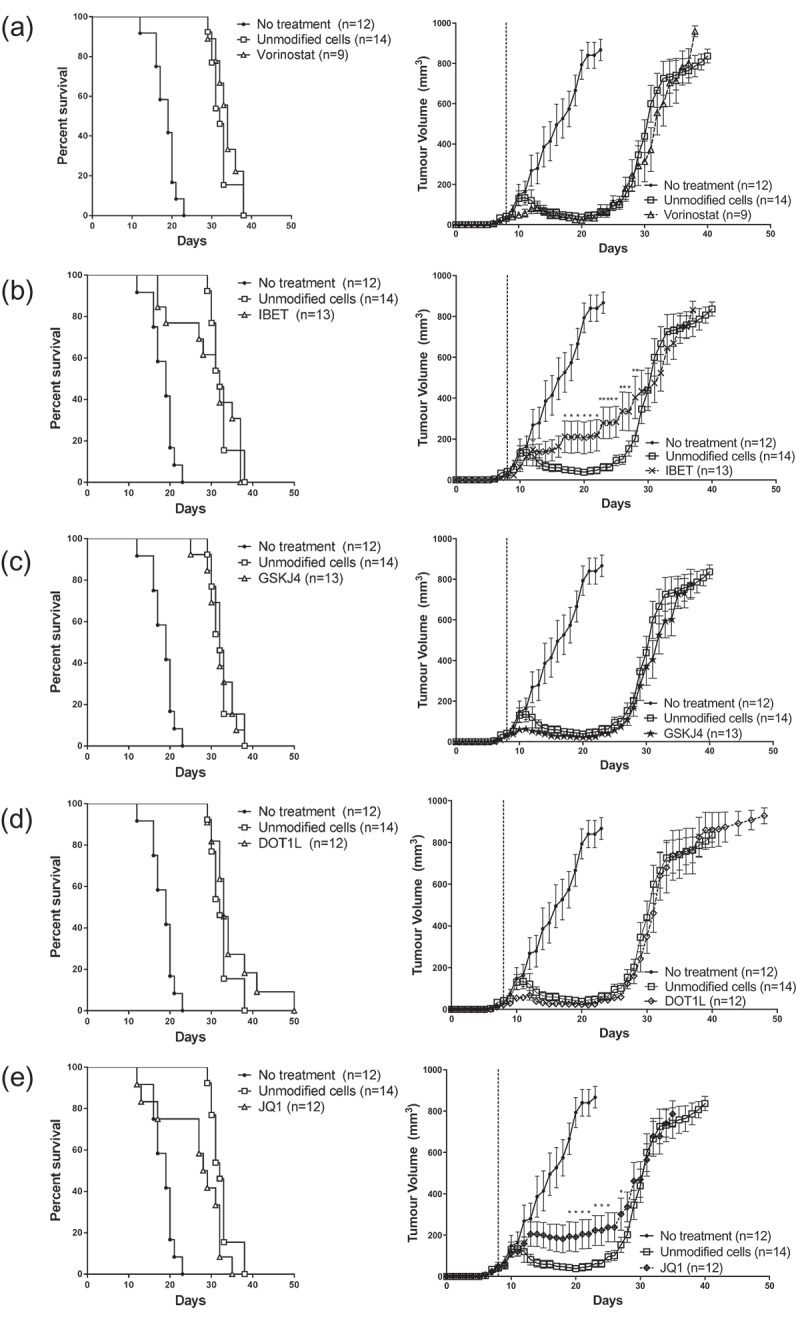
Activation of naïve CD8+ T cells stimulates proliferation and differentiation into cytotoxic T-lymphocytes (CTLs). Adoptive T Cell Therapy (ACT) involves multiple rounds of ex vivo activation to generate enough CTLs for reinfusion into patients, but this drives differentiation into terminal effector T cells. Less differentiated CTL populations, such as stem cell memory T cells, are more ideal candidates for ACT because of increased self-renewal and persistent properties. Ex vivo targeting of T cell differentiation with epigenetic modifiers is a potential strategy to improve cytotoxic T-lymphocyte (CTL) generation for ACT. We established a pipeline to assess the effects of epigenetic modifiers on CD8+ T cell proliferation, differentiation, and efficacy in a preclinical melanoma model. Single treatment with epigenetic modifiers inhibited T cell proliferation in vitro, producing CD44hiCD62Lhi effector-like T cells rather than a stem cell memory T cell phenotype. Most epigenetic modifying agents had no significant effect on ACT efficacy with the notable exception of the bromodomain and extraterminal (BET)-inhibitor JQ1 which was associated with a decrease in efficacy compared to unmodified T cells. These findings reveal the complexity of epigenetic targeting of T cell differentiation, highlighting the need to precisely define the epigenetic targeting strategies to improve CTL generation for ACT.

Keywords: BET inhibition; Cancer immunotherapy; T cell differentiation; adoptive cell therapy; cytotoxic T lymphocytes; melanoma.


Figures

Figure 1.
Figure 1.
T cell proliferation inhibition by epigenetic modifiers in vitro. (a) Representative FACS diagram depicting gBT-I T cell proliferation measured by CFSE dilution 72-h post-activation, in the presence of titrated concentrations of TWS119. Plots are gated on live, CD8+ cells. (b) Dot plots representing the percentage of proliferating (CFSElo) gBT-I T cells in the presence of titrated concentration of epigenetic modifiers, 72 h after activation in a 384-well plate. Each graph represents treatment with one drug. Each datapoint represents the mean ± SD proliferation of three separate experiments.
Figure 2.
Figure 2.
T cell phenotype of activated T cells in the presence of epigenetic modifiers. (a) Representative FACS diagram depicting expression of CD44 and CD62L on activated gBT-I T cells, in the presence or absence of different drugs. Plots are gated on live, CD8+ cells, and are representative of three to four separate experiments. (b) CD44 and CD62L expression represented at dot plots.
Figure 3.
Figure 3.
TWS119 treatment failed to generate a Tscm phenotype on gBT-I T cells. (a) Dot plot representing CFSElo proliferating gBT-I T cells in the presence of TWS119, 72 h after activation in a 6-well plate. Each datapoint represents the mean ± SD proliferation. (b) CD44 and CD62L expression of CFSElo T cells post-activation (top row). CFSE profile of CD44loCD62Lhi cells demonstrates that most cells in this population remain naïve (bottom row). Plots are representative for three separate experiments. (c) TWS119-treated cells were sorted into CD44loCD62Lhi, CD44hiCD62Lhi populations, and untreated CD44loCD62Lhi populations (1 x 10^5 cells) were transferred into a lymphodepleted animal. CD44 and CD62L expression on transferred (CD45.1) cells 4 weeks post-transfer are represented as FACS diagrams, and in (d) a dot plot.
Figure 4.
Figure 4.
ACT with epigenetic modified gBT-I T cells did not improve tumour regression and survival. Tumour growth and survival curves of mice inoculated subcutaneously with B16.gB prior to receiving ACT consisting of activated gBT-I T cells treated with (a) vorinostat, (b) IBET, (c) GSKJ4, (d) DOT1L and (e) JQ1. Graphs represent three separate experiments. Tumour growth between unmodified group and each modified group were compared with a Mixed Model ANOVA. Time points with significant differences in mean tumour volumes are represented with an asterisk (*<0.05, **<0.01). Survival curves were compared with a Log-Rank (Mantel-Cox) test and there was a significant difference between unmodified T cells and untreated animals (p < 0.001). There was no significant difference between each treatment and unmodified T cells, with the exception of JQ1 treated cells. (JQ1 vs unmodified, p = 0.0237).

References

    1. Rosenberg SA, Restifo NP.. Adoptive cell transfer as personalized immunotherapy for human cancer. Science. 2015. April 3;348(6230):62–68. PubMed PMID: 25838374. - PMC - PubMed
    1. Verdegaal EM. Adoptive cell therapy: a highly successful individualized therapy for melanoma with great potential for other malignancies. Curr Opin Immunol. 2016. April;39:90–95. PubMed PMID: 26829458. - PubMed
    1. Khammari A, Labarriere N, Vignard V, et al. Treatment of metastatic melanoma with autologous Melan-A/MART-1-specific cytotoxic T lymphocyte clones. J Invest Dermatol. 2009. December;129(12):2835–2842. PubMed PMID: 19554023. - PubMed
    1. Tran E, Robbins PF, Lu YC, et al. T-cell transfer therapy targeting mutant KRAS in cancer. N Engl J Med. 2016. December 8;375(23):2255–2262. PubMed PMID: 27959684; PubMed Central PMCID: PMCPMC5178827. - PMC - PubMed
    1. Robbins PF, Dudley ME, Wunderlich J, et al. Cutting edge: persistence of transferred lymphocyte clonotypes correlates with cancer regression in patients receiving cell transfer therapy. J Iimmunol. 2004. December 15;173(12):7125–7130. PubMed PMID: 15585832; PubMed Central PMCID: PMCPMC2175171 - PMC - PubMed