Review
. 2019;19(18):1621-1649.
doi: 10.2174/1568026619666190712204603.

Discovery and Development of Anti-HIV Therapeutic Agents: Progress Towards Improved HIV Medication

Affiliations
Review

Discovery and Development of Anti-HIV Therapeutic Agents: Progress Towards Improved HIV Medication

Kenji Maeda et al. Curr Top Med Chem. 2019.

Abstract

The history of the human immunodeficiency virus (HIV)/AIDS therapy, which spans over 30 years, is one of the most dramatic stories of science and medicine leading to the treatment of a disease. Since the advent of the first AIDS drug, AZT or zidovudine, a number of agents acting on different drug targets, such as HIV enzymes (e.g. reverse transcriptase, protease, and integrase) and host cell factors critical for HIV infection (e.g. CD4 and CCR5), have been added to our armamentarium to combat HIV/AIDS. In this review article, we first discuss the history of the development of anti-HIV drugs, during which several problems such as drug-induced side effects and the emergence of drug-resistant viruses became apparent and had to be overcome. Nowadays, the success of Combination Antiretroviral Therapy (cART), combined with recently-developed powerful but nonetheless less toxic drugs has transformed HIV/AIDS from an inevitably fatal disease into a manageable chronic infection. However, even with such potent cART, it is impossible to eradicate HIV because none of the currently available HIV drugs are effective in eliminating occult "dormant" HIV cell reservoirs. A number of novel unique treatment approaches that should drastically improve the quality of life (QOL) of patients or might actually be able to eliminate HIV altogether have also been discussed later in the review.

Keywords: AIDS; Combination antiretroviral therapy; HIV; Integrase inhibitors; Protease inhibitors; Reverse transcriptase inhibitors..


Figures

Fig. (1)
Fig. (1)
HIV-1 replication cycle and anti-HIV-1 agents that target its several steps. Molecular mechanisms of replication cycle (life cycle) are well understood from entry of HIV to generation of new matured viral particles; (i) adsorption and membrane fusion, (ii) reverse transcription, (iii) integration, (iv) processing, (v) assembly, (vi) budding, (vii) maturation, etc. Several anti-HIV drugs have been reported in the last three decades: reverse transcriptase (RT) inhibitors including nucleoside reverse transcriptase inhibitors (NRTIs) and non-nucleoside reverse transcriptase inhibitors (NNRTIs), protease inhibitors (PIs), integrase inhibitors (INIs), entry/fusion inhibitors, etc.
Fig. (2)
Fig. (2)
Chemical structures of HIV-1 nucleoside/nucleotide reverse transcriptase inhibitors (NRTIs).
Fig. (3)
Fig. (3)
Chemical structures of HIV-1 non-nucleoside reverse transcriptase inhibitors (NNRTIs).
Fig. (4)
Fig. (4)
Structure of wild-type HIV-1 RT with EFdA-TP. A: EFdA shown in CPK mode, selected RT residues are shown as thick sticks. Some of these RT residues are implicated in drug resistance, and some (Ala114, Phe160, and Asp185) are in the active site responsible for tight interaction with EFdA [77, 78]. The finger region of the RT is shown as a yellow ribbon, and the palm region is shown in orange. Parts of the other RT domains are shown as white ribbons. B: EFdA-TP in the active site cavity of HIV-1 RT (PDB: 5J2M) [243]. The hydrophobic pocket of the wild-type HIV-1 RT active site and EFdA-TP are shown. The 4ʹ-ethynyl group of EFdA showed good vdW interactions with several residues, such as A114, Y115, F160, M184, and D185, in the active site cavity of RT. EFdA-TP strongly interacts with these residues and shifts positions inside the active site, thus terminating DNA polymerization [78, 244]. C-E: Detailed vdW interactions between the 4ʹ-ethynyl group of EFdA and residues in the active site cavity of HIV-1 RT, namely, Met184 (C), Ala114 (D), and Phe160 (E). These amino acids show good interaction with the 4ʹ-ethynyl group of EFdA. Surface colors: 4ʹ-ethynyl, white; A114, magenta; Y115, green; F160, red; M184, yellow; D185, orange. RT residues are shown as ball and stick, EFdA shown as thick sticks.
Fig. (5)
Fig. (5)
Chemical structures of HIV-1 protease inhibitors (PIs).
Fig. (6)
Fig. (6)
A: Binding of polypeptides in the HIV-1 protease active site. Hydrogen bonds shown as yellow dotted lines. Polypeptide substrates bind to the enzyme in an extended conformation with a minimum of seven amino acid residues interacting with the enzyme, denoted P4 to P1 and P1′ to P4′ by standard nomenclature [14]. B: Structure of HIV-1 protease and an inhibitor (DRV) bound to the active site. Protease (PDB code 4HLA) ribbons in orange and maroon, DRV in green carbons, selected protease residues in gray carbons. Polar interactions are shown by yellow dashed lines.
Fig. (7)
Fig. (7)
Structure of HIV-1 integrase and an inhibitor (DTG) bound to the active site. Figure made from PDB ID: 3S3M. The figure has purple carbons for DNA, gray carbons for protein (yellow ribbons), and green carbons for DTG. Fluorine, phosphorous, oxygen and nitrogen atoms are shown in cyan, orange, red and blue respectively. Magnesium is shown as pink spheres. DTG is represented as thick sticks, the protein and DNA atoms are represented as thin sticks.
Fig. (8)
Fig. (8)
Chemical structures of HIV-1 integrase inhibitors (INSTIs, strand-transfer inhibitors).
Fig. (9)
Fig. (9)
Brief mechanisms of HIV-1 entry and fusion. The HIV-1 envelope protein gp120 interacts with a cell surface protein CD4, resulting in a conformational change of gp120, which subsequently binds to a second receptor, CCR5 or CXCR4. The conformational change of gp120 causes the exposure of another envelope protein gp41 and penetration of its N-terminus through cell membranes, and then the formation of the gp41 trimer-of-hairpins structure, which is referred as a six-helical bundle structure. This bundle structure is formed by a central parallel trimer of the N-terminal helical region (HR1 region) surrounded by the C-terminal helical region (HR2 region) that is oriented in an antiparallel and hairpin fashion. This bundle formation causes fusion of HIV/cell-membranes and results in completion of the infection.
Fig. (10)
Fig. (10)
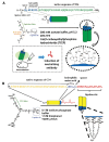
A: Schematic representation of HIV-1 gp41 and amino acid sequences of HR2 region peptides (HIV-1NL4-3 strain). B: Helical wheel representation of the C34 peptide. Numbering of amino acid residues is based on gp41 of the HIV-1NL4-3 strain. C: The design concept of introduction of the Glu-Lys pairs to the solvent-accessible site.
Fig. (11)
Fig. (11)
A: Artificial remodeling of dynamic structures of HR1 regions leads to synthetic antigen molecules inducing neutralizing antibodies. B: Three equivalent HR2 region peptides lead to synthetic fusion inhibitors.
Fig. (12)
Fig. (12)
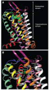
Structure of CCR5 and a CCR5 inhibitor (MVC) bound in the hydrophobic cavity (PDB ID 4MBS). A: Bird’s eye view of MVC (CPK mode) bound within CCR5. B: The interaction of MVC with selected active site residues of CCR5 as determined from a crystal structure. The hydrogen bond interactions are shown by yellow dotted lines. MVC is shown in thick sticks with green carbons. CCR5 residues are shown in ball and stick representation with gray carbons. Nitrogens, oxygens, polar hydrogens and fluorines are in blue, red, white and cyan respectively. The different CCR5 domains in ribbon representation are colored as follows: TM1 (transmembrane domain 1), aquamarine; TM2, Yellow; TM3, Orange; TM4, White; TM5, Pink; TM6, Purple; TM7, Red; N-term, Gray; ECL1 (extracellular domain 1), Blue; ECL2, Maroon; ECL3, Plum. Figure generated using Maestro version 10.7.015.
Fig. (13)
Fig. (13)
Chemical structures of CCR5 inhibitors.
Fig. (14)
Fig. (14)
Structures of Capsid inhibitors.
Fig. (15)
Fig. (15)
Structures of representative small CD4 mimic molecules.
Fig. (16)
Fig. (16)
Chemical structures of HIV-1 latency-reversing agents (LRAs).

References

    1. Barré-Sinoussi F., Chermann J.C., Rey F., Nugeyre M.T., Chamaret S., Gruest J., Dauguet C., Axler-Blin C., Vézinet-Brun F., Rouzioux C., Rozenbaum W., Montagnier L. Isolation of a T-lymphotropic retrovirus from a patient at risk for acquired immune deficiency syndrome (AIDS). Science. 1983;220(4599):868–871. doi: 10.1126/science.6189183. - DOI - PubMed
    1. Gallo R.C., Sarin P.S., Gelmann E.P., Robert-Guroff M., Richardson E., Kalyanaraman V.S., Mann D., Sidhu G.D., Stahl R.E., Zolla-Pazner S., Leibowitch J., Popovic M. Isolation of human T-cell leukemia virus in acquired immune deficiency syndrome (AIDS). Science. 1983;220(4599):865–867. doi: 10.1126/science.6601823. - DOI - PubMed
    1. Mitsuya H., Weinhold K.J., Furman P.A., St Clair M.H., Lehrman S.N., Gallo R.C., Bolognesi D., Barry D.W., Broder S. 3′-Azido-3′-deoxythymidine (BW A509U): An antiviral agent that inhibits the infectivity and cytopathic effect of human T-lymphotropic virus type III/lymphadenopathy-associated virus in vitro. Proc. Natl. Acad. Sci. USA. 1985;82(20):7096–7100. doi: 10.1073/pnas.82.20.7096. - DOI - PMC - PubMed
    1. Yarchoan R., Klecker R.W., Weinhold K.J., Markham P.D., Lyerly H.K., Durack D.T., Gelmann E., Lehrman S.N., Blum R.M., Barry D.W. Administration of 3′-azido-3′-deoxythymidine, an inhibitor of HTLV-III/LAV replication, to patients with AIDS or AIDS-related complex. Lancet. 1986;1(8481):575–580. doi: 10.1016/S0140-6736(86)92808-4. - DOI - PubMed
    1. Graves M.C., Lim J.J., Heimer E.P., Kramer R.A. An 11-kDa form of human immunodeficiency virus protease expressed in Escherichia coli is sufficient for enzymatic activity. Proc. Natl. Acad. Sci. USA. 1988;85(8):2449–2453. doi: 10.1073/pnas.85.8.2449. - DOI - PMC - PubMed

Substances